

Abstract
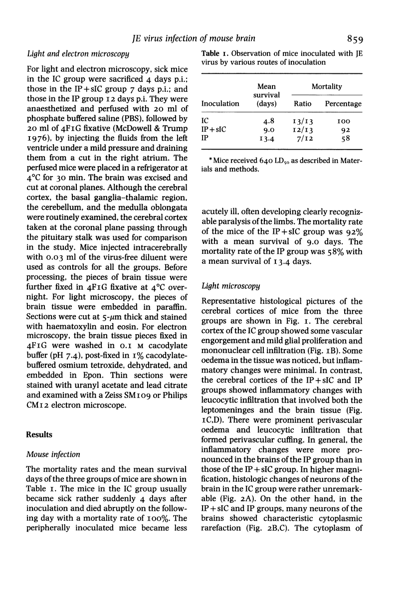
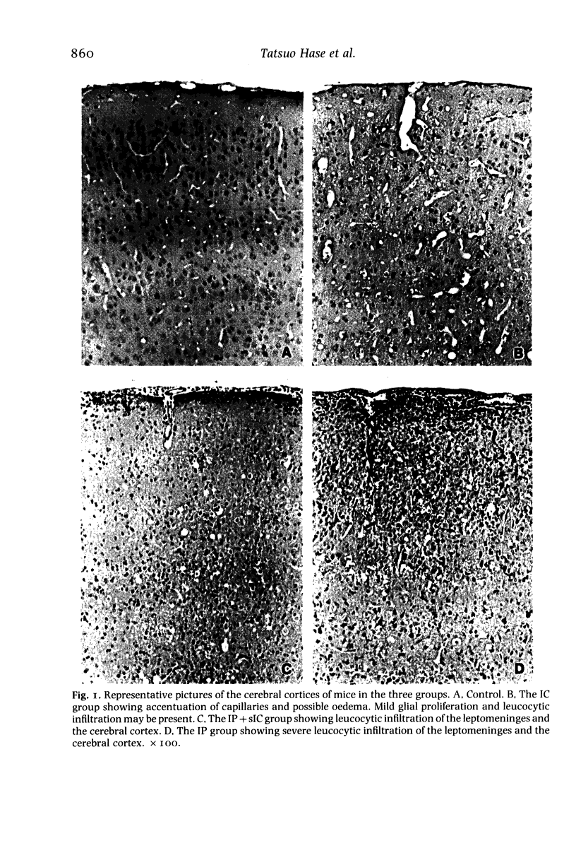
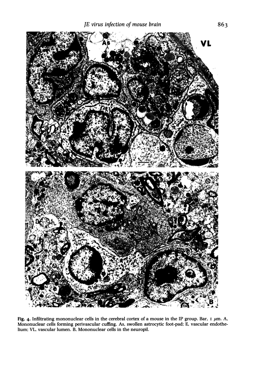
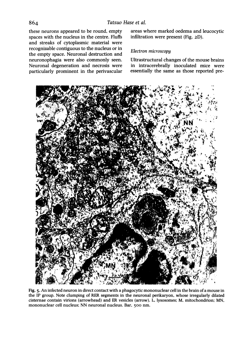
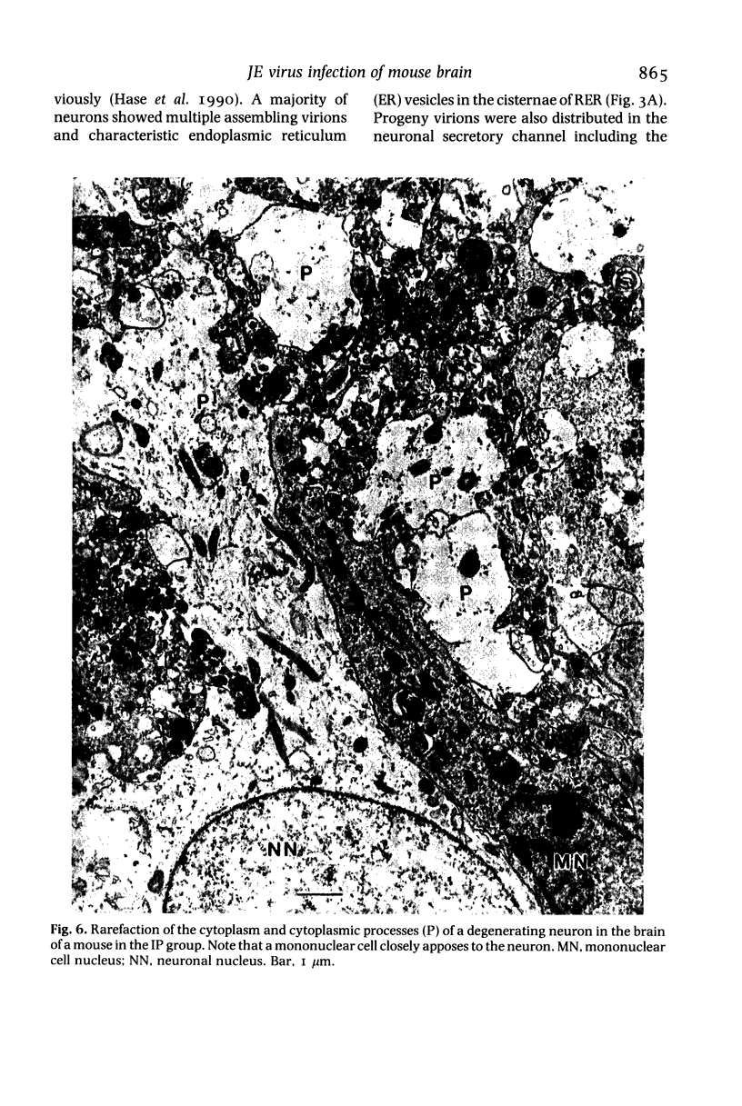
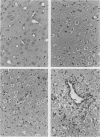
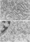
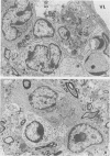
The brains of mice infected with Japanese encephalitis (JE) virus by intracerebral inoculation (IC), intraperitoneal inoculation with sham intracerebral inoculation (IP+sIC), and intraperitoneal inoculation (IP) were studied by light and electron microscopy. The mortality rates and mean survival days were 100% and 4.8 days for the IC group, 92% and 9.0 days for the IP+sIC group, and 58% and 13.4 days for the IP group. Accordingly, the brain samples of sick mice were examined by light and electron microscopy 4 days post-inoculation (p.i.) for the IC group, 7 days p.i. for the IP+sIC group and 12 days p.i. for the IP group. In light microscopy, the mouse brains in the IC group showed little inflammatory change with only mild generalized glial-cell proliferation and mononuclear cell infiltration. In electron microscopy, however, a majority of neurons in the brain were seen to be infected with virus that replicated exclusively in the neuronal secretory system, including rough endoplasmic reticulum (RER) and the Golgi apparatus. In contrast, light microscopic observation of the brains from the IP+sIC and the IP groups showed prominent inflammatory changes with leucocytic infiltration and perivascular cuffing. Neuronal degeneration and neuronophagia were also prominent. In electron microscopy, neurons were infected in the same manner as in the IC group, but showed more advanced degenerative changes with marked cytoplasmic rarefaction and frequent neuronal disintegration. Mononuclear cells were frequently found in direct contact with degenerating and disintegrating neurons. The results showed that (a) the basic process of JE virus replication in brain neurons was present in the three groups of mice, (b) in the peripherally inoculated mice the process was accompanied by inflammatory reaction with resultant neuronal destruction, and (c) breach in the blood-brain barrier at the time of peripheral viral inoculation played an important role in the viral invasion of the CNS.
Full text
PDF


Images in this article


Selected References
These references are in PubMed. This may not be the complete list of references from this article.
- Eckels K. H., Yu Y. X., Dubois D. R., Marchette N. J., Trent D. W., Johnson A. J. Japanese encephalitis virus live-attenuated vaccine, Chinese strain SA14-14-2; adaptation to primary canine kidney cell cultures and preparation of a vaccine for human use. Vaccine. 1988 Dec;6(6):513–518. doi: 10.1016/0264-410x(88)90103-x. [DOI] [PubMed] [Google Scholar]
- Hase T., Summers P. L., Eckels K. H., Baze W. B. Maturation process of Japanese encephalitis virus in cultured mosquito cells in vitro and mouse brain cells in vivo. Arch Virol. 1987;96(3-4):135–151. doi: 10.1007/BF01320956. [DOI] [PubMed] [Google Scholar]
- Johnson R. T., Mims C. A. Pathogenesis of viral infections of the nervous system. N Engl J Med. 1968 Jan 4;278(1):23–contd. doi: 10.1056/NEJM196801042780106. [DOI] [PubMed] [Google Scholar]
- MIYAKE M. THE PATHOLOGY OF JAPANESE ENCEPHALITIS. A REVIEW. Bull World Health Organ. 1964;30:153–160. [PMC free article] [PubMed] [Google Scholar]
- McDowell E. M., Trump B. F. Histologic fixatives suitable for diagnostic light and electron microscopy. Arch Pathol Lab Med. 1976 Aug;100(8):405–414. [PubMed] [Google Scholar]
- Monath T. P., Cropp C. B., Harrison A. K. Mode of entry of a neurotropic arbovirus into the central nervous system. Reinvestigation of an old controversy. Lab Invest. 1983 Apr;48(4):399–410. [PubMed] [Google Scholar]
- Webb H. E., Smith C. E. Relation of immune response to development of central nervous system lesions in virus infections of man. Br Med J. 1966 Nov 12;2(5523):1179–1181. doi: 10.1136/bmj.2.5523.1179. [DOI] [PMC free article] [PubMed] [Google Scholar]
- YASUZUMI G., TSUBO I. ANALYSIS OF THE DEVELOPMENT OF JAPANESE B ENCEPHALITIS (JBE) VIRUS. II. ELECTRON MICROSCOPE STUDIES OF NEURONS INFECTED WITH JBE VIRUS. J Ultrastruct Res. 1965 Apr;12:304–316. doi: 10.1016/s0022-5320(65)80101-0. [DOI] [PubMed] [Google Scholar]