Abstract
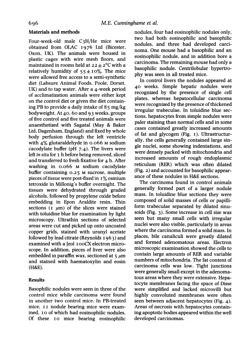
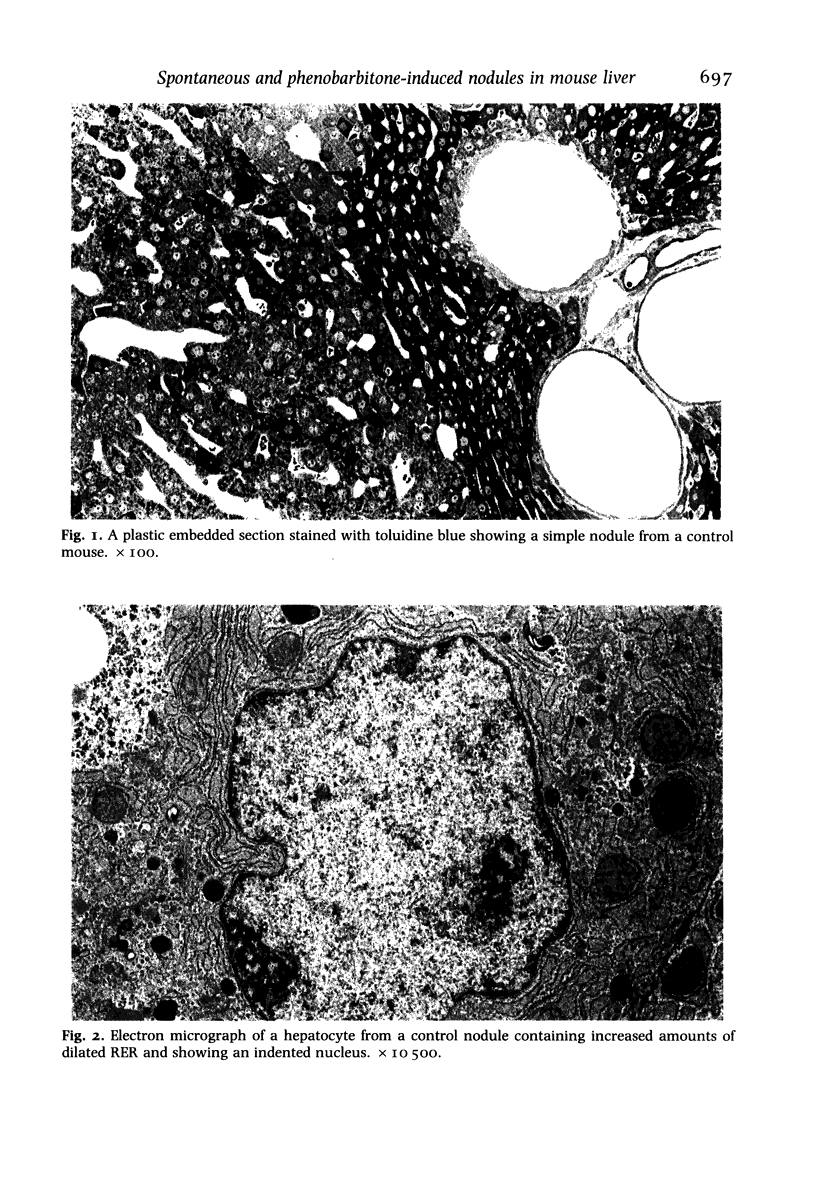
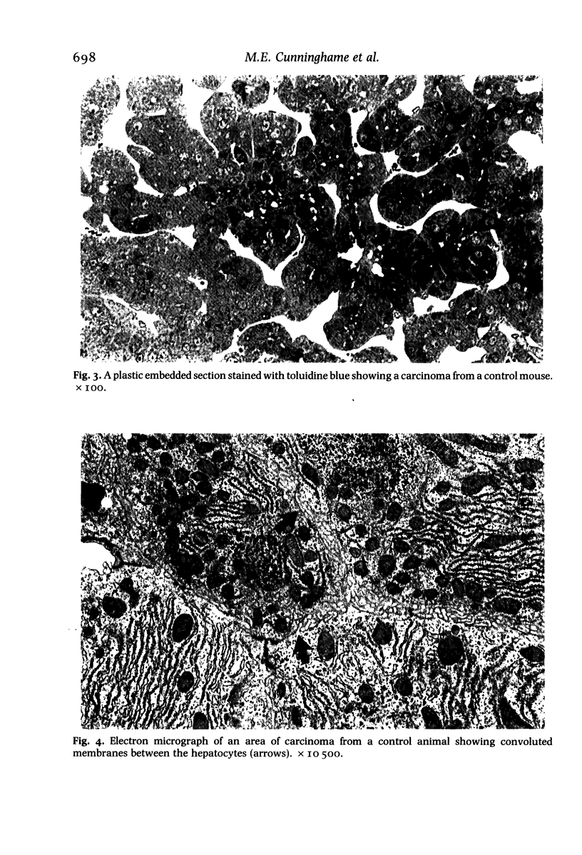
Male C3H/He mice were given 0 (control) or 85 mg/kg/day phenobarbitone (PB) in the diet. At 40, 60 and 93 weeks, groups of mice were killed and the ultrastructure of spontaneous and PB-induced liver nodules was examined. Treated mice showed typical centrilobular hypertrophy and eosinophilic nodules which may be considered as an end stage lesion. The nodule cells were similar in appearance to those in areas of centrilobular hypertrophy except for the presence of convoluted membranes which are considered to be indicative of proliferation. The incidence of carcinoma was not increased by PB treatment. The carcinomas from control and treated animals differed in their ultrastructure in that increased levels of smooth endoplasmic reticulum (SER) were seen in the carcinomas of the PB animals. The presence of SER proliferation in the carcinomas of PB animals suggests that carcinoma may respond to the enzyme-inducing effects of PB.
Full text
PDF
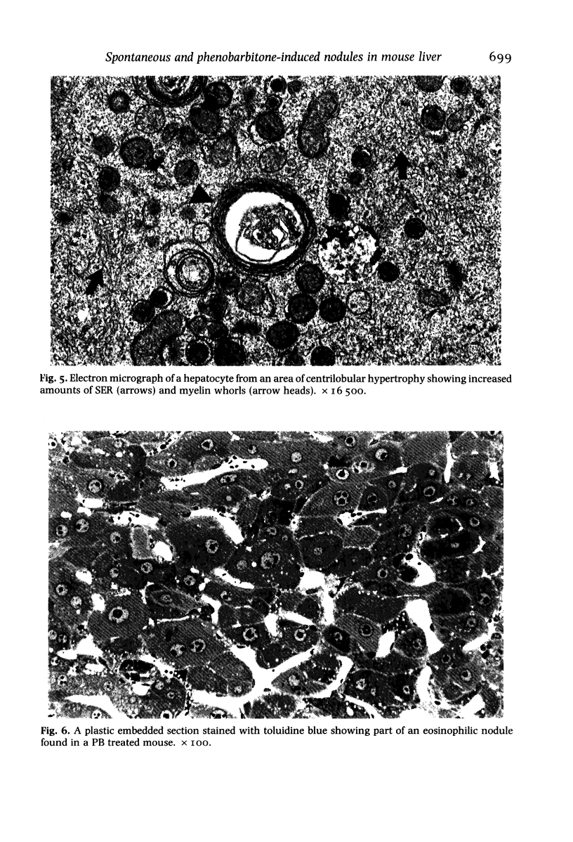
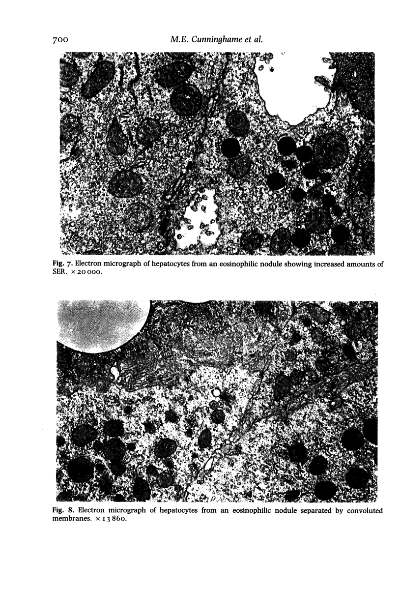
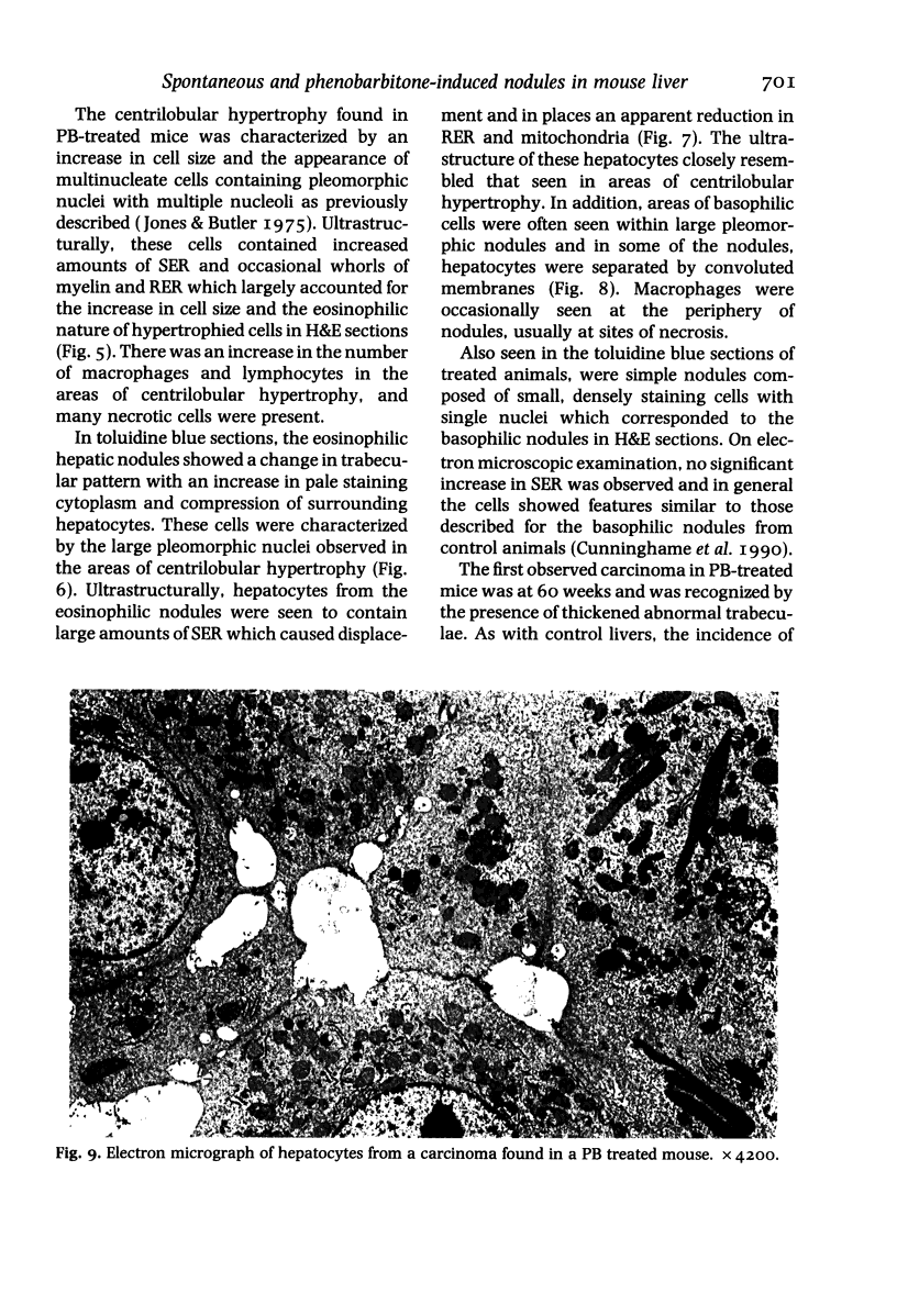
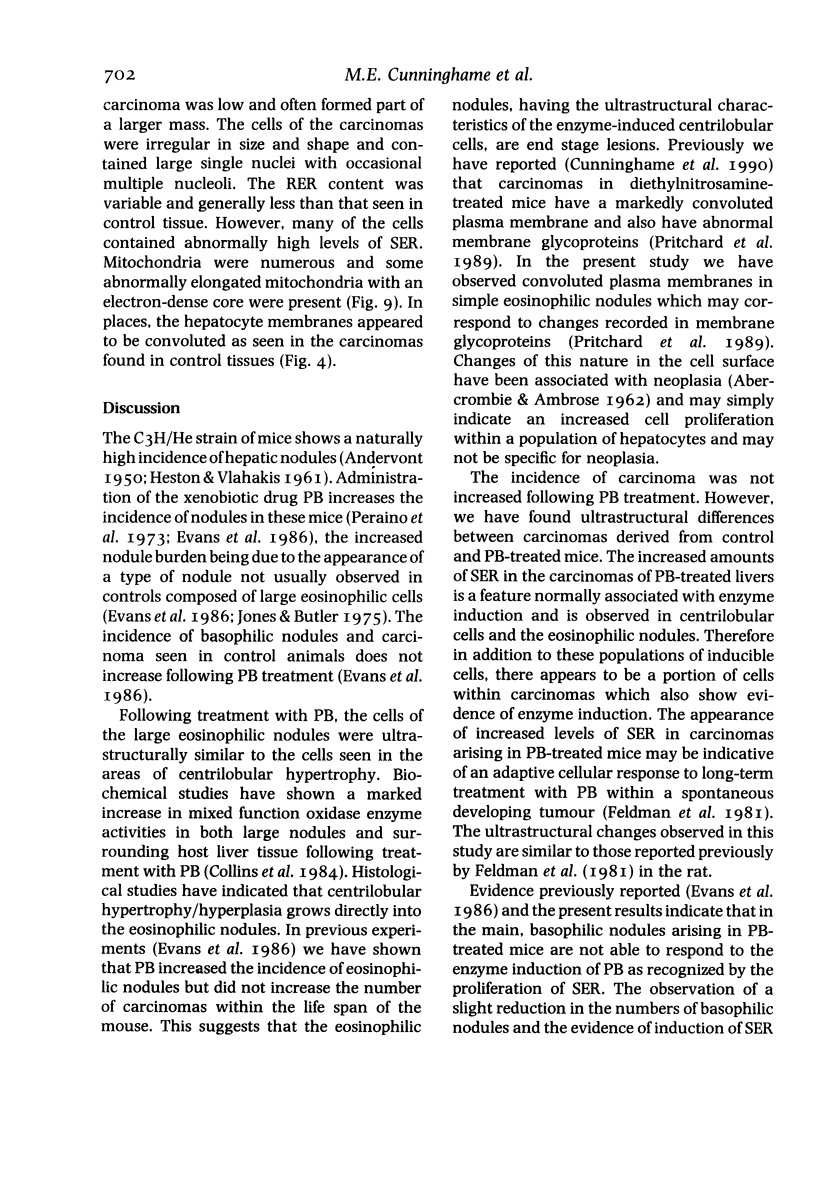
Images in this article
Selected References
These references are in PubMed. This may not be the complete list of references from this article.
- ANDERVONT H. B. Studies on the occurrence of spontaneous hepatomas in mice of strains C3H and CBA. J Natl Cancer Inst. 1950 Dec;11(3):581–592. [PubMed] [Google Scholar]
- Collins M. A., Lake B. G., Evans J. G., Walker R., Gangolli S. D., Conning D. M. Sustained induction of hepatic xenobiotic metabolising enzyme activities by phenobarbitone in C3H/He mice: relevance to nodule formation. Toxicology. 1984 Nov;33(2):129–144. doi: 10.1016/0300-483x(84)90068-4. [DOI] [PubMed] [Google Scholar]
- Cunninghame M. E., Evans J. G., Butler W. H. Ultrastructural features of diethylnitrosamine-induced lesions in the mouse liver. Int J Exp Pathol. 1990 Oct;71(5):677–687. [PMC free article] [PubMed] [Google Scholar]
- Evans J. G., Collins M. A., Savage S. A., Lake B. G., Butler W. H. The histology and development of hepatic nodules in C3H/He mice following chronic administration of phenobarbitone. Carcinogenesis. 1986 Apr;7(4):627–631. doi: 10.1093/carcin/7.4.627. [DOI] [PubMed] [Google Scholar]
- HESTON W. E., VLAHAKIS G. Influence of the Ay gene on mammary-gland tumors, hepatomas, and normal growth in mice. J Natl Cancer Inst. 1961 Apr;26:969–983. [PubMed] [Google Scholar]
- Pritchard D. J., Evans J. G., Lake B. G., Butler W. H. The lectin binding characteristics of spontaneous and phenobarbitone induced hepatic lesions in C3H/He mice. Carcinogenesis. 1989 Sep;10(9):1737–1742. doi: 10.1093/carcin/10.9.1737. [DOI] [PubMed] [Google Scholar]
- Vesselinovitch S. D., Mihailovich N., Rao K. V. Morphology and metastatic nature of induced hepatic nodular lesions in C57BL x C3H F1 mice. Cancer Res. 1978 Jul;38(7):2003–2010. [PubMed] [Google Scholar]


