Abstract
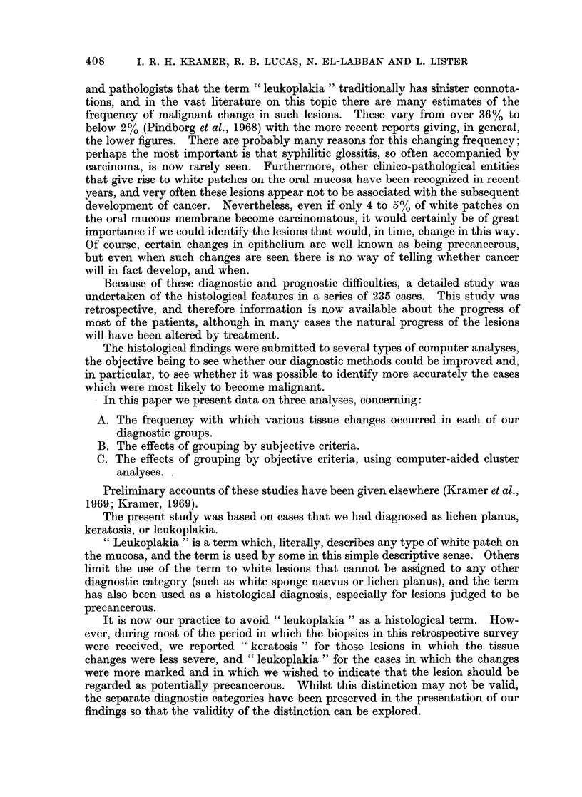
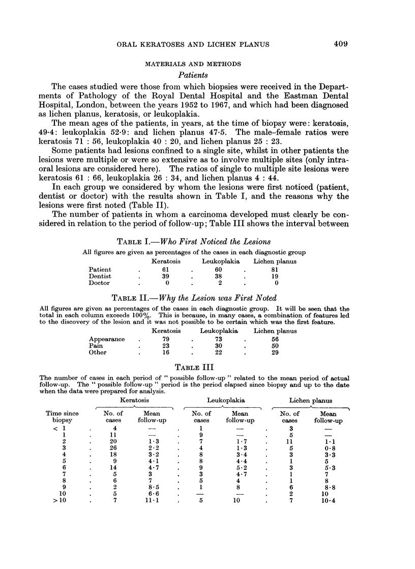
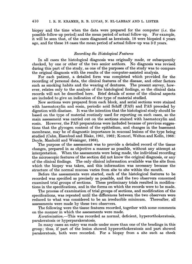
In a retrospective survey of 235 cases in which the diagnosis on biopsy was lichen planus, keratosis or leukoplakia, the histological features were re-assessed in as objective a manner as possible. For each case, the tissue changes were recorded under 37 headings, without any attempt at interpretation. The information was then transferred to punched cards, and used for various types of computer-aided analysis. Firstly, the frequency with which each tissue change occurred was assessed for each diagnostic group, and the data presented also show how often each change occurred in those cases that subsequently developed a carcinoma.
Re-analysis of the cases on the basis of short lists of tissue changes that were thought to characterize each diagnostic category showed, as expected, that many other factors must have played a part in the original diagnosis.
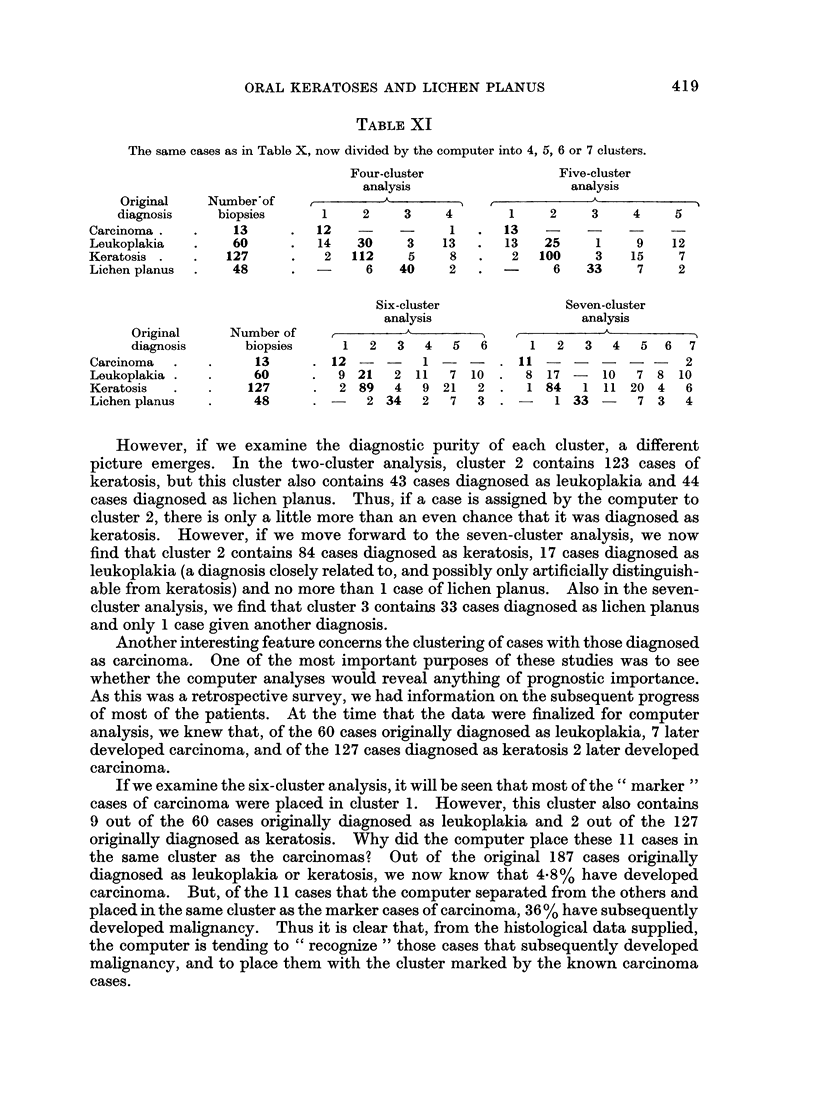
Analysis of the histological findings on an objective basis, using a cluster analysis programme, provided an encouraging degree of separation into the diagnostic groups. When a number of known carcinoma cases were included in the cluster analysis as “markers”, a small number of leukoplakia and keratosis cases were placed by the computer into the same cluster as these “markers”. Of the original 187 cases originally diagnosed as leukoplakia or keratosis, 4.8% are known to have developed a carcinoma, but of the 11 cases the computer placed in the same cluster as the “marker” cases of carcinoma, 36% have subsequently developed carcinoma. Thus, in the cluster analyses, the computer is tending to “recognize” those cases that later developed carcinoma, and to separate them from the bulk of the cases in which malignant change has not occurred.
Full text
PDF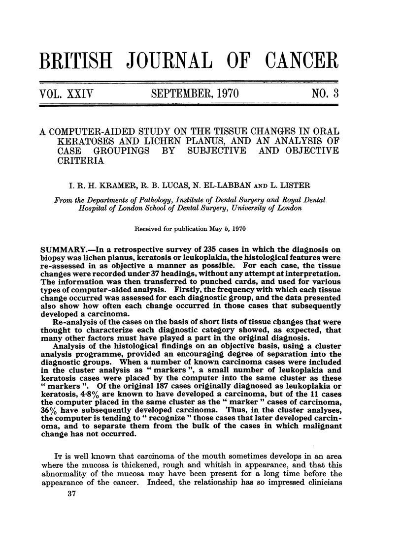
Selected References
These references are in PubMed. This may not be the complete list of references from this article.
- Anderson J. A., Boyle J. A. Computer diagnosis: statistical aspects. Br Med Bull. 1968 Sep;24(3):230–235. doi: 10.1093/oxfordjournals.bmb.a070641. [DOI] [PubMed] [Google Scholar]
- CAHN L. R., EISENBUD L., BLAKE M. N. Histochemical analyses of white lesions of the mouth. I. The basement membrane. Oral Surg Oral Med Oral Pathol. 1961 May;14:596–602. doi: 10.1016/0030-4220(61)90038-x. [DOI] [PubMed] [Google Scholar]
- Carlsson J. A numerical taxonomic study of human oral streptococci. Odontol Revy. 1968;19(2):137–160. [PubMed] [Google Scholar]
- Colman G. The application of computers to the classification of streptococci. J Gen Microbiol. 1968 Jan;50(1):149–158. doi: 10.1099/00221287-50-1-149. [DOI] [PubMed] [Google Scholar]
- Drucker D. B., Melville T. H. Computer classification of streptococci, mostly of oral origin. Nature. 1969 Feb 15;221(5181):664–664. doi: 10.1038/221664a0. [DOI] [PubMed] [Google Scholar]
- Kramer I. R. Precancerous conditions of the oral mucosa. A computer-aided study. Ann R Coll Surg Engl. 1969 Dec;45(6):340–356. [PMC free article] [PubMed] [Google Scholar]
- LEVIN H. L. Oral lichen planus and leukoplakia; differential diagnosis and treatment. U S Armed Forces Med J. 1957 Feb;8(2):198–208. [PubMed] [Google Scholar]
- Pindborg J. J., Jolst O., Renstrup G., Roed-Petersen B. Studies in oral leukoplakia: a preliminary report on the period pervalence of malignant transformation in leukoplakia based on a follow-up study of 248 patients. J Am Dent Assoc. 1968 Apr;76(4):767–771. doi: 10.14219/jada.archive.1968.0127. [DOI] [PubMed] [Google Scholar]
- Radden B. G., Reade P. C. Oral lichen planus. Med J Aust. 1966 Mar 12;1(11):441–445. doi: 10.5694/j.1326-5377.1966.tb72469.x. [DOI] [PubMed] [Google Scholar]


