Abstract
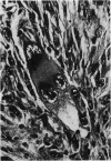
A total of 85 spontaneous rat fibrohistiocytic tumours were evaluated histologically and assessed for the presence or absence of metastases. The overall incidence in controls from 2-year carcinogenicity studies was 2.7%. The tumours occurred principally in the subcutaneous and deep soft tissues, and generally appeared after 18 months of age. Four histological types were recognized: histiocytic (17%), pleomorphic (33%), cellular (17%) and very fibrous (33%). Histiocytic tumours were highly malignant, and most produced metastases. Pleomorphic and cellular neoplasms occasionally produced metastases and must be regarded as potentially malignant. Very fibrous lesions were essentially benign. The close resemblance, both histologically and biologically, between rat and human fibrohistiocytic neoplasms supports the use of the fibrohistiocytic concept in laboratory-animal pathology. Study of these rat tumours may provide insight into the development of human fibrohistiocytic neoplasms.
Full text
PDF
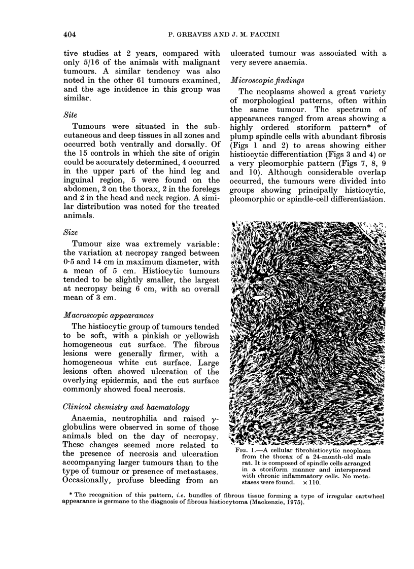
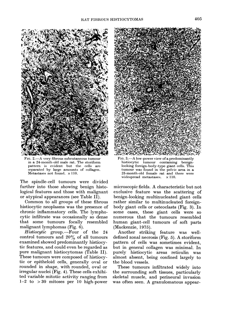
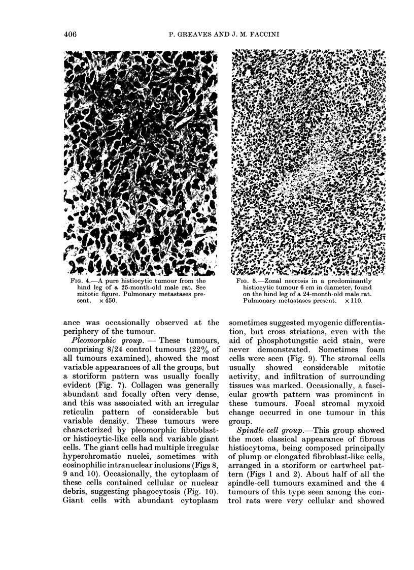
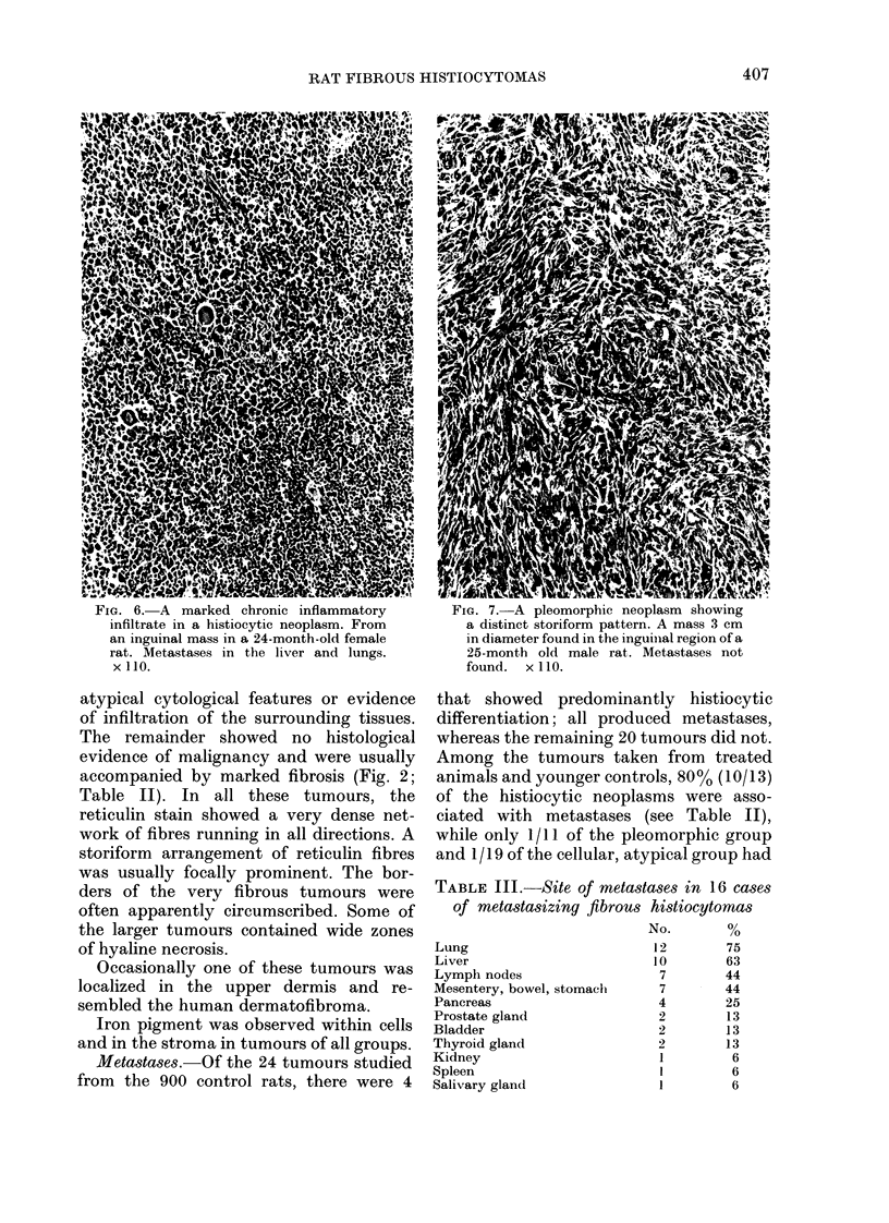
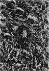
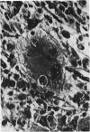
Images in this article
Selected References
These references are in PubMed. This may not be the complete list of references from this article.
- Fu Y. S., Gabbiani G., Kaye G. I., Lattes R. Malignant soft tissue tumors of probable histiocytic origin (malignant fibrous histiocytomas): general considerations and electron microscopic and tissue culture studies. Cancer. 1975 Jan;35(1):176–198. doi: 10.1002/1097-0142(197501)35:1<176::aid-cncr2820350123>3.0.co;2-n. [DOI] [PubMed] [Google Scholar]
- Guccion J. G., Enzinger F. M. Malignant giant cell tumor of soft parts. An analysis of 32 cases. Cancer. 1972 Jun;29(6):1518–1529. doi: 10.1002/1097-0142(197206)29:6<1518::aid-cncr2820290616>3.0.co;2-#. [DOI] [PubMed] [Google Scholar]
- Harris M. The ultrastructure of benign and malignant fibrous histiocytomas. Histopathology. 1980 Jan;4(1):29–44. doi: 10.1111/j.1365-2559.1980.tb02895.x. [DOI] [PubMed] [Google Scholar]
- Johnson K. H., Ghobrial H. K., Buoen L. C., Brand I., Brand K. G. Nonfibroblastic origin of foreign body sarcomas implicated by histological and electron microscopic studies. Cancer Res. 1973 Dec;33(12):3139–3154. [PubMed] [Google Scholar]
- Katenkamp D., Stiller D. Cellular composition of the so-called dermatofibroma (histiocytoma cutis). Virchows Arch A Pathol Anat Histol. 1975 Sep 18;367(4):325–336. doi: 10.1007/BF01239339. [DOI] [PubMed] [Google Scholar]
- Merkow L. P., Frich J. C., Jr, Slifkin M., Kyreages C. G., Pardo M. Ultrastructure of a fibroxanthosarcoma (malignant fibroxanthoma). Cancer. 1971 Aug;28(2):372–383. doi: 10.1002/1097-0142(197108)28:2<372::aid-cncr2820280217>3.0.co;2-u. [DOI] [PubMed] [Google Scholar]
- O'BRIEN J. E., STOUT A. P. MALIGNANT FIBROUS XANTHOMAS. Cancer. 1964 Nov;17:1445–1455. doi: 10.1002/1097-0142(196411)17:11<1445::aid-cncr2820171112>3.0.co;2-g. [DOI] [PubMed] [Google Scholar]
- Pradhan S. N., Chung E. B., Ghosh B., Paul B. D., Kapadia G. J. Potential carcinogens. I. Carcinogenicity of some plant extracts and their tannin-containing fractions in rats. J Natl Cancer Inst. 1974 May;52(5):1579–1582. doi: 10.1093/jnci/52.5.1579. [DOI] [PubMed] [Google Scholar]
- Soule E. H., Enriquez P. Atypical fibrous histiocytoma, malignant fibrous histiocytoma, malignant histiocytoma, and epithelioid sarcoma. A comparative study of 65 tumors. Cancer. 1972 Jul;30(1):128–143. doi: 10.1002/1097-0142(197207)30:1<128::aid-cncr2820300119>3.0.co;2-w. [DOI] [PubMed] [Google Scholar]
- Weiss S. W., Enzinger F. M. Malignant fibrous histiocytoma: an analysis of 200 cases. Cancer. 1978 Jun;41(6):2250–2266. doi: 10.1002/1097-0142(197806)41:6<2250::aid-cncr2820410626>3.0.co;2-w. [DOI] [PubMed] [Google Scholar]
- Westwood F. R., Longstaff E., Butler W. H. Cellular progression of neoplasia in the subcutis of mice after implantation of 3,4-benzpyrene. Br J Cancer. 1979 Jun;39(6):761–772. doi: 10.1038/bjc.1979.130. [DOI] [PMC free article] [PubMed] [Google Scholar]


