Abstract
Francisella tularensis is a category A bioterror pathogen which in some cases can cause a severe and fatal human infection. Very few virulence factors are known in this species due to the difficulty in working with it as well as the lack of tools for genetic manipulation. This work describes the construction of a shuttle vector that can replicate in Escherichia coli and F. tularensis as well as two distinct promoter trap constructs based on the shuttle vector backbone. Replication in F. tularensis is based on the promiscuous origin of replication from the Staphylococcus aureus plasmid pC194. We demonstrate the novel plasmids can coexist with established F. tularensis vectors based on the pFNL10 plasmid, the current workhorse of F. tularensis genetics. Our promoter trap can identify promoters that are activated during intracellular growth and survival. These new vectors provide additional tools for the genetic manipulation of F. tularensis.
Keywords: Francisella tularensis, Shuttle vector, Promoter trap
1. Introduction
Francisella tularensis is a Gram-negative γ-Prote-obacteria that has been classified as a category A pathogen by the Centers for Disease control (Ellis et al., 2002). Little is known about the virulence factors required or pathogenicity of this important bio-defense species (Oyston et al., 2004). Both the former USSR and United States studied the weaponization of this species in the 1950s and 1960s (Dennis et al., 2001), prompting renewed interest in the species from a biodefense point of view in the new century. What makes F. tularensis attractive as a bioweapon is that the infectious dose has been determined to be less than 25 colony-forming units, the species is readily aerosolized and there is no licensed vaccine available (Oyston et al., 2004). There are four recognized subspecies of F. tularensis, two of which cause disease in humans. The four subspecies can be separated based on virulence characteristics, geographic distribution, and lipopolysaccharide carbohydrate content; F. tularensis subsp. tularensis is distributed mainly in North America and is considered to be the most virulent of the species; F. tularensis subsp. holartica is present in mainly Europe, North America and Siberia and causes a mild form of tularemia in humans not usually fatal; the two species that do not infect healthy humans are F. tularensis subsp. mediasiatica, found in Central Asia and the former Soviet Union, and F. tularensis subsp. novicida that can be found in Australia and more recent introductions into North America have been noted (Johansson et al., 2004; Oyston et al., 2004). It has been demonstrated using multi locus variable number tandem repeat analysis (MLVA) that the F. tularensis subsp. tularensis and F. tularensis subsp. holartica in North America are physically separated and the geographic distribution is similar to that of tick and animal distributions suggesting that human infection is an accidental component of the F. tularensis lifecycle (Farlow et al., 2005).
Human infection by F. tularensis progresses via the entry and survival within macrophages (Anthony et al., 1991). There have been three proteins definitively demonstrated to be involved with F. tularensis macrophage survival and virulence; AcpA, thought to inhibit the respiratory burst of the macrophage (Mohapatra et al., 2007; Reilly et al., 1996) and MglAB, which are regulatory factors thought to control the F. tularensis pathogenicity island containing the igl and pdp gene clusters (Lauriano et al., 2004). The only other confirmed virulence factors of F. tularensis are the lipopolysacchride (Prior et al., 2003; Vinogradov et al., 1991) and a putative capsule (Sandstrom et al., 1988). While it is known that these factors are involved in F. tularensis infection and survival, the exact mechanisms are still unclear.
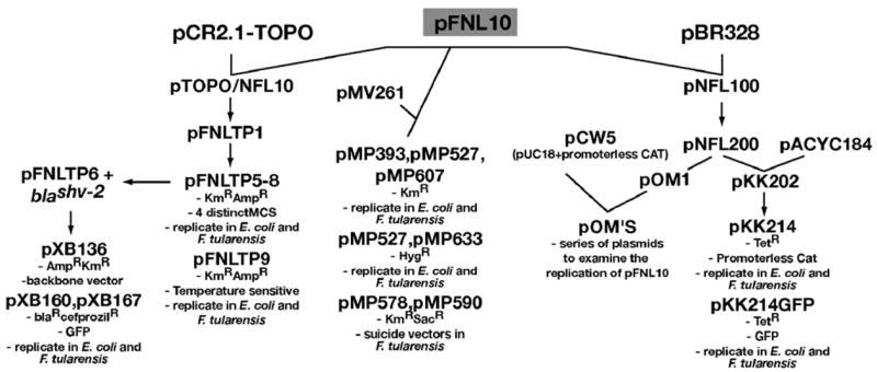
Efforts to identify virulence factors have been hampered by the lack of tools for genetic manipulation of this species as well as the restrictions for working with the highly virulent strains of F. tularensis. There are few naturally occurring plasmids within F. tularensis and those that have been used in the literature are of a single origin type derived from pFNL10 and thus incompatible with each other (Pomerantsev et al., 2001a, 2001b) (Fig. 1). The literature in the last year has contained descriptions of a new transposon mutagenesis system for F. tularensis (Maier et al., 2006) as well as the description of the plasmids available for the use in F. tularensis (Lovullo et al., 2006), however, both were variations of the existing technologies. To date the known shuttle vectors for use in F. tularensis and Escherichia coli are all based on the F. novicida cryptic plasmid pFNL10 (Lovullo et al., 2006; Maier et al., 2004; Norqvist et al., 1996; Pavlov et al., 1996).
Fig. 1.

pFNL10 based vectors for F. tularensis. One series of plasmids based on the combination of pFNL10 and pCR2.1-TOPO includes the shuttle vectors pFNLTP5-9 (Maier et al., 2004, 2006). The pFNLTP vectors are now being modified for other uses such as the examination the F. tularensis β lactamases which expand the range of useful antimicrobial markers (Bina et al., 2006). Other plasmids were used to study the replication mechanism of pFNL10 as well as a few shuttle vectors each of which has utilized origins of replication from other plasmids in combination with pFNL10 (Kuoppa et al., 2001; Norqvist et al., 1996; Pavlov et al., 1996; Pomerantsev et al., 2001a, 2001b). Examination of hygromycin resistance and a suicide vector system in F. tularensis was examined by combining pMV261 and pFNL10 (Lovullo et al., 2006). These plasmids represent the full arsenal of plasmids currently available for F. tularensis work.
In this work, we describe the construction of two plasmid vectors for use in F. tularensis that are not based on the pFNL10 plasmid and thus can work in concert with these established F. tularensis vectors for complementation and or multiple gene replacements (Lovullo et al., 2006; Maier et al., 2004; Norqvist et al., 1996; Pavlov et al., 1996). We have constructed three novel plasmids named pCU18, pCUG1, and pCUG2. The plasmid pCU18 is an E. coli/F. tularensis shuttle vector whereas pCUG1 and pCUG2 are promoter-trap constructs that utilize a promoterless green-fluorescent gene activated by F. tularensis promoter regions while in macrophages. We demonstrate that these plasmids are compatible and stably maintained with the pFNL10-based plasmids as well as demonstrating that pCUG1 can function as a promoter trap. The introduction of these new tools provides additional mechanisms for the identification of F. tularensis virulence factors and regulatory studies.
2. Materials and methods
2.1. Strains and growth conditions
Francisella tularensis type B live vaccine strain (LVS) was obtained from Karen Elkins (Center for Biologics Evaluation and Research, Food and Drug Administration, Rockville, MD). BSL2 containment conditions were utilized when working with the LVS strain. F. tularensis stock cultures were grown at 37 °C with 5% CO2 on Mueller–Hinton agar supplemented with 2.5% (vol/vol) donor calf serum (Mediatech, Herndon, VA), 2% (vol/vol) IsoVitaleX (Becton Dickinson, Franklin Lakes, NJ), 0.1% (wt/vol) glucose, and 0.025% (wt/vol) iron pyrophosphate (Huntley et al., 2007). Following 48–72 h of growth on agar, individual colonies were inoculated into Mueller–Hinton broth medium supplemented with 1.23 mM calcium chloride dihydrate, 1.03 mM magnesium chloride hexahydrate, 0.1% (wt/vol) glucose, 0.025% (wt/vol) iron pyrophosphate, and 2% (vol/vol) IsoVitaleX. Broth cultures were grown at 37 °C for 12–16 h with shaking. Freezer stocks of F. tularensis were prepared in modified Mueller-Hinton medium with 10% (wt/vol) sucrose.Escherichia coli DH5α (Stratagene, La Jolla, CA) was routinely cultivated in Luria-Bertani (LB) broth or on LB agar plates at 37 °C. Antibiotics were used at the following concentrations; ampicillin—100 μg/ml, chloramphenicol—30 μg/ml.
2.2. Plasmid construction
The E. coli/Fransicella shuttle vector known as pCU18 was created by the combination of the pUC18 vector (Yanischperron et al., 1985) commonly used in E. coli and pC194 a broad host range plasmid from Staphylococcus aureus (Horinouchi and Weisblum, 1982). Each parent plasmid was digested with NdeI and SphI and the resulting fragments were purified using QIAquick Gel Extraction Kit (Catalog number 27106; Qiagen, Valencia CA). The purified fragments were joined via ligation according to established molecular biology protocols (Sambrook et al., 1989) and the resulting constructs were transformed into E. coli DH5α. Transformants were selected on LB agar containing ampicillin and chloramphenicol at previously mentioned concentrations to select for constructs containing the resistance marker of each parent plasmid. Restriction profiles were used to confirm the pCU18 plasmid.
The pCUG vectors were created by combining the promoterless green fluorescent protein (GFP) gene from pFPV25 (Valdivia and Falkow, 1996), the broad host range origin of replication and chloramphenicol cassette from Staphylococcus aureus pC194 (Horinouchi and Weisblum, 1982) with the origin of replication from pUC18 (Yanischperron et al., 1985). The GFP gene was digested from pFPV25 by using restriction enzymes EcoRI and HindIII. The same enzymatic digest was performed on pUC18 while only HindIII was used to digest pC194 (Fig. 2). The plasmid fragments were purified using QIAquick Gel Extraction Kit, ligated and the resulting constructs were transformed into E. coli DH5α according to established molecular biology protocols (Sambrook et al., 1989). Transformants were selected on LB agar media containing ampicillin and chloramphenicol at previously mentioned concentrations. Restriction analyses lead to the identification of the pC194 fragment in two orientations, thus forming two distinct vectors, pCUG1 and pCUG2.
Fig. 2.

Construction of the novel shuttle vectors and promoter trap plasmid for F. tularensis. Descriptions of restriction enzymes utilized to create each of pCUG1, pCUG2, and pCU18 are described in the text. Relevant features are highlighted as follows: antibiotic cassettes are represented by black arrows; other open reading frames are grey arrows; relevant features of the origins of replication are either in hollow arrows or hollow boxes. Note: pCUG1 and pCUG2 is essentially the same vector with the pC194 segment cloned in opposite orientation. pCU18 is a shuttle vector that can coexist in E. coli and F. tularensis.
2.3. Electroporation of F. tularensis LVS
Francisella tularensis LVS cultures were grown for 48 h at 37 °C with 5% CO2 on Mueller-Hinton agar described above. The cell growth was then resuspended in Mueller-Hinton broth media to an optical density of 0.1 @ 600 nm and grown until the OD600 was between 0.23 and 0.25 (approximately 4 h). The F. tularensis were then pelleted by centrifugation at 8000 rpm for 10 min using a Sorvall SS-34 rotor. Cells were washed three times by centrifugation and resuspension in 0.5 M sucrose. The cells were resuspended in a final volume of 0.5 M sucrose to provide 20 OD600 units per milliliter. These electrocompetent cells were separated into the 100 μl aliquots and mixed with 1 μg of plasmid DNA and allowed to incubate at room temperature for 30 min. Cells were electroporated in a Bio-Rad GenePulser (2500 V, 100 ohm, 50 μF). Immediately after electroporation 0.9 ml of Mueller-Hinton broth was added to the transformation mixture and allowed to incubate 3–5 h at 37 °C with agitation. The mixture was then plated on Mueller-Hinton agar containing the appropriate antibiotics.
2.4. Plasmid stability
Francisella tularensis LVS cultures containing the single plasmids (pCUG1 or pKK214GFP) or both of the plasmids were grown for 48 h at 37 °C with 5% CO2 on Mueller-Hinton agar with appropriate antibiotics as described above. The cell growth was then resuspended in Mueller-Hinton broth media containing antibiotics and allowed to grow overnight. Multiple parallel cultures were established so that removal of an aliquot and decreased growth volume would not affect the outcome of the experiment. An aliquot was removed at 24 h intervals, diluted and plated on both Mueller-Hinton agar containing antibiotics and Mueller-Hinton agar lacking antibiotics. The plates were then incubated for an additional for 48 h at 37 °C with 5% CO2. Colony forming units were determined and the percentage of cells retaining the plasmids was obtained from the difference of colony forming units on Mueller-Hinton agar with and without antibiotics.
2.5. Cloning of genomic DNA
Genomic DNA was extracted from F. tularensis using Easy-DNA genomic DNA isolation kit (Invitrogen, Carlsbad, CA). Genomic DNA was incompletely digested with AluI and fragments of 500–1000 base pairs were selected and cloned into pCUG1 in front of the promoterless green fluorescent protein coding sequence. AluI was selected as it cut the genomic DNA at a rate of ~1 cut/148 bases, suggesting that we could isolate a number of overlapping promoter regions. A total of 20,000 individual E. coli clones were obtained and pooled. The resulting plasmid pool was isolated from E. coli using the GenElute™ HP Plasmid Kits (Catalog number, NA0300S, Sigma–Aldrich, St. Louis MO) and subsequently transformed into F. tularensis LVS using the method described above. The F. tularensis LVS transformants were also pooled prior to infection of murine macrophage.
2.6. Expression of green fluorescent protein from native F. tularensis promoters
We optimized the macrophage infection process so that each macrophage contained approximately a single transformed F. tularensis LVS cell. Murine macrophage J774.1 cells (ATCC TIB67) were grown at 37 °C with 5% CO2 in DMEM media (Mediatech, Herndon, VA) supplemented with 1% Glutamax (Invitrogen, Carlsbad, CA) and 20 mM sodium pyruvate (Mediatech, Herndon, VA). Cells were resuspended and counted with a hemocytometer to seed a 25 cm2 flask with ~1.5 × 10⁶ J774.1 cells. These cells were allowed to adhere and grown overnight at 37 °C with 5% CO2. The following day 1.5 ml of a 0.1 OD600 culture of F. tularensis LVS containing the pCUG1 with genomic fragment plasmid pool was added to each of the flasks containing macrophages. The flasks were then centrifuged briefly (1000 rpm for 5 min) to ensure interaction of the transformed F. tularensis LVS and macrophage after which they were incubated for 2 h at 37 °C. The media was then decanted, the cell monolayer was washed once with fresh media to remove non-adherent F. tularensis, fresh media was added and the incubation was allowed to proceed for an additional 2 h. This corresponded to a multiplicity of infection of ~ 30 bacteria per murine macrophage and through empirical testing resulted in infection of approximately one F. tularensis LVS cell per macrophage as quantified by hemocytometer (for the macrophage) and colony forming unit counts (for F. tularensis). The macrophages were then resuspended using a cell scarper and taken to the flow cytometry core facility for analysis. Cells containing fluorescent signal were separated from those not containing signal, the macrophages were then lysed with 0.2% sodium deoxycholate and the intracellular F. tularensis LVS were plated on Mueller-Hinton agar. The plasmids were isolated from the F. tularensis LVS, subjected to sequence analysis and compared to the sequenced F. tularensis LVS genome (GenBank Accession No. AM233362) to identify putative promoter sequences.
3. Results and discussion
In constructing these plasmid vectors, we took advantage of the promiscuous nature of the pC194 origin of replication (Horinouchi and Weisblum, 1982) to overcome the problem of the pFNL10 based plasmid vectors that are commonly used in F. tularensis work (Pavlov et al., 1996; Pomerantsev et al., 2001a, 2001b). The pC194 origin can replicate within F. tularensis LVS. In addition to the promiscuous nature of the origin we utilized the pUC18 vector to obtain a multiple cloning site with many unique restriction sites. The combination of these two factors resulted in the formation of the E. coli/F. tularensis shuttle vector named pCU18 (Fig. 2). The plasmid can replicate stably in both species (data not shown) with the addition of the appropriate antibiotics.
Construction of the promoter trap vectors took advantage of the same features as pCU18, the promiscuous origin of replication of pC194 and the E. coli origin of replication from pCU18 (Fig. 2). However, the addition of the promoterless green fluorescent protein gene from pFPV25 (Valdivia and Falkow, 1996) allowed identification of the promoters that were activated under certain stress or stimuli by the presence or absence of a visual measurable signal. A promoter trap method for F. tularensis was devised and implemented by Kuoppa et al. (Kuoppa et al., 2001) using a promoterless antibiotic cassette, however, this method did not allow for the selection of promoters that were activated upon intracellular growth and survival. While this previous method is excellent for the identification of constitutive strong promoters, we wanted to examine promoters that were activated upon the addition of stimuli. Cloning of small fragments of genomic DNA allowed us to identify putative modulatory promoter sequences in F. tularensis LVS.
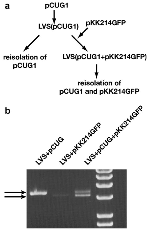
Once the vectors were constructed we wanted to examine their stability within F. tularensis LVS individually, as well as if they were placed within the same bacterium with a pFNL10-based plasmid, in this case pKK214GFP (Fig. 1). We anticipated that the different origins of replication would allow both plasmids to coexist within a single host. To test this we transformed pCUG1 and pKK214GFP into F. tularensis LVS individually as well as together and isolated transformants on MH agar with appropriate antibiotics. When grown under selective pressure we were able to recover the pCUG1 and pKK214GFP from the single transformants, as well as from the doubly transformed cells (Fig. 3). We did not note any significant differences in the electroporation efficiencies of each of these plasmids (data not shown). Additionally, the coexistence of the plasmids did not appear to cause any large aberrations in the plasmid sequence or structure as is evident by the identical plasmid profiles (Fig. 3). This data taken together suggests that the plasmids can exist together in a single bacterium.
Fig. 3.

(a) Schema of analysis of both plasmids within a single F. tularensis LVS isolate. pCUG1 was electroporated into F. tularensis LVS and stably maintained with selective pressure and subsequently re-isolated. Additionally, pKK214GFP was transformed into the F. tularensis LVS containing pCUG1 and also stably maintained with selective pressure. (b) Analysis of the plasmid profiles of F. tularensis containing pCUG1 and or pKK214 demonstrating that both plasmids can be stably maintained together.
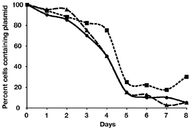
To further examine the stability of these plasmids in a single bacterium, we grew these cultures without any selective pressure and examined the loss of the plasmid from the population over time (Fig. 4). Without selective pressure the plasmids were lost from F. tularensis LVS rapidly, however the bacterium containing both pCUG1 and pKK214GFP did not lose the plasmids at an increased rate when compared to the rate of plasmid loss in the bacterium harboring a single plasmid. This suggests that neither of the plasmids were significantly negatively impacting the replication and maintenance of the other plasmid. This again confirms that the origins of replication are not competing with one another and thus can coexist in the same cell over extended periods of time without detrimental effects.
Fig. 4.

Examination of the plasmid stability in the absence of selective pressure. Over time each of the plasmids, pCUG1 (boxes) and pKK214GFP (triangles), are rapidly lost. When the rate of loss for the singly maintained plasmids is compared to the rate of the cells containing both plasmids (circles) there does not appear to be an increase in the rate of plasmid loss.
We then decided to test the promoter trap system by digesting genomic DNA and inserting it into pCUG1, transforming that pool of potential promoter constructs into F. tularensis LVS and infecting murine macrophages. The digestion of the genomic DNA was preformed with AluI, as it was determined bioinformatically that this enzyme cut approximately once every 148 bases. This would allow us to select incomplete digests to identify larger or smaller promoter constructs over time. Additionally, AluI provides a blunt end preventing any restriction enzyme cloning bias that may occur. We isolated 20,000 individual E. coli colonies and purified this promoter construct pool and then transformed F. tularensis LVS. The transformed LVS were allowed to infect J774.1 murine macrophages and any bacteria not internalized were removed. The resulting cultures were sorted by FACS analysis and those strains with fluorescent signal were plated to re-isolate the promoter containing plasmid construct (Fig. 5a). When a select number of the promoter construct plasmids were screened for the inserts we observed a range of sizes (Fig. 5b). This suggested that the promoters were not the result of a single successful clone that had grown out either in the transformation stage or during macrophage infection. Of note Fig. 5b, lane 6 appears to contain two distinct inserts. We have attempted to minimize these cloning events, however, we recognize that this is a possibility and the screening after re-isolation should rapidly identify any of these double inserts. The sequencing of the inserts did not reveal any consensus sequence to be utilized as an intracellular activated promoter and the majority of the promoters examined are upstream of hypothetical proteins or proteins that contain little or no functional annotation. This confirms what little, we know of the virulence and regulation within F. tularensis (McLendon et al., 2006; Oyston et al., 2004; Titball et al., 2003; Titball, 2003). Further larger studies identifying more promoters will be required to delineate an intracellular activated promoter consensus sequence and we plan on extending these studies to establish a regulatory network within F. tularensis. The availability of multiple F. tularensis genomes (Larsson et al., 2005; Petrosino et al., 2006) and the genomes of close relatives will allow further studies of these processes to identify potential mechanisms of intervention in these regulatory pathways leading to potential therapies.
Fig. 5.

Identification of F. tularensis promoters activated during intracellular growth in macrophage. (a) depicts the experimental method used in the identification of intracellular activated promoter regions. (b) An example of nine fragments obtained from the promoter screen assay, demonstrating a range of insert sizes, suggesting that the screen is functional.
Overall this work describes the construction of three novel vectors; one that can act as a shuttle vector between E. coli and F. tularensis and an additional two vectors which can function as promoter traps for intracellular activated F. tularensis promoters. These plasmids have an advantage over existing shuttle or promoter trap vectors in that they are not based on the pFNL10 plasmid and thus can be used in tandem with those vectors that have been described in the literature and are truly the gold standard for F. tularensis vectors (Pavlov et al., 1996; Pomerantsev et al., 2001a, 2001b). Our promoter trap strategy allows the identification of promoters that can be activated by defined stimuli or during intracellular growth. This methodology has allowed us to identify a group of candidate promoters activated during F. tularensis growth within macrophage.
The existence of two compatible plasmid systems in F. tularensis will now allow a number of classical genetic screens to be utilized. With similar modifications that have occurred in the pFNL10-based plasmids we can design systems to examine the interactions of multiple proteins that may play roles in metabolism or pathogenesis. We can clone the predicted promoter regions into one vector system and directly examine the effects of potential binding proteins, cloned onto compatible plasmids. There are multiple applications were an additional, compatible vector system will be useful if not absolutely required.
Acknowledgments
The authors thank Karl Klose (University of Texas San Antonio) and Mark Smeltzer (University of Arkansas Medical School) for generously providing plasmids pKK214GFP and pC194, respectively. This work is supported by the National Institute of Allergy and Infectious Diseases, National Institutes of Health Grant PO1-AI055637-03.
References
- Anthony LD, et al. Growth of Francisella spp. in rodent macrophages Infect Immun. 1991;59:3291–3296. doi: 10.1128/iai.59.9.3291-3296.1991. [DOI] [PMC free article] [PubMed] [Google Scholar]
- Bina XWR, et al. The Bla2 beta-lactamase from the livevaccine strain of Francisella tularensis encodes a functional protein that is only active against penicillin-class beta-lactam antibiotics. Arch Microbiol. 2006;186:219–228. doi: 10.1007/s00203-006-0140-6. [DOI] [PubMed] [Google Scholar]
- Dennis DT, et al. Tularemia as a biological weapon: medical and publichealth management. JAMA. 2001;285:2763–2773. doi: 10.1001/jama.285.21.2763. [DOI] [PubMed] [Google Scholar]
- Ellis J, Oyston PCF, Green M, Titball RW. Tularemia. Clin Microbiol Rev. 2002;15:631–646. doi: 10.1128/CMR.15.4.631-646.2002. [DOI] [PMC free article] [PubMed] [Google Scholar]
- Farlow J, et al. Francisella tularensis in the United States. Emerging Infect Dis. 2005;11:1835–1841. doi: 10.3201/eid1112.050728. [DOI] [PMC free article] [PubMed] [Google Scholar]
- Horinouchi S, Weisblum B. Nucleotide sequence and functional map of pC194, a plasmid that specifies inducible chloramphenicol resistance. J Bacteriol. 1982;150:815–825. doi: 10.1128/jb.150.2.815-825.1982. [DOI] [PMC free article] [PubMed] [Google Scholar]
- Huntley JF, et al. Characterization of Francisella tularensis outer membrane proteins. J Bacteriol. 2007;189:561–574. doi: 10.1128/JB.01505-06. [DOI] [PMC free article] [PubMed] [Google Scholar]
- Johansson A, et al. Worldwide genetic relationships among Francisella tularensis isolates determined by multiplelocus variable-number tandem repeat analysis. J Bacteriol. 2004;186:5808–5818. doi: 10.1128/JB.186.17.5808-5818.2004. [DOI] [PMC free article] [PubMed] [Google Scholar]
- Kuoppa K, et al. Construction of a reporter plasmid for screening in vivo promoter activity in Francisella tularensis. FEMS Microbiol Lett. 2001;205:77–81. doi: 10.1111/j.1574-6968.2001.tb10928.x. [DOI] [PubMed] [Google Scholar]
- Larsson P, et al. The complete genome sequence of Francisella tularensis, the causative agent of tularemia. Nat Genet. 2005;37:153–159. doi: 10.1038/ng1499. [DOI] [PubMed] [Google Scholar]
- Lauriano CM, et al. MglA regulates transcription of virulence factors necessary for Francisella tularensis intraamoebae and intramacrophage survival. Proc Natl Acad Sci USA. 2004;101:4246–4249. doi: 10.1073/pnas.0307690101. [DOI] [PMC free article] [PubMed] [Google Scholar]
- Lovullo ED, et al. Genetic tools for highly pathogenic Francisella tularensis subsp tularensis. Microbiology. 2006;152:3425–3435. doi: 10.1099/mic.0.29121-0. [DOI] [PubMed] [Google Scholar]
- Maier TM, et al. Construction and characterization of a highly efficient Francisella shuttle plasmid. Appl Environ Microbiol. 2004;70:7511–7519. doi: 10.1128/AEM.70.12.7511-7519.2004. [DOI] [PMC free article] [PubMed] [Google Scholar]
- Maier TM, et al. In vivo Himar1-based transposon mutagenesis of Francisella tularensis. Appl Environ Microbiol. 2006;72:1878–1885. doi: 10.1128/AEM.72.3.1878-1885.2006. [DOI] [PMC free article] [PubMed] [Google Scholar]
- McLendon MK, et al. Francisella tularensis: taxonomy, genetics, and immunopathogenesis of a potential agent of biowarfare. Annu Rev Microbiol. 2006;60:167–185. doi: 10.1146/annurev.micro.60.080805.142126. [DOI] [PMC free article] [PubMed] [Google Scholar]
- Mohapatra NP, et al. AcpA is a Francisella acid phosphatase that affects intramacrophage survival and virulence. Infect Immun. 2007;75:390–396. doi: 10.1128/IAI.01226-06. [DOI] [PMC free article] [PubMed] [Google Scholar]
- Norqvist A, et al. Construction of a shuttle vector for use in Francisella tularensis. FEMS Immunol Med Microbiol. 1996;13:257–260. doi: 10.1111/j.1574-695X.1996.tb00248.x. [DOI] [PubMed] [Google Scholar]
- Oyston PC, et al. Tularaemia: bioterrorism defence renews interest in Francisella tularensis. Nat Rev Microbiol. 2004;2:967–978. doi: 10.1038/nrmicro1045. [DOI] [PubMed] [Google Scholar]
- Pavlov VM, et al. Cryptic plasmid pFNL10 from Francisella novicida-like F6168: the base of plasmid vectors for Francisella tularensis. FEMS Immunol Med Microbiol. 1996;13:253–256. doi: 10.1111/j.1574-695X.1996.tb00247.x. [DOI] [PubMed] [Google Scholar]
- Petrosino JF, et al. Chromosome rearrangement and diversification of Francisella tularensis revealed by the type B (OSU18) genome sequence. J Bacteriol. 2006;188:6977–6985. doi: 10.1128/JB.00506-06. [DOI] [PMC free article] [PubMed] [Google Scholar]
- Pomerantsev AP, et al. Genetic organization of the Francisella plasmid pFNL10. Plasmid. 2001a;46:210–222. doi: 10.1006/plas.2001.1548. [DOI] [PubMed] [Google Scholar]
- Pomerantsev AP, et al. Nucleotide sequence, structural organization, and functional characterization of the small recombinant plasmid pOM1 that is specific for Francisella tularensis. Plasmid. 2001b;46:86–94. doi: 10.1006/plas.2001.1538. [DOI] [PubMed] [Google Scholar]
- Prior JL, et al. Characterization of the O antigen gene cluster and structural analysis of the O antigen of Francisella tularensis subsp tularensis. J Med Microbiol. 2003;52:845–851. doi: 10.1099/jmm.0.05184-0. [DOI] [PubMed] [Google Scholar]
- Reilly TJ, et al. Characterization and sequencing of a respiratory burst-inhibiting acid phosphatase from Francisella tularensis. J Biol Chem. 1996;271:10973–10983. doi: 10.1074/jbc.271.18.10973. [DOI] [PubMed] [Google Scholar]
- Sambrook J, et al. Molecular Cloning: A Laboratory Manual. Cold Spring Harbor Laboratory; Cold Spring Harbor, NY: 1989. [Google Scholar]
- Sandstrom G, et al. A capsule-deficient mutant of Francisella tularensis LVS exhibits enhanced sensitivity to killing by serum but diminished sensitivity to killing by polymorphonuclear leukocytes. Infect Immun. 1988;56:1194–1202. doi: 10.1128/iai.56.5.1194-1202.1988. [DOI] [PMC free article] [PubMed] [Google Scholar]
- Titball RW, et al. Will the enigma of Francisella tularensis virulence soon be solved? Trends Microbiol. 2003;11:118–123. doi: 10.1016/s0966-842x(03)00020-9. [DOI] [PubMed] [Google Scholar]
- Titball RW. Francisella tularensis: an overview. ASM News. 2003;69:558–563. [Google Scholar]
- Valdivia RH, Falkow S. Bacterial genetics by flow cytometry: rapid isolation of Salmonella typhimurium acid-inducible promoters by differential fluorescence induction. Mol Microbiol. 1996;22:367–378. doi: 10.1046/j.1365-2958.1996.00120.x. [DOI] [PubMed] [Google Scholar]
- Vinogradov EV, et al. Structure of the O-antigen of Francisella tularensis strain 15. Carbohydr Res. 1991;214:289–297. doi: 10.1016/0008-6215(91)80036-m. [DOI] [PubMed] [Google Scholar]
- Yanischperron C, et al. Improved M13 phage cloning vectors and host strains-nucleotide-sequences of the M13mp18 and pUC19 vectors. Gene. 1985;33:103–119. doi: 10.1016/0378-1119(85)90120-9. [DOI] [PubMed] [Google Scholar]


