

Abstract
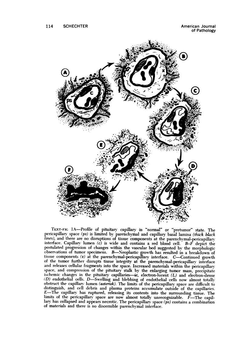
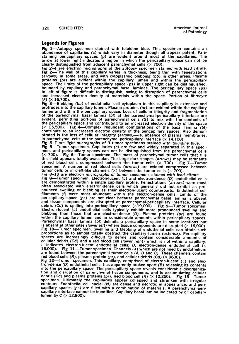
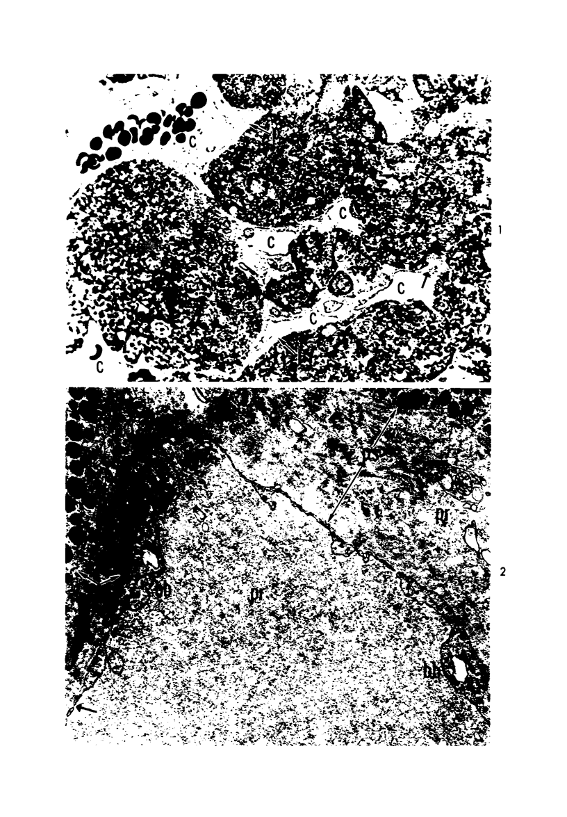
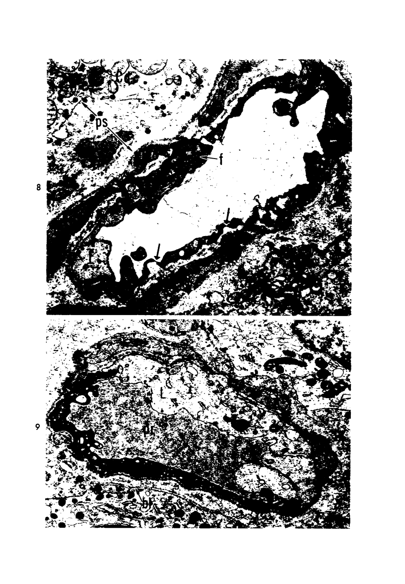
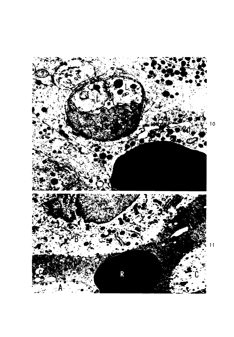
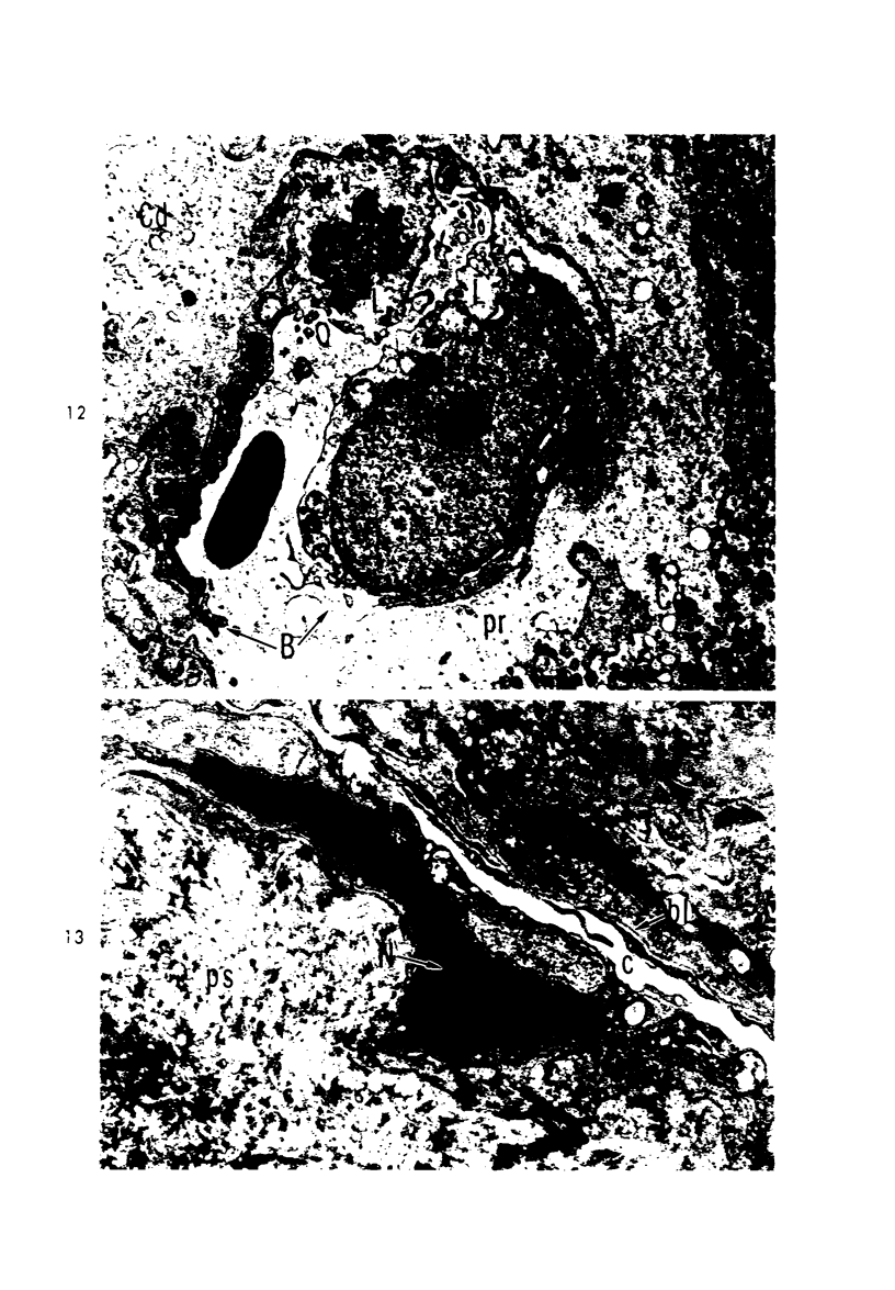
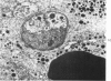
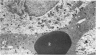
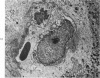
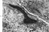
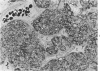
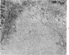
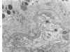
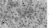
The fine structure of the capillary bed of the anterior pituitary has been studied in 19 cases of pituitary tumor and 1 autopsy specimen. Tumor specimens were less well vascularized than the autopsy specimen. Endothelial cells within tumor specimens were often observed with swollen portions of cytoplasm, or cytoplasmic blebs, projecting into the capillary lumen. Blebbing, in many cases, nearly obstructed the capillary lumen and was most often associated with endothelial cells that had an electron-lucent cytoplasm, in contrast to endothelial cells that had an electron-dense cytoplasm, even within the same capillary profile. Compared with electron-lucent cells, electron-dense cells contained more endothelial filaments. Also observed were capillaries that had apparently broken apart releasing their contents into the pericapillary space and shrunken remnants of capillaries. A number of abnormal features were observed in the pericapillary spaces—ie, disruption of the parenchymal-pericapillary interface, disorganization of basal laminae, increased amounts of plasma proteins and cellular debris within the space and, ultimately, complete loss of the normal limits and characteristics of the space. Similar, though less pronounced, changes were observed in the autopsy specimen. The hypothesis is advanced that changes observed in the capillary bed are, in large part, a result of tumor growth, which increasingly disrupts tissue organization at the parenchymal-pericapillary interface and, because the tumor mass in the sella turcica can only enlarge upwards, compresses the pituitary stalk and portal veins. The result is ischemia and eventual necrosis. The observations are then in good agreement with recent reports on changes in capillary fine structure associated with ischemia.
Full text
PDF


Images in this article

Selected References
These references are in PubMed. This may not be the complete list of references from this article.
- BUERGER M., HEVELKE G. Do human beings have the age of their blood vessels? Angiology. 1956 Apr;7(2):137–151. doi: 10.1177/000331975600700203. [DOI] [PubMed] [Google Scholar]
- Becker C. G., Murphy G. E. Demonstration of contractile protein in endothelium and cells of the heart valves, endocardium, intima, arteriosclerotic plaques, and Aschoff bodies of rheumatic heart disease. Am J Pathol. 1969 Apr;55(1):1–37. [PMC free article] [PubMed] [Google Scholar]
- Bergland R. M., Torack R. M. An ultrastructural study of follicular cells in the human anterior pituitary. Am J Pathol. 1969 Nov;57(2):273–297. [PMC free article] [PubMed] [Google Scholar]
- Berkowitz L. R., Fiorello O., Kruger L., Maxwell D. S. Selective staining of nervous tissue for light microscopy following preparation for electron microscopy. J Histochem Cytochem. 1968 Dec;16(12):808–814. doi: 10.1177/16.12.808. [DOI] [PubMed] [Google Scholar]
- CARDELL R. R., Jr THE CYTOPHYSIOLOGY OF THE ANTERIOR PITUITARY GLAND. Henry Ford Hosp Med Bull. 1963 Dec;11:409–430. [PubMed] [Google Scholar]
- Cardell R. R., Jr, Knighton R. S. The cytology of a human pituitary tumor: an electron microscopic study. Trans Am Microsc Soc. 1966 Jan;85(1):58–78. [PubMed] [Google Scholar]
- Chiang J., Kowada M., Ames A., 3rd, Wright R. L., Majno G. Cerebral ischemia. III. Vascular changes. Am J Pathol. 1968 Feb;52(2):455–476. [PMC free article] [PubMed] [Google Scholar]
- FARQUHAR M. G. Fine structure and function in capillaries of the anterior pituitary gland. Angiology. 1961 Jul;12:270–292. doi: 10.1177/000331976101200704. [DOI] [PubMed] [Google Scholar]
- Fujita H., Kataoka K. Capillary endothelial cells of the anterior pituitary should be excluded from the reticulo-endothelial system. Light and electron microscopic observations on rabbits and rats. Z Anat Entwicklungsgesch. 1969;128(4):318–328. doi: 10.1007/BF00522530. [DOI] [PubMed] [Google Scholar]
- GUSEK W. [Comparative light and electron microscope studies on pituitray adenoma in acromegaly]. Endokrinologie. 1962 Jul;42:257–283. [PubMed] [Google Scholar]
- Hills C. P. Ultrastructural changes in the capillary bed of the rat cerebral cortex in anoxic-ischemic brain lesions. Am J Pathol. 1964 Apr;44(4):531–551. [PMC free article] [PubMed] [Google Scholar]
- Kent S. P. Diffusion of plasma proteins into cells: a manifestation of cell injury in human myocardial ischemia. Am J Pathol. 1967 Apr;50(4):623–637. [PMC free article] [PubMed] [Google Scholar]
- Kornblum R. N., Fisher R. S. Pituitary lesions in craniocerebral injuries. Arch Pathol. 1969 Sep;88(3):242–248. [PubMed] [Google Scholar]
- Kuromatsu C. The fine structure of the human pituitary chromophobe adenoma with special reference to the classification of this tumor. Arch Histol Jpn. 1968 Feb;29(1):41–61. doi: 10.1679/aohc1950.29.41. [DOI] [PubMed] [Google Scholar]
- Paiz C., Hennigar G. R. Electron microscopy and histochemical correlation of human anterior pituitary cells. Am J Pathol. 1970 Apr;59(1):43–74. [PMC free article] [PubMed] [Google Scholar]
- Porcile E., Racadot J. Ultrastructure des cellules de Crooke observées dans l'hypophyse humaine au cours de la maladie de Cushing. C R Acad Sci Hebd Seances Acad Sci D. 1966 Oct 3;263(14):948–951. [PubMed] [Google Scholar]
- RINEHART J. F., FARQUHAR M. G. The fine vascular organization of the anterior pituitary gland; an electron microscopic study with histochemical correlations. Anat Rec. 1955 Feb;121(2):207–239. doi: 10.1002/ar.1091210206. [DOI] [PubMed] [Google Scholar]
- Rosenquist T. H., Bernick S. Histochemistry of renal basal laminae. Adolescent compared with senescent rats. J Gerontol. 1971 Apr;26(2):176–185. doi: 10.1093/geronj/26.2.176. [DOI] [PubMed] [Google Scholar]
- Willms-Kretschmer K., Majno G. Ischemia of the skin. Electron microscopic study of vascular injury. Am J Pathol. 1969 Mar;54(3):327–353. [PMC free article] [PubMed] [Google Scholar]