

Abstract
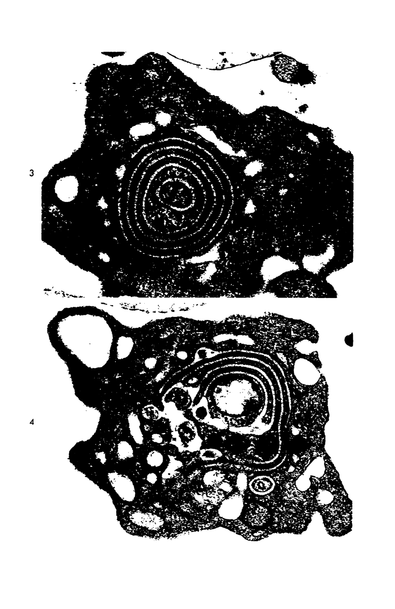
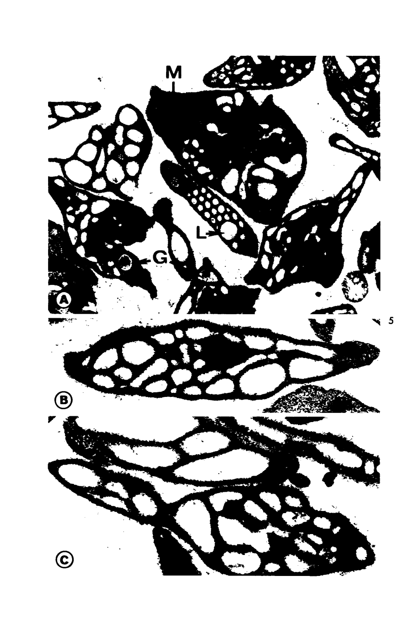
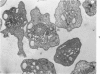
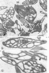
Platelet degranulation is a characteristic feature of platelet response to aggregating agents, but the mechanism and route by which secretory organelles are transferred to plasma are still uncertain. In the present study, human platelets were incubated with cytochalasin B, an agent which stabilizes discoid shape, and trypsin, which is known to cause release reaction and degranulation. Platelets treated in this manner retained their disc form, but were nearly devoid of granules and dense bodies. Electron-dense tracers indicated that degranulation was accomplished by fusion of secretory organelles with channels of the open canalicular system. The degranulated discoid platelet appears to survive exposure to cytochalasin B and trypsin and may prove to be a useful model for in vivo and in vitro experimental studies.
Full text
PDF


Images in this article


Selected References
These references are in PubMed. This may not be the complete list of references from this article.
- Day H. J., Holmsen H. Concepts of the blood platelet release reaction. Ser Haematol. 1971;4(1):3–27. [PubMed] [Google Scholar]
- Estensen R. D. Cytochalasin B. I. Effect on cytokinesis of Novikoff hepatoma cells. Proc Soc Exp Biol Med. 1971 Apr;136(4):1256–1260. doi: 10.3181/00379727-136-35470. [DOI] [PubMed] [Google Scholar]
- GRETTE K. Studies on the mechanism of thrombin-catalyzed hemostatic reactions in blood platelets. Acta Physiol Scand Suppl. 1962;195:1–93. [PubMed] [Google Scholar]
- HASLAM R. J. ROLE OF ADENOSINE DIPHOSPHATE IN THE AGGREGATION OF HUMAN BLOOD-PLATELETS BY THROMBIN AND BY FATTY ACIDS. Nature. 1964 May 23;202:765–768. doi: 10.1038/202765a0. [DOI] [PubMed] [Google Scholar]
- HOVIG T. THE EFFECT OF VARIOUS ENZYMES ON THE ULTRASTRUCTURE, AGGREGATION, AND CLOT RETRACTION ABILITY OF RABBIT BLOOD PLATELETS. Thromb Diath Haemorrh. 1965 Mar 15;13:84–113. [PubMed] [Google Scholar]
- Novikoff A. B., Goldfischer S. Visualization of peroxisomes (microbodies) and mitochondria with diaminobenzidine. J Histochem Cytochem. 1969 Oct;17(10):675–680. doi: 10.1177/17.10.675. [DOI] [PubMed] [Google Scholar]
- SCHMID H. J., JACKSON D. P., CONLEY C. L. Mechanism of action of thrombin on platelets. J Clin Invest. 1962 Mar;41:543–553. doi: 10.1172/JCI104508. [DOI] [PMC free article] [PubMed] [Google Scholar]
- Schroeder T. E. The contractile ring. I. Fine structure of dividing mammalian (HeLa) cells and the effects of cytochalasin B. Z Zellforsch Mikrosk Anat. 1970;109(4):431–449. [PubMed] [Google Scholar]
- Webber A. J., Johnson S. A. Platelet participation in blood coagulation aspects of hemostasis. Am J Pathol. 1970 Jul;60(1):19–42. [PMC free article] [PubMed] [Google Scholar]
- Wessells N. K., Spooner B. S., Ash J. F., Bradley M. O., Luduena M. A., Taylor E. L., Wrenn J. T., Yamada K. Microfilaments in cellular and developmental processes. Science. 1971 Jan 15;171(3967):135–143. doi: 10.1126/science.171.3967.135. [DOI] [PubMed] [Google Scholar]
- White J. G. A search for the platelet secretory pathway using electron dense tracers. Am J Pathol. 1970 Jan;58(1):31–49. [PMC free article] [PubMed] [Google Scholar]
- White J. G. Effects of ethylenediamine tetracetic acid (EDTA) on platelet structure. Scand J Haematol. 1968;5(4):241–254. doi: 10.1111/j.1600-0609.1968.tb01743.x. [DOI] [PubMed] [Google Scholar]
- White J. G. Interaction of membrane systems in blood platelets. Am J Pathol. 1972 Feb;66(2):295–312. [PMC free article] [PubMed] [Google Scholar]
- White J. G., Krivit W. The ultrastructural localization and release of platelet lipids. Blood. 1966 Feb;27(2):167–186. [PubMed] [Google Scholar]
- White J. G. The interplatelet zone. Am J Pathol. 1970 Jan;58(1):19–29. [PMC free article] [PubMed] [Google Scholar]