Abstract
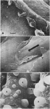
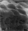
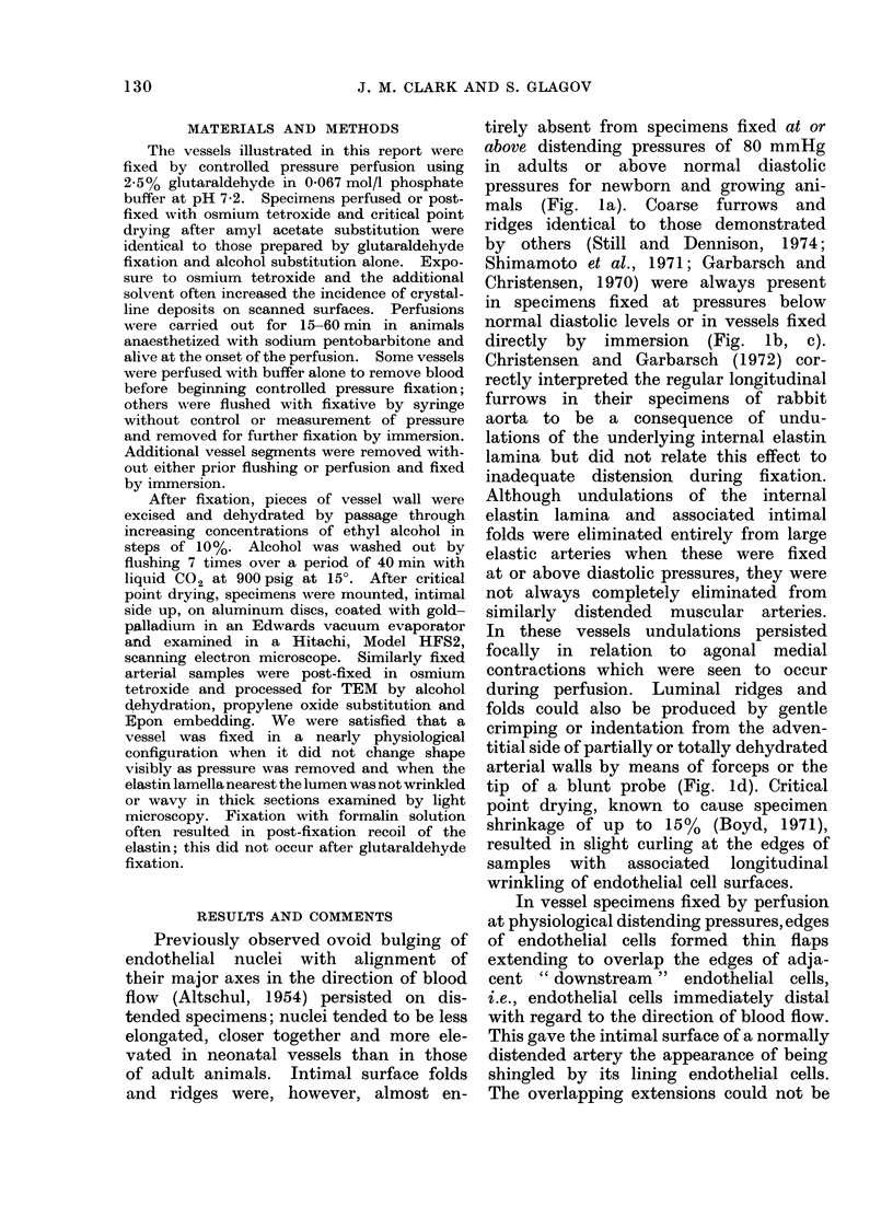
Perfusion fixation at physiological pressures, careful tissue handling, adequate drying and reduced beam exposure time eliminated many of the intimal surface projections, ridges and bridges which have been taken for normal structures on scanning electron microscopy. In the normal distended vessel, ovoid endothelial nuclei bulged into the lumen with their major axes aligned in the direction of flow: adjacent cell margins overlapped consistently in the direction of flow, with each cell overlapping the edge of its downstream neighbour. Regular longitudinal furrows associated with undulations of the internal elastin lamina were entirely eliminated from elastic arteries when distending pressures exceeded diastolic levels during fixation.
Full text
PDF
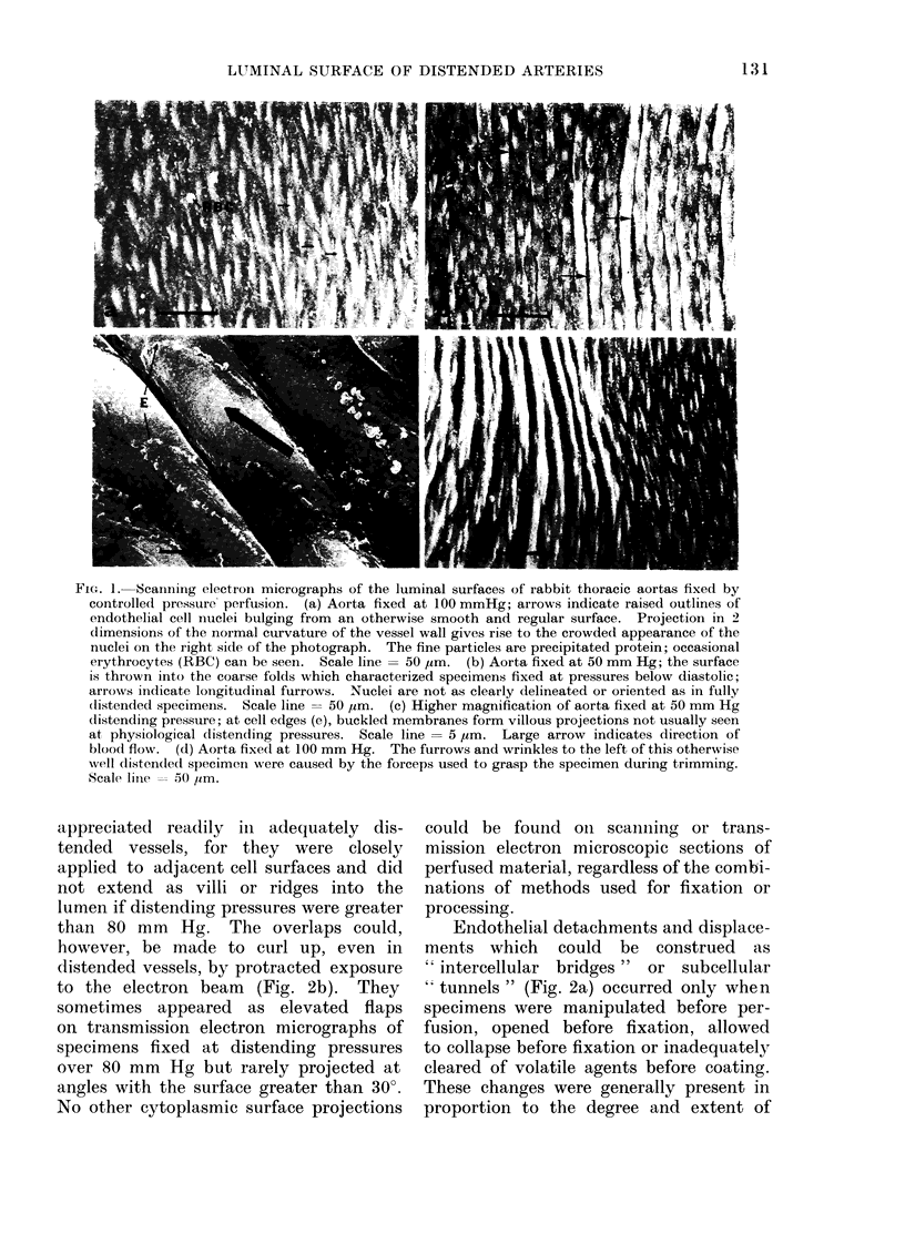
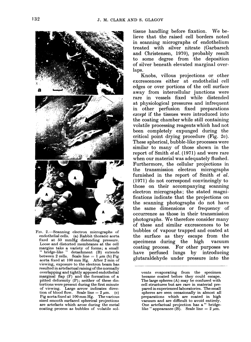
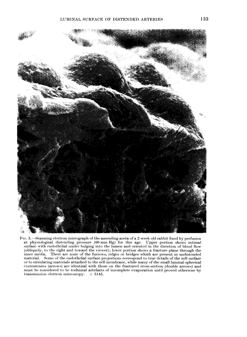
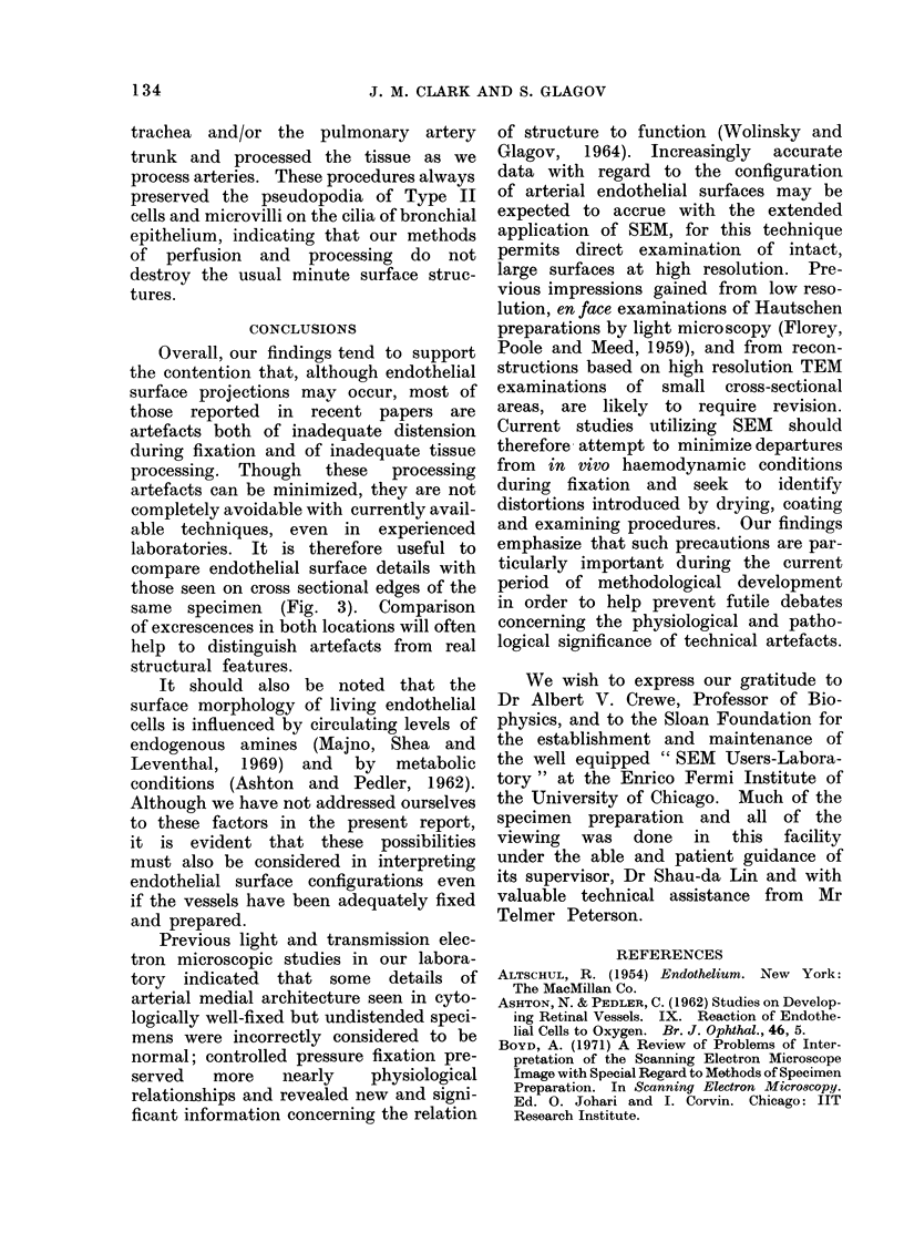
Images in this article
Selected References
These references are in PubMed. This may not be the complete list of references from this article.
- Christensen B. C., Garbarsch C. A scanning electron microscopic (SEM) study on the endothelium of the normal rabbit aorta. Angiologica. 1972;9(1):15–26. doi: 10.1159/000157911. [DOI] [PubMed] [Google Scholar]
- Garbarsch C., Christensen B. C. Scanning electron microscopy of aortic endothelial cell boundaries after staining with silver nitrate. Angiologica. 1970;7(6):365–373. doi: 10.1159/000157852. [DOI] [PubMed] [Google Scholar]
- Smith U., Ryan J. W., Michie D. D., Smith D. S. Endothelial projections as revealed by scanning electron microscopy. Science. 1971 Sep 3;173(4000):925–927. doi: 10.1126/science.173.4000.925. [DOI] [PubMed] [Google Scholar]
- Still W. J., Dennison S. The arterial endothelium of the hypertensive rat: a scanning and transmission electron microscopical study. Arch Pathol. 1974 Jun;97(6):337–342. [PubMed] [Google Scholar]
- WOLINSKY H., GLAGOV S. STRUCTURAL BASIS FOR THE STATIC MECHANICAL PROPERTIES OF THE AORTIC MEDIA. Circ Res. 1964 May;14:400–413. doi: 10.1161/01.res.14.5.400. [DOI] [PubMed] [Google Scholar]


