Abstract
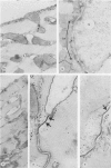
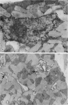
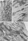
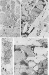
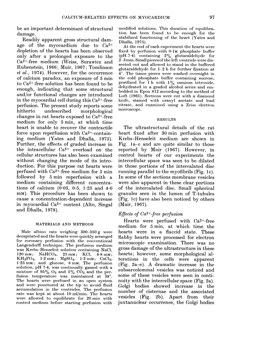
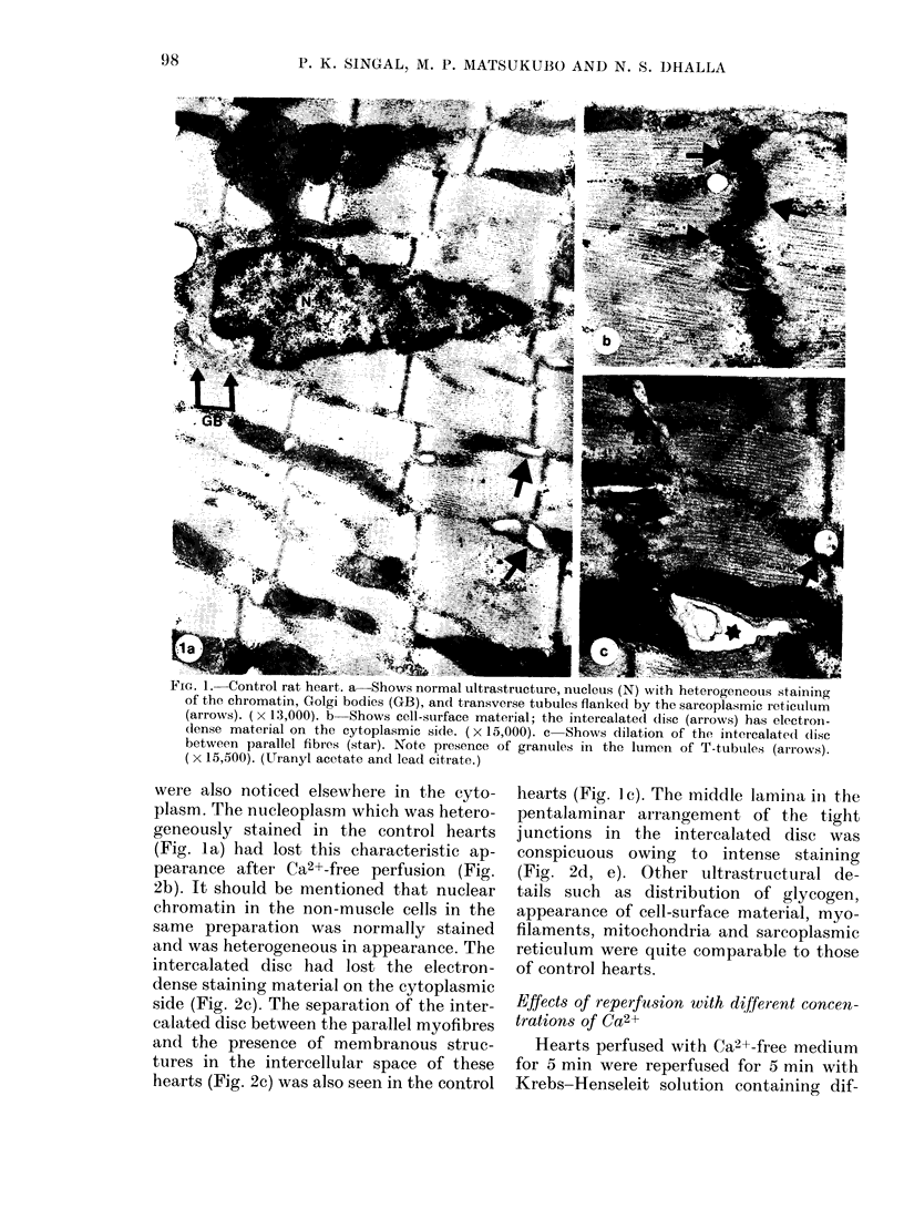
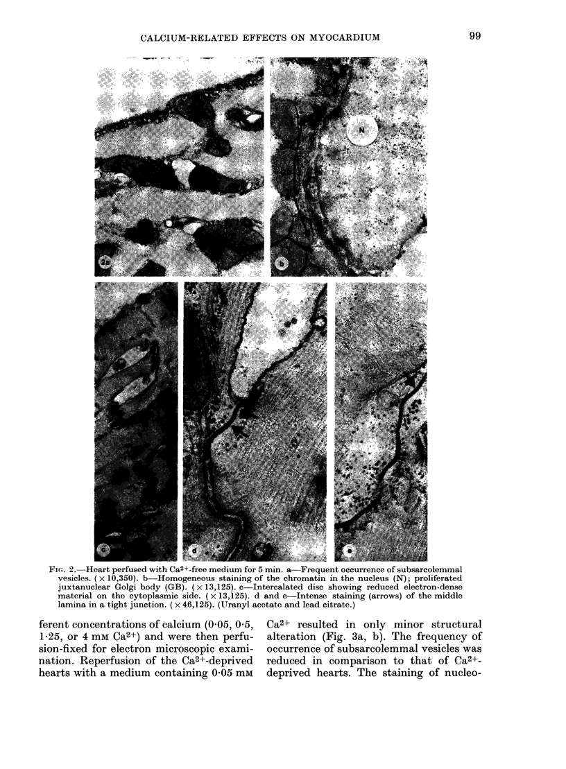
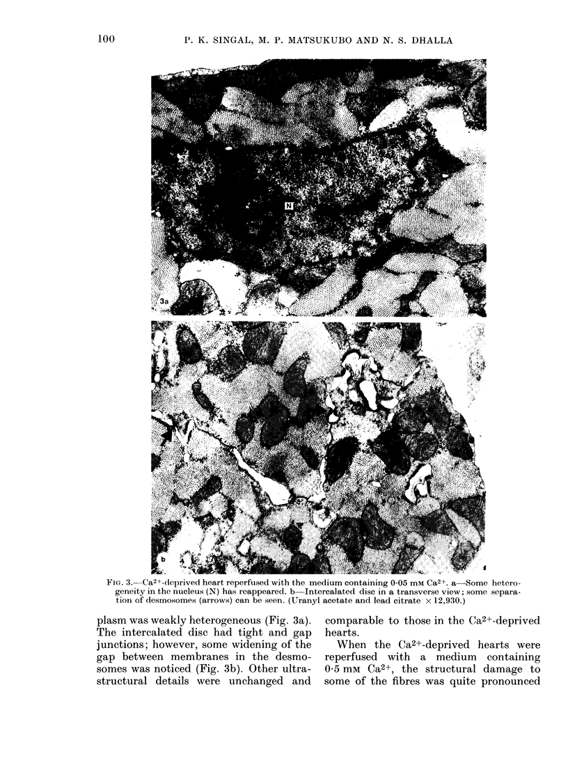
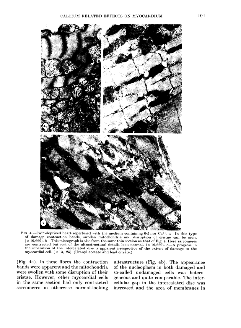
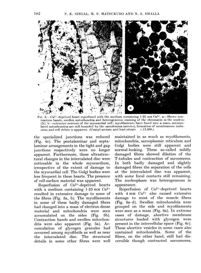
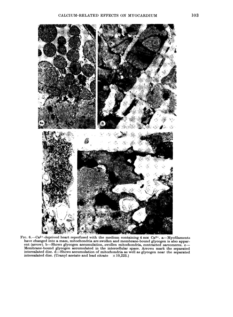
Isolated rat hearts, perfused with Ca2+-free medium for 5 min, were perfusion-fixed for ultrastructural studies. Increased pinocytotoic activity and characteristic changes in the staining of the intercalated disc coupled with conspicuous staining of the middle lamina in the tight junctions in these hearts indicated an alteration in the sarcolemmal activity as well as cell--cell relationship. The intracellular effects of Ca2+-free perfusion were indicated by the presence of an active Golgi body as well as the loss of heterogenic staining of the nucleoplasm. The latter appears to be a direct consequence of Ca2+ depletion of the cell, as heterogeneity reappeared upon reintroduction of the calcium in the perfusion medium. The extent of structural damage upon reintroduction of calcium was dependent upon the extracellular calcium concentration. These observations suggest that perfusion with Ca2+-free medium causes some membrane changes in the heart which probably make it more vulnerable to the reintroduction of calcium. Furthermore, the structural damage observed upon reintroduction of calcium appeared to be related to the amount of calcium overload.
Full text
PDF
Images in this article
Selected References
These references are in PubMed. This may not be the complete list of references from this article.
- Ashraf M., White F., Bloor C. M. Ultrastructural influence of reperfusing dog myocardium with calcium-free blood after coronary artery occlusion. Am J Pathol. 1978 Feb;90(2):423–434. [PMC free article] [PubMed] [Google Scholar]
- Bionk A. B., Ruigrok T. J., Maas A. H., Zimmerman A. N. Changes in high-energy phosphate compounds of isolated rat hearts during Ca2+-free perfusion and reperfusion with Ca2+. J Mol Cell Cardiol. 1976 Dec;8(12):973–979. doi: 10.1016/0022-2828(76)90078-x. [DOI] [PubMed] [Google Scholar]
- Fleckenstein A., Janke J., Döring H. J., Pachinger O. Ca overload as the determinant factor in the production of catecholamine-induced myocardial lesions. Recent Adv Stud Cardiac Struct Metab. 1973;2:455–466. [PubMed] [Google Scholar]
- Hearse D. J. Reperfusion of the ischemic myocardium. J Mol Cell Cardiol. 1977 Aug;9(8):605–616. doi: 10.1016/s0022-2828(77)80357-x. [DOI] [PubMed] [Google Scholar]
- Jennings R. B., Ganote C. E., Reimer K. A. Ischemic tissue injury. Am J Pathol. 1975 Oct;81(1):179–198. [PMC free article] [PubMed] [Google Scholar]
- LUFT J. H. Improvements in epoxy resin embedding methods. J Biophys Biochem Cytol. 1961 Feb;9:409–414. doi: 10.1083/jcb.9.2.409. [DOI] [PMC free article] [PubMed] [Google Scholar]
- Lee S. L., Dhalla N. S. Subcellular calcium transport in failing hearts due to calcium deficiency and overload. Am J Physiol. 1976 Oct;231(4):1159–1165. doi: 10.1152/ajplegacy.1976.231.4.1159. [DOI] [PubMed] [Google Scholar]
- Muir A. R. The effects of divalent cations on the ultrastructure of the perfused rat heart. J Anat. 1967 Apr;101(Pt 2):239–261. [PMC free article] [PubMed] [Google Scholar]
- Puri P. S., Varley K. G., Kim S. W., Barwinsky J., Cohen M., Dhalla N. S. Alterations in energy metabolism and ultrastructure upon reperfusion of the ischemic myocardium after coronary occlusion. Am J Cardiol. 1975 Aug;36(2):234–243. [PubMed] [Google Scholar]
- Shen A. C., Jennings R. B. Kinetics of calcium accumulation in acute myocardial ischemic injury. Am J Pathol. 1972 Jun;67(3):441–452. [PMC free article] [PubMed] [Google Scholar]
- Shen A. C., Jennings R. B. Myocardial calcium and magnesium in acute ischemic injury. Am J Pathol. 1972 Jun;67(3):417–440. [PMC free article] [PubMed] [Google Scholar]
- Yates J. C., Dhalla N. S. Structural and functional changes associated with failure and recovery of hearts after perfusion with Ca2+-free medium. J Mol Cell Cardiol. 1975 Feb;7(2):91–103. doi: 10.1016/0022-2828(75)90011-5. [DOI] [PubMed] [Google Scholar]


