Abstract
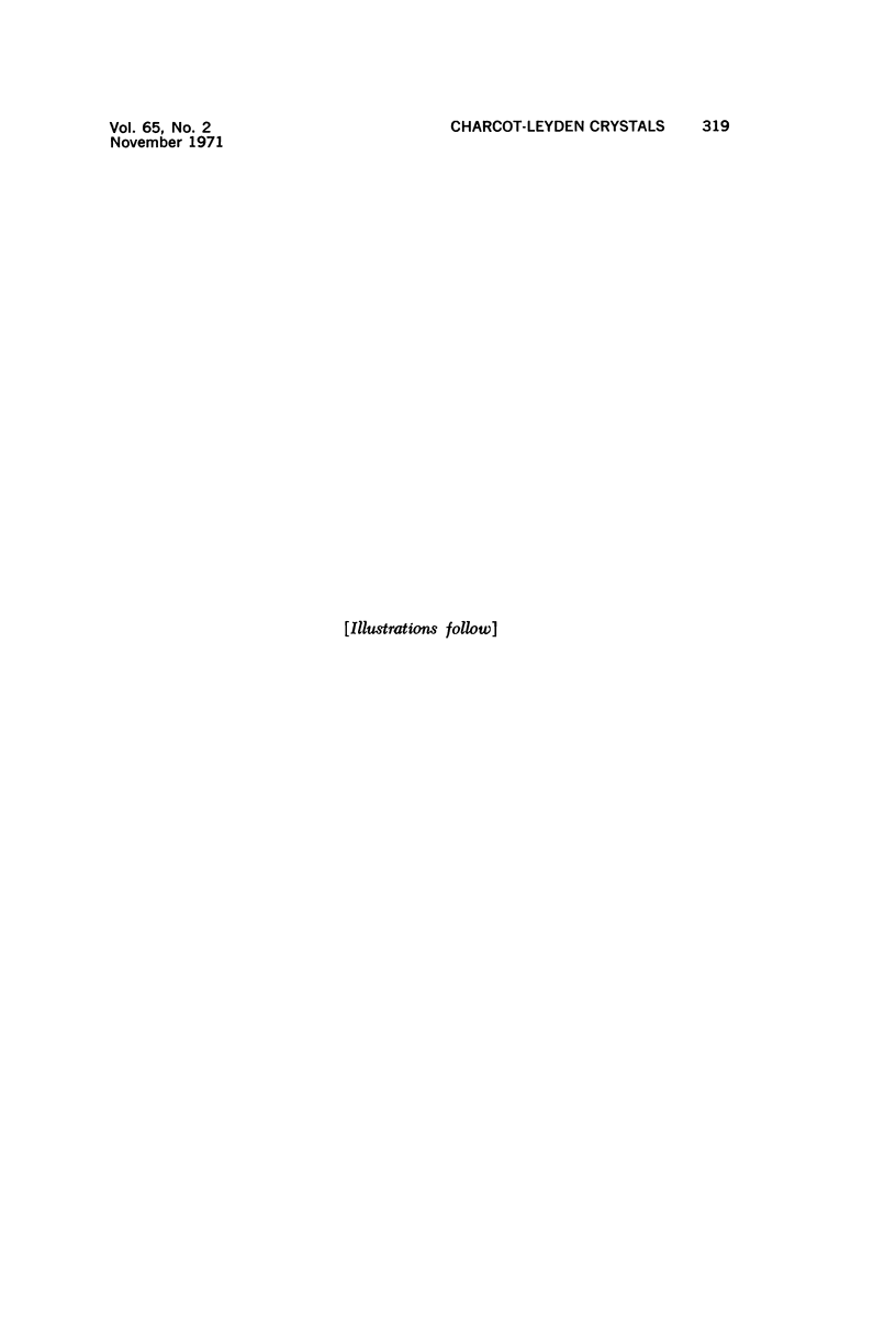
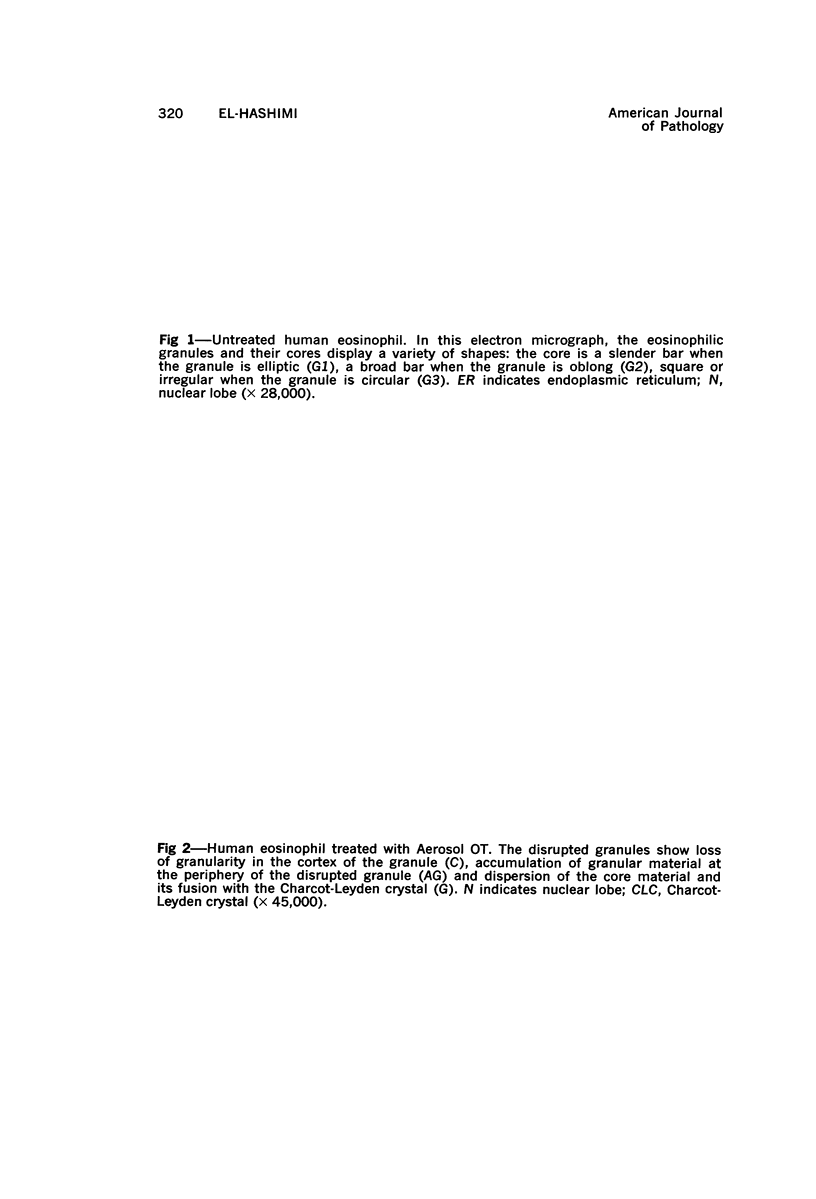
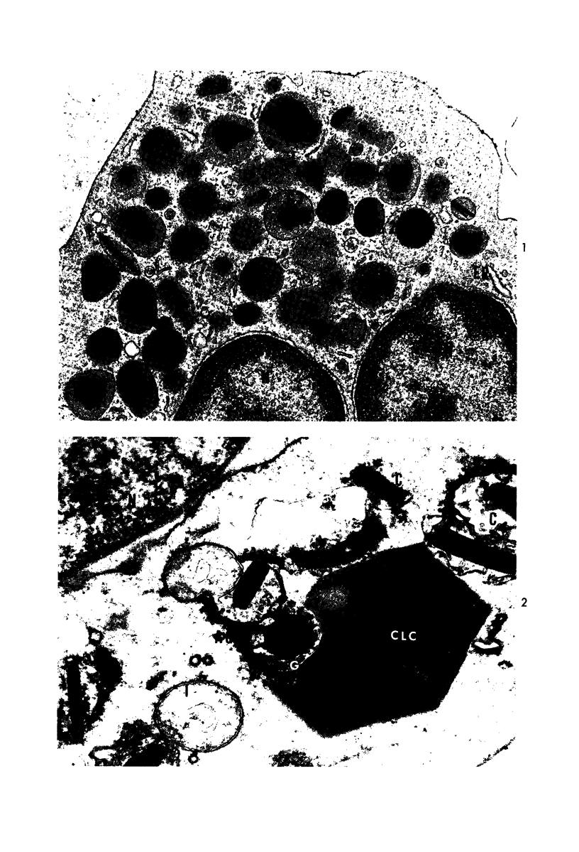
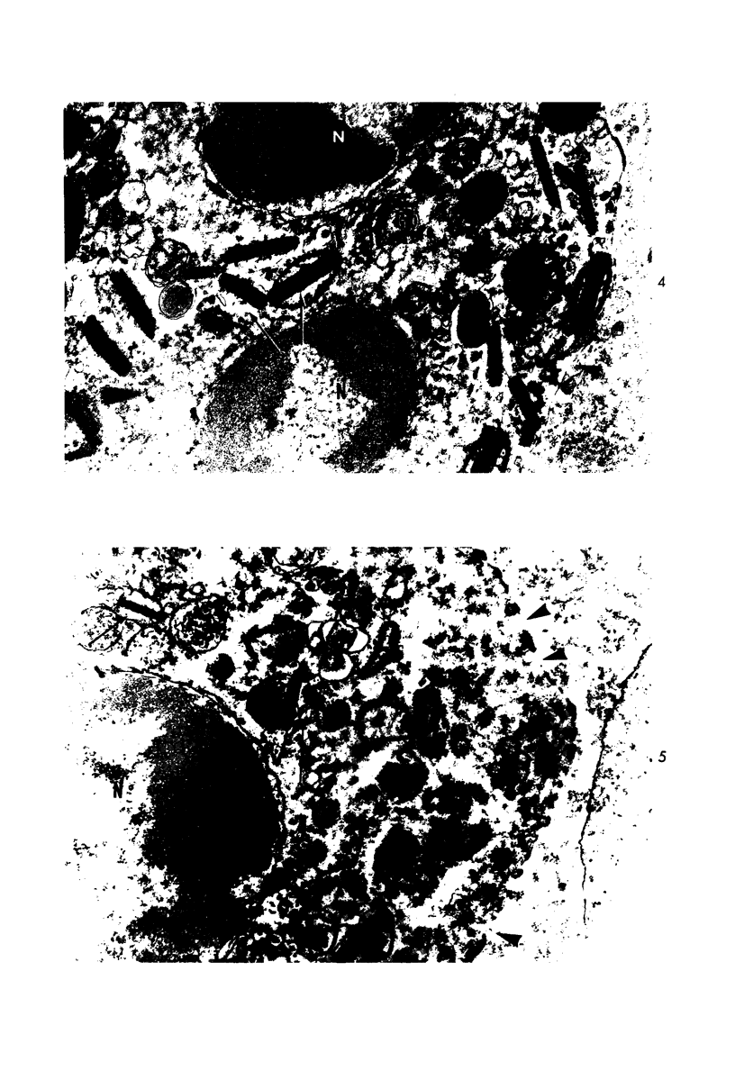
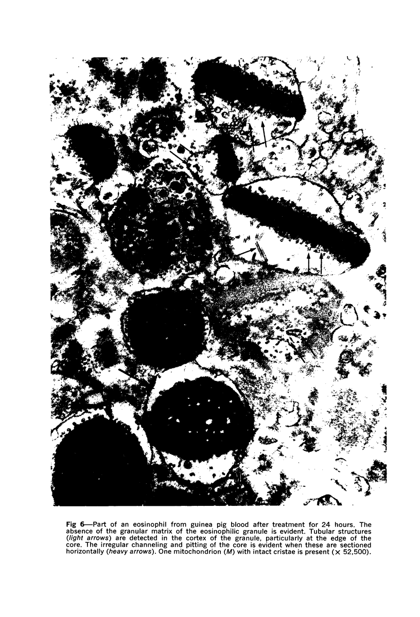
Charcot-Leyden crystals develop spontaneously in certain diseases of man and can be formed within minutes from eosinophils lysed with a surface-active agent (Aerosol-OT). Treated human eosinophils examined by electron microscopy showed the general features of cell lysis. After disruption of the eosinophilic granule of man, there remained an insoluble crystalline core, which was now more dispersed, a few tubular structures resembling microtubules and a fine granular accumulation around the periphery of the granule. It is suggested that such dispersion represents rearrangement of the crystalline structure allowing the material of the core to be incorporated into a Charcot-Leyden crystal. In guinea pig eosinophils, Charcot-Leyden crystals were not found even after prolonged lysis. By electron microscopy, the cellular changes in guinea pig eosinophils were generally similar to, but developed more slowly than, those in human eosinophils. Tubular structures detected in the cortical region of the granule were more numerous than thosenoted in the disrupted eosinophil granule of man. The different reaction to injury of the eosinophils of man and those of the guinea pig appears to be characteristic for each of the two species.
Full text
PDF
Images in this article
Selected References
These references are in PubMed. This may not be the complete list of references from this article.
- ARCHER G. T., BLACKWOOD A. FORMATION OF CHARCOT-LEYDEN CRYSTALS IN HUMAN EOSINOPHILS AND BASOPHILS AND STUDY OF THE COMPOSITION OF ISOLATED CRYSTALS. J Exp Med. 1965 Jul 1;122:173–180. doi: 10.1084/jem.122.1.173. [DOI] [PMC free article] [PubMed] [Google Scholar]
- ARCHER G. T., HIRSCH J. G. ISOLATION OF GRANULES FROM EOSINOPHIL LEUCOCYTES AND STUDY OF THEIR ENZYME CONTENT. J Exp Med. 1963 Aug 1;118:277–286. doi: 10.1084/jem.118.2.277. [DOI] [PMC free article] [PubMed] [Google Scholar]
- Anderson D. R. A method of preparing peripheral leucocytes for electron microscopy. J Ultrastruct Res. 1965 Oct;13(3):263–268. doi: 10.1016/s0022-5320(65)80075-2. [DOI] [PubMed] [Google Scholar]
- Anderson D. R. Ultrastructure of normal and leukemic leukocytes in human peripheral blood. J Ultrastruct Res. 1966 Sep;9:1–42. doi: 10.1016/s0022-5320(66)90002-5. [DOI] [PubMed] [Google Scholar]
- BARGMANN W., KNOOP A. Uber das Granulum des Eosinophilen. Z Zellforsch Mikrosk Anat. 1958;48(1):130–136. [PubMed] [Google Scholar]
- BEAVER P. C. Biological and physiopathological ideas on eosinophilia. Observations on the nature of Charcot-Leyden crystals. Bull Soc Pathol Exot Filiales. 1962 Jul-Aug;55:471–476. [PubMed] [Google Scholar]
- BEAVER P. C., DANARAJ T. J. Pulmonary ascariasis resembling eosinophilic lung; autopsy report with description of larvae in the bronchioles. Am J Trop Med Hyg. 1958 Jan;7(1):100–111. [PubMed] [Google Scholar]
- BESSIS M., THIERY J. P. Electron microscopy of human white blood cells and their stem cells. Int Rev Cytol. 1961;12:199–241. doi: 10.1016/s0074-7696(08)60541-0. [DOI] [PubMed] [Google Scholar]
- BUDDECKE E., ESSELLIER A. F., MARTI H. R. Uber die chemische Natur der Charcot-Leydenschen Kristalle. Hoppe Seylers Z Physiol Chem. 1956 Sep 25;305(4-6):203–206. [PubMed] [Google Scholar]
- Bainton D. F., Farquhar M. G. Segregation and packaging of granule enzymes in eosinophilic leukocytes. J Cell Biol. 1970 Apr;45(1):54–73. doi: 10.1083/jcb.45.1.54. [DOI] [PMC free article] [PubMed] [Google Scholar]
- Cotran R. S., Litt M. The entry of granule-associated peroxidase into the phagocytic vacuoles of eosinophils. J Exp Med. 1969 Jun 1;129(6):1291–1306. doi: 10.1084/jem.129.6.1291. [DOI] [PMC free article] [PubMed] [Google Scholar]
- FREEMAN J. A., SPURLOCK B. O. A new epoxy embedment for electron microscopy. J Cell Biol. 1962 Jun;13:437–443. doi: 10.1083/jcb.13.3.437. [DOI] [PMC free article] [PubMed] [Google Scholar]
- Georgsson G., Wessel W. Elektronenmikroskopische und enzymatisch-analytische Untersuchung eines eosinophilen Granuloms. Virchows Arch Pathol Anat Physiol Klin Med. 1967;343(2):176–188. [PubMed] [Google Scholar]
- HORNUNG M. Preservation recrystallization and preliminary biochemical characterization of Charcot-Leyden crystals. Proc Soc Exp Biol Med. 1962 May;110:119–124. doi: 10.3181/00379727-110-27442. [DOI] [PubMed] [Google Scholar]
- Hudson G. Eosinophil granules and phosphotungstic acid: an electron microscope study of guinea-pig bone marrow. Exp Cell Res. 1966 Feb;41(2):265–273. doi: 10.1016/s0014-4827(66)80134-9. [DOI] [PubMed] [Google Scholar]
- LITT M. STUDIES IN EXPERIMENTAL EOSINOPHILIA. VI. UPTAKE OF IMMUNE COMPLEXES BY EOSINOPHILS. J Cell Biol. 1964 Nov;23:355–361. doi: 10.1083/jcb.23.2.355. [DOI] [PMC free article] [PubMed] [Google Scholar]
- OSHIMA T. Standardization of techniques for infecting mice with Toxocara canis and observations on the normal migration routes of the larvae. J Parasitol. 1961 Aug;47:652–656. [PubMed] [Google Scholar]
- Scott R. E., Horn R. G. Fine structural features of eosinophile granulocyte development in human bone marrow. Evidence for granule secretion. J Ultrastruct Res. 1970 Oct;33(1):16–28. doi: 10.1016/s0022-5320(70)90116-4. [DOI] [PubMed] [Google Scholar]
- Seeman P. M., Palade G. E. Acid phosphatase localization in rabbit eosinophils. J Cell Biol. 1967 Sep;34(3):745–756. doi: 10.1083/jcb.34.3.745. [DOI] [PMC free article] [PubMed] [Google Scholar]
- Spurlock B. O., Skinner M. S., Kattine A. A. A simple rapid method for staining epoxy-embedded specimens for light microscopy with the polychromatic stain paragon-1301. Am J Clin Pathol. 1966 Aug;46(2):252–258. doi: 10.1093/ajcp/46.2.252. [DOI] [PubMed] [Google Scholar]
- VERCAUTEREN R. The properties of the isolated granules from blood eosinophiles. Enzymologia. 1953 Apr 15;16(1):1–13. [PubMed] [Google Scholar]
- WELSH R. A. The genesis of the Charcot-Leyden crystal in the eosinophilic leukocyte of man. Am J Pathol. 1959 Nov-Dec;35:1091–1103. [PMC free article] [PubMed] [Google Scholar]
- ZUCKER-FRANKLIN D., HIRSCH J. G. ELECTRON MICROSCOPE STUDIES ON THE DEGRANULATION OF RABBIT PERITONEAL LEUKOCYTES DURING PHAGOCYTOSIS. J Exp Med. 1964 Oct 1;120:569–576. doi: 10.1084/jem.120.4.569. [DOI] [PMC free article] [PubMed] [Google Scholar]
- Zucker-Franklin D., Davidson M., Thomas L. The interaction of mycoplasmas with mammalian cells. I. HeLa cells, neutrophils, and eosinophils. J Exp Med. 1966 Sep 1;124(3):521–532. doi: 10.1084/jem.124.3.521. [DOI] [PMC free article] [PubMed] [Google Scholar]


