Abstract
Pacemaker migration is a rare, but important, complication of pacemaker insertion mainly documented in children. We report the case of a 60-year-old woman who was admitted with right iliac fossa pain thought to be caused by appendicitis. She was noted to have both an epicardial and endocardial pacemaker in situ. Imaging and laparoscopy revealed migration of the epicardial pacemaker to the right iliac fossa. We describe the possible mechanisms of pacemaker migration.
Keywords: Pacemaker, Migration, Laparotomy
A 60-year-old Caucasian female presented with a 3-day history of right lower quadrant abdominal pain, vomiting and pyrexia.
She had a past medical history of congenital aortic stenosis complicated by aortic regurgitation for which she underwent aortic valve replacement (AVR) at Harefield Hospital with a fresh homograft in 1990. She developed third-degree heart block following the valve replacement and, thus, an endocardial pacemaker was inserted. Ten months prior to her current admission, the homograft was replaced with a metallic AVR due to worsening symptoms of cardiac failure and an epicardial pacemaker was inserted deep to the posterior rectus sheath in the epigastric region. This epicardial pacemaker would only be activated once the endocardial pacemaker loses function. Her only medication on admission was warfarin.
On examination, her blood pressure was 153/96 mmHg, pulse was 95 beats/min in sinus rhythm and her oxygen saturations was 98% on air. She was pyrexial with a temperature of 37.5°C. Her cardiorespiratory examination was unremarkable. Abdominal examination revealed tenderness in the right iliac fossa but there was no rebound tenderness or guarding and bowel sounds were normal.
Her initial blood tests were normal: haemoglobin 135 g/l, white cell count 4.7 × 10⁹/l, platelets 362 g/l, and C-reactive protein < 5.0. However her INR was 3.5.
Her ECG revealed a paced rhythm of 95 beats/min. Her chest radiograph revealed one pacemaker sited in the left pectoral region and on the abdominal radiograph an epicardial pacemaker device in the right lower abdomen.
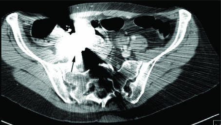
The initial impression was that of appendicitis. Her warfarin was stopped and she was commenced on i.v. heparin. She was started on ciprofloxacin. An abdominal and pelvic computerised tomography (CT) scan was performed which revealed an intraperitoneal pacing device in the right iliac fossa which produced substantial artefact. The appendix looked prominent but there was no associated fluid collection or mass lesion (Fig. 1).
Figure 1.
CT abdomen showing intraperitoneal migration of the pacemaker. Arrow shows site of pacemaker in the right iliac fossa.
She continued to have right iliac fossa pain and so a diagnostic laparoscopy was performed 3 days after admission. The findings were that of the epicardial pacemaker lying in the right iliac fossa adjacent to the caecum with its wires intimately related to the caecum. The gross appearance of the appendix and pelvic organs was normal. The decision was made to terminate the operation and the patient was transferred to Harefield Hospital where she underwent a laparotomy. At surgery, the pacemaker was situated postero-inferiorly to the caecum. The pacemaker wires entered near the epigastric region immediately beneath the cutaneous scar. The pacemaker wires were divided flush with the peritoneal surface and the pacemaker with wires were removed. Her symptoms improved and she was discharged 4 days later after adequate warfarinisation.
Discussion
Cardiac pacemaker migration is an uncommon complication with most cases observed in the paediatric population.1–3 Pacemaker migration in adults has been rarely observed.
The patient in this report had undergone insertion of the abdominal pacemaker only 10 months prior to her presentation with abdominal pain. Pacemaker migration in this short duration has not been previously described in the literature. The location of the pacing device adjacent to the caecum was the cause of the patient's right iliac fossa pain which was thought initially to be due to appendicitis. CT scanning was invaluable in confirming the intraperitoneal location of the pacemaker and laparoscopy was useful in excluding appendicitis. The presence of the epicardial pacing leads adjacent to the caecum and lack of cardiothoracic services at our hospital prompted transfer of the patient to a cardiothoracic centre for her laparotomy.
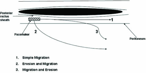
There are three mechanisms of pacemaker migration as illustrated in Figure 2. These include simple migration, migration and erosion and erosion followed by migration. In this case, we believe the pacemaker first eroded into the peritoneal cavity and then migrated into the right lower quadrant fossa as the pacing wires were at the site of the skin incision for the abdominal pacemaker in the right upper quadrant.
Figure 2.
Paramedian sagittal section through abdominal wall demonstrating the different mechanisms of pacemaker migration.
There are two techniques for abdominal implantation of epicardial pacemakers. They may be inserted in the subcutaneous tissues when the patient has adequate adipose tissue to prevent the pacemaker eroding through the skin or alternatively in the retrofascial preperitoneal plane as with our patient.
Pacemaker migration in adults has been rarely observed.4 A 77-year-old woman had an epicardial electrode implanted in the diaphragmatic surface of the right ventricle which was connected to a pacemaker inserted behind the rectus abdominus muscle. Eight years after placement of the pacemaker, the patient was admitted with abdominal pain and an absence of pacemaker activity. The abdominal radiograph demonstrated the pulse generator to have migrated into the pelvis and laparotomy revealed it to be located behind the urinary bladder.
Cases of pacemaker migration in children have also been described despite insertion in the retrofascial abdominal plane. In one case, the pacemaker had eroded through the parietal peritoneum and into large bowel and had passed through the rectum; in a second case, the pacemaker was found floating inside the peritoneum cavity at laparotomy.1,2 In both cases, the time from cardiac pacemaker insertion to symptoms was nearly 2 years.
Conclusions
Pacemaker migration must be considered in all patients presenting with abdominal pain and a history of an abdominally placed pacemaker. Careful evaluation by way of detailed history, examination and CT scanning allows safe management of this uncommon problem. The need for careful attention to the insertion and fixation of pacemakers must also be stressed to help prevent this rare and potentially fatal complication.
References
- 1.Manikoth P, Qora HT, Sajwani MJ, Valliattu J, Pawar N, Venkatraman M. A mysterious journey of a cardiac pacemaker. Int J Cardiol. 2006;107:287–8. doi: 10.1016/j.ijcard.2005.01.054. [DOI] [PubMed] [Google Scholar]
- 2.Salim MA, DiSessa TG, Watson DC. The wandering pacemaker: intraperitoneal migration of an epicardially placed pacemaker and femoral nerve stimulation. Pediatr Cardiol. 1999;20:164–6. doi: 10.1007/s002469900430. [DOI] [PubMed] [Google Scholar]
- 3.Abrams S, Peart I. Twiddler's syndrome in children: an unusual cause of pacemaker failure. Br Heart J. 1995;73:190–2. doi: 10.1136/hrt.73.2.190. [DOI] [PMC free article] [PubMed] [Google Scholar]
- 4.Garcia-Bengochea J, Rubio J, Sierra J, Fernandez A. Pacemaker migration into the pouch of Douglas. Texas Heart Inst J. 2003;30:83. [PMC free article] [PubMed] [Google Scholar]


