Abstract
Although lesions in the organs of hamsters resemble those of monkeys and guinea-pigs, the extension of the pathological process to the brain is a new feature, so far found only in hamsters.
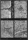
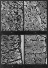
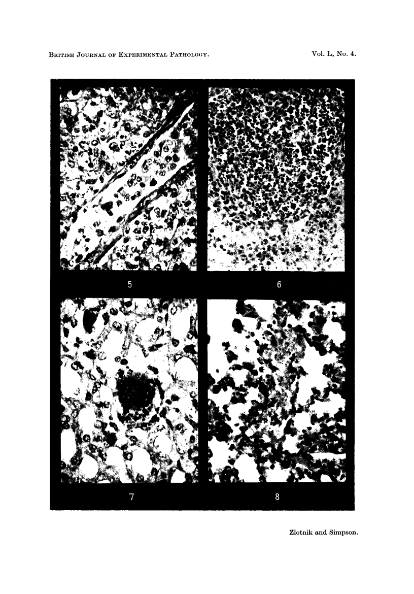
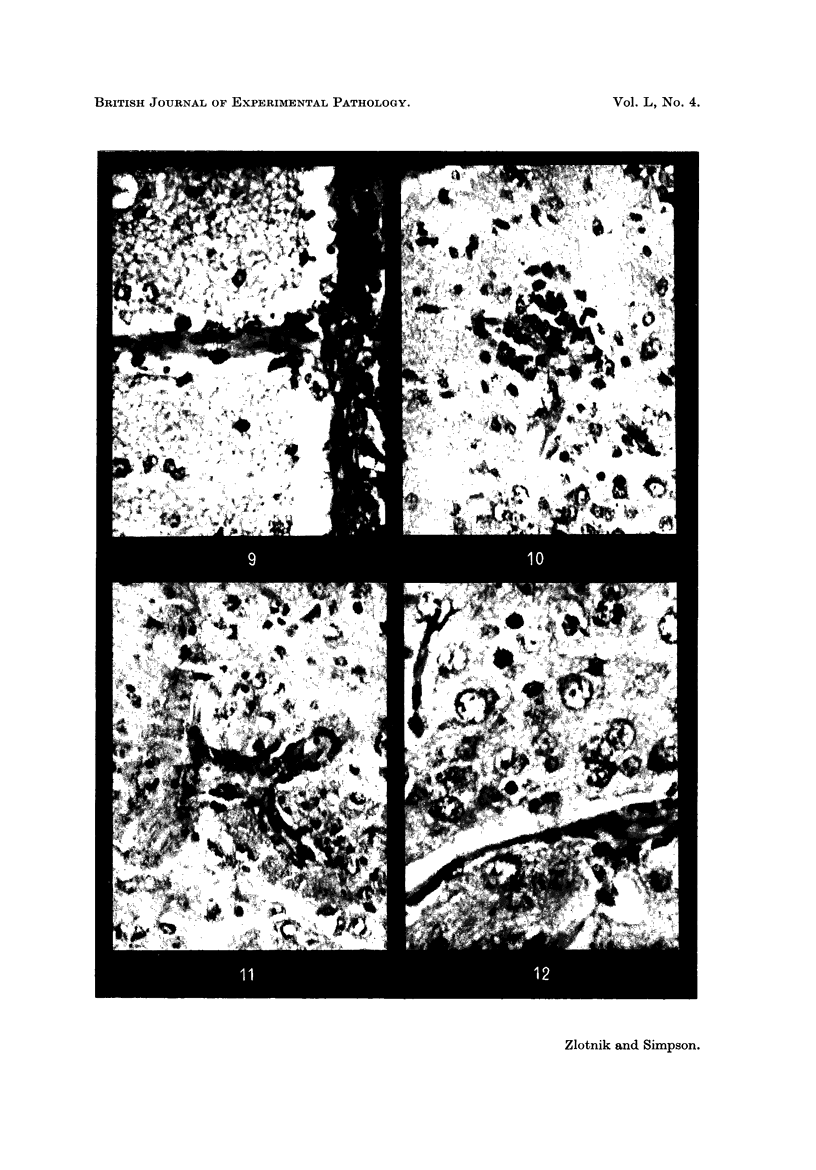
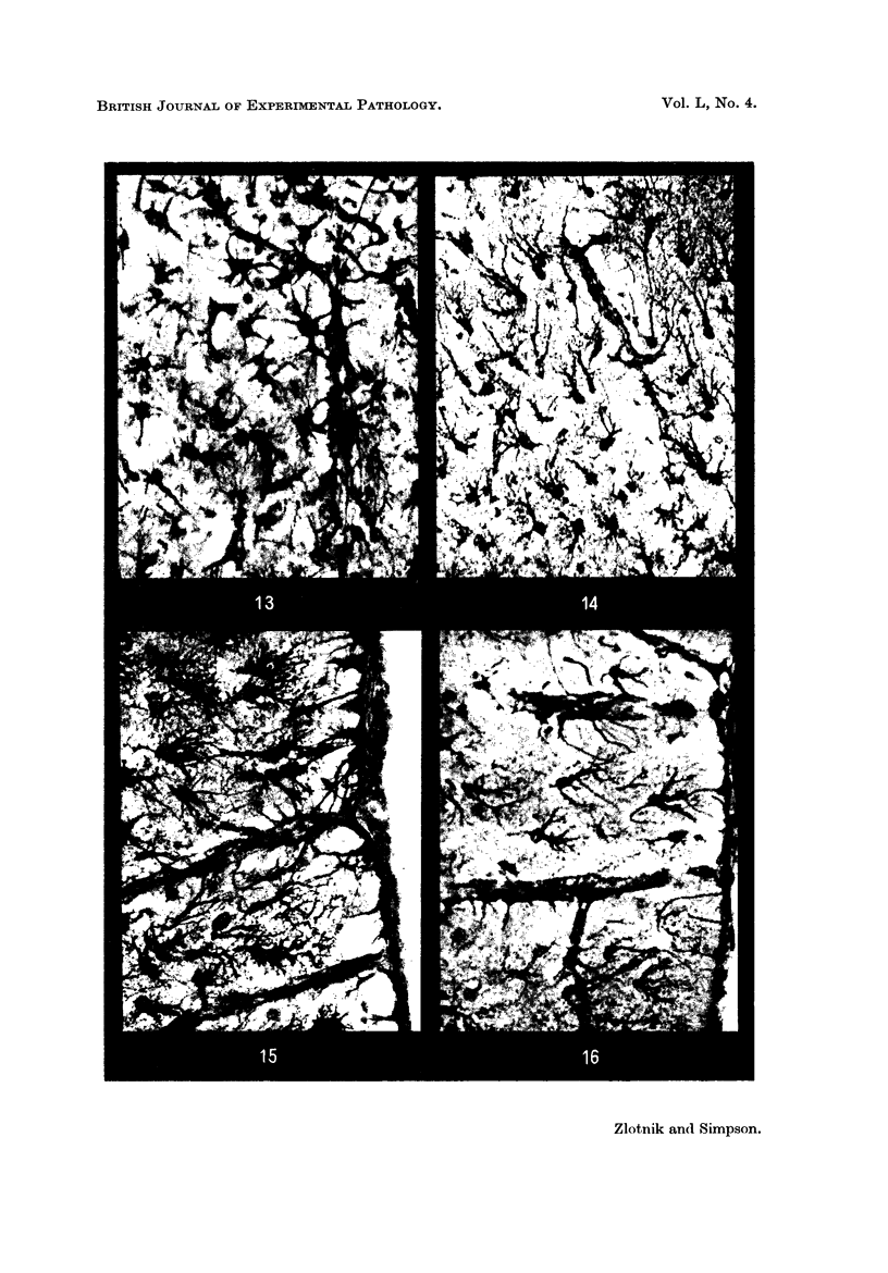
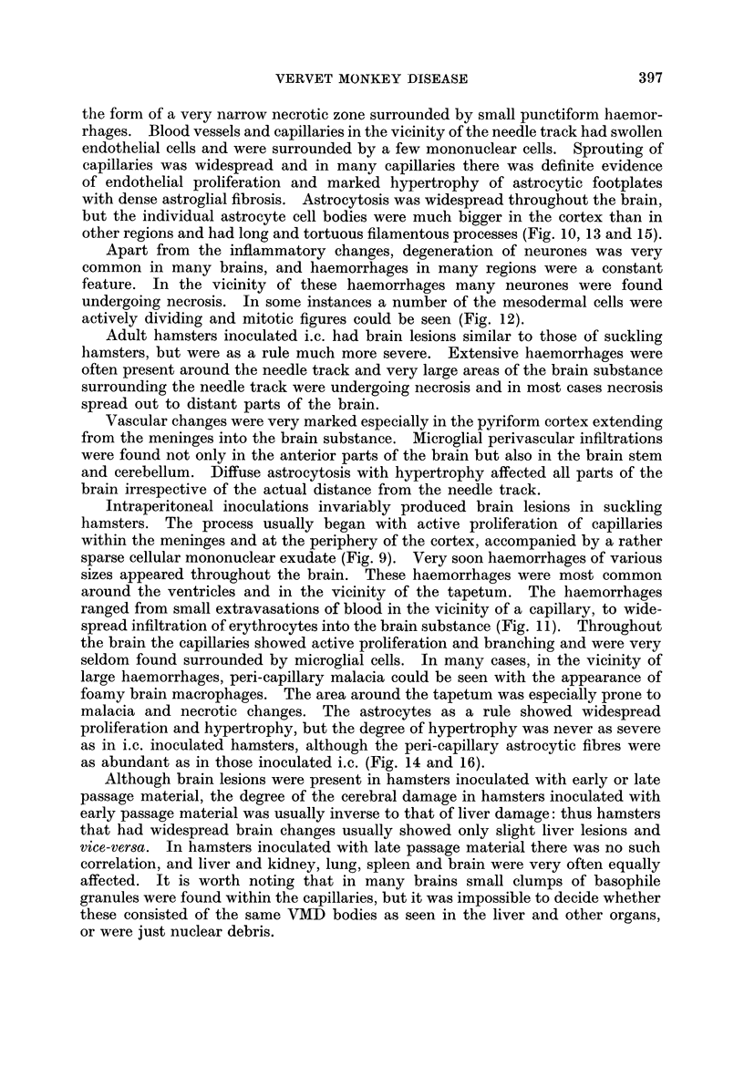
The brain lesions in the hamster consist of capillary proliferations, haemorrhages, microglial changes and very severe astrogliosis. Necrosis of the needle track in i.c. inoculated hamsters is usually very pronounced.
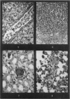
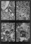
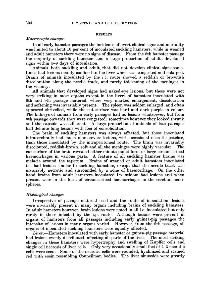
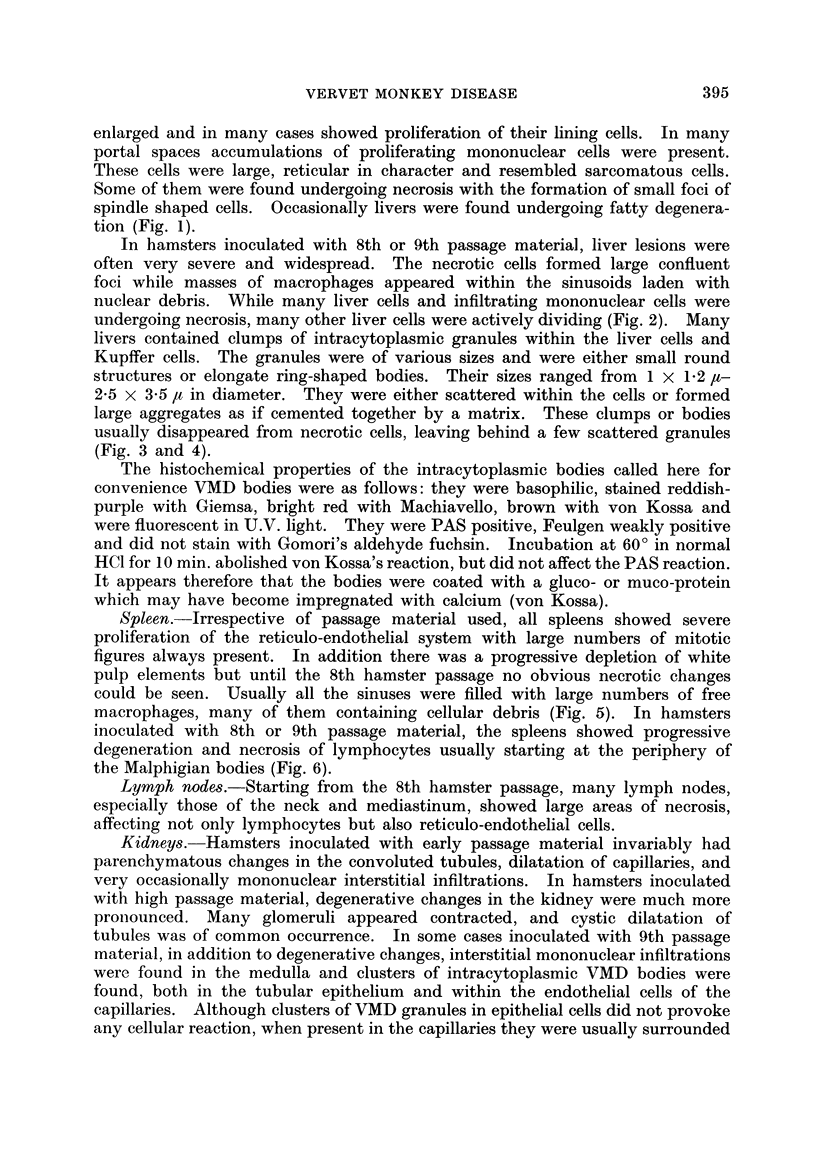
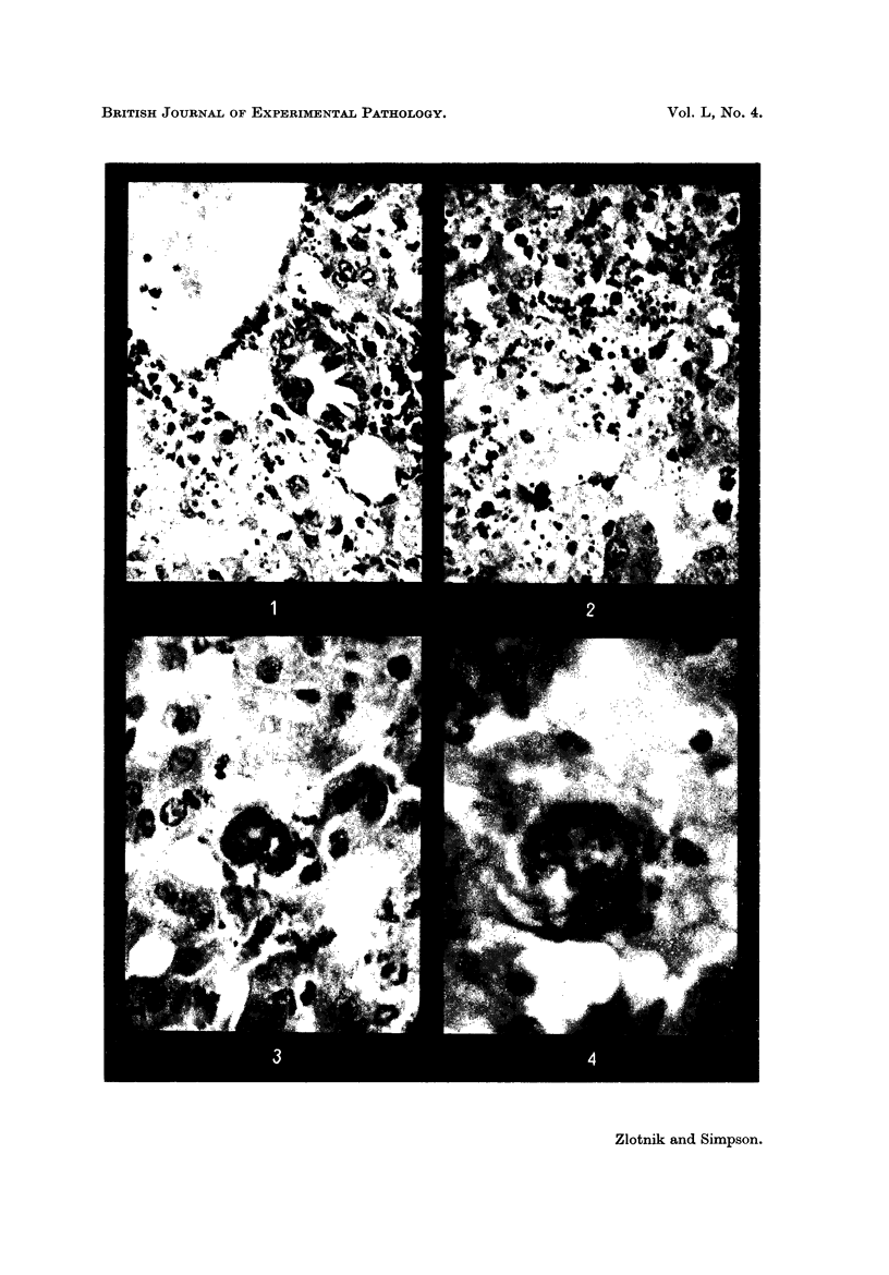
Intracytoplasmic bodies believed to be the infective agent are found in hamsters not only in the liver but also in the kidneys and lung. These bodies are pleomorphic vary in size from 1-3·5 μ in diameter and they are either round or elliptical, and very commonly appear in the liver as ring-shaped structures.
Full text
PDF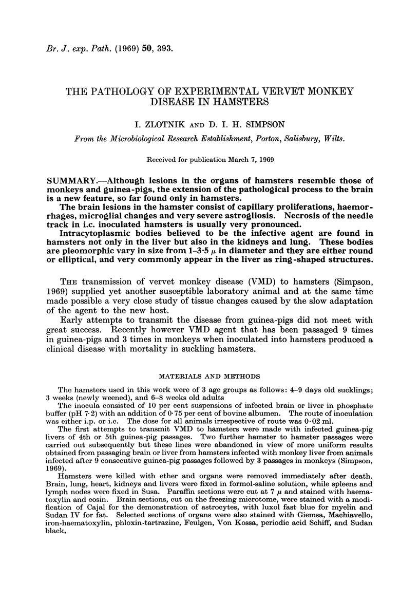

Images in this article
Selected References
These references are in PubMed. This may not be the complete list of references from this article.
- Haas R., Maass G., Oehlert W. Untersuchungen zur Tierpathogenität eines von Cercopithecus aethiops übertragenen menschenpathogenen Erregers. Med Klin. 1968 Aug 30;63(35):1359–passim. [PubMed] [Google Scholar]
- Kissling R. E., Robinson R. Q., Murphy F. A., Whitfield S. G. Agent of disease contracted from green monkeys. Science. 1968 May 24;160(3830):888–890. doi: 10.1126/science.160.3830.888. [DOI] [PubMed] [Google Scholar]
- Simpson D. I., Zlotnik I., Rutter D. A. Vervet monkey disease. Experiment infection of guinea pigs and monkeys with the causative agent. Br J Exp Pathol. 1968 Oct;49(5):458–464. [PMC free article] [PubMed] [Google Scholar]
- Zlotnik I. The reaction of astrocytes to acute virus infections of the central nervous system. Br J Exp Pathol. 1968 Dec;49(6):555–564. [PMC free article] [PubMed] [Google Scholar]


