Abstract
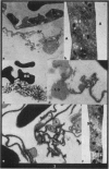
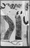
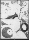
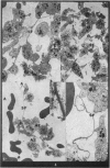
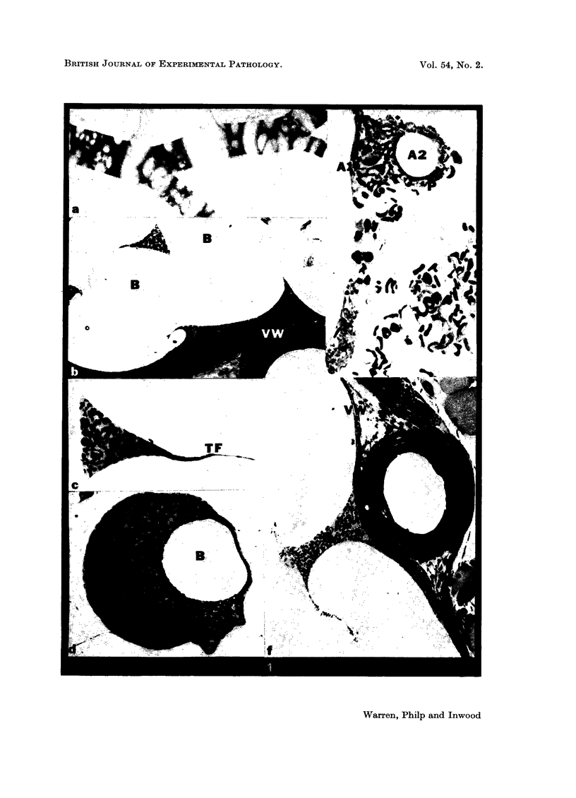
The pathogenesis of the ill effects following air embolism cannot be attributed solely to the space occupying and surface tension effects of the air bubbles altering the normal flow of blood through the vasculature. Decompression sickness was induced in rats and the following features of this process observed by electron microscopy in the vessels of the mesentery: imprisonment of blood elements (especially platelets) took place within the various enclosures created by the boundaries set up by different sized air bubbles between the layer of blood and the vessel walls, and the air/blood interface. Air bubble size and the thickness of the film of blood between bubbles varied enormously.
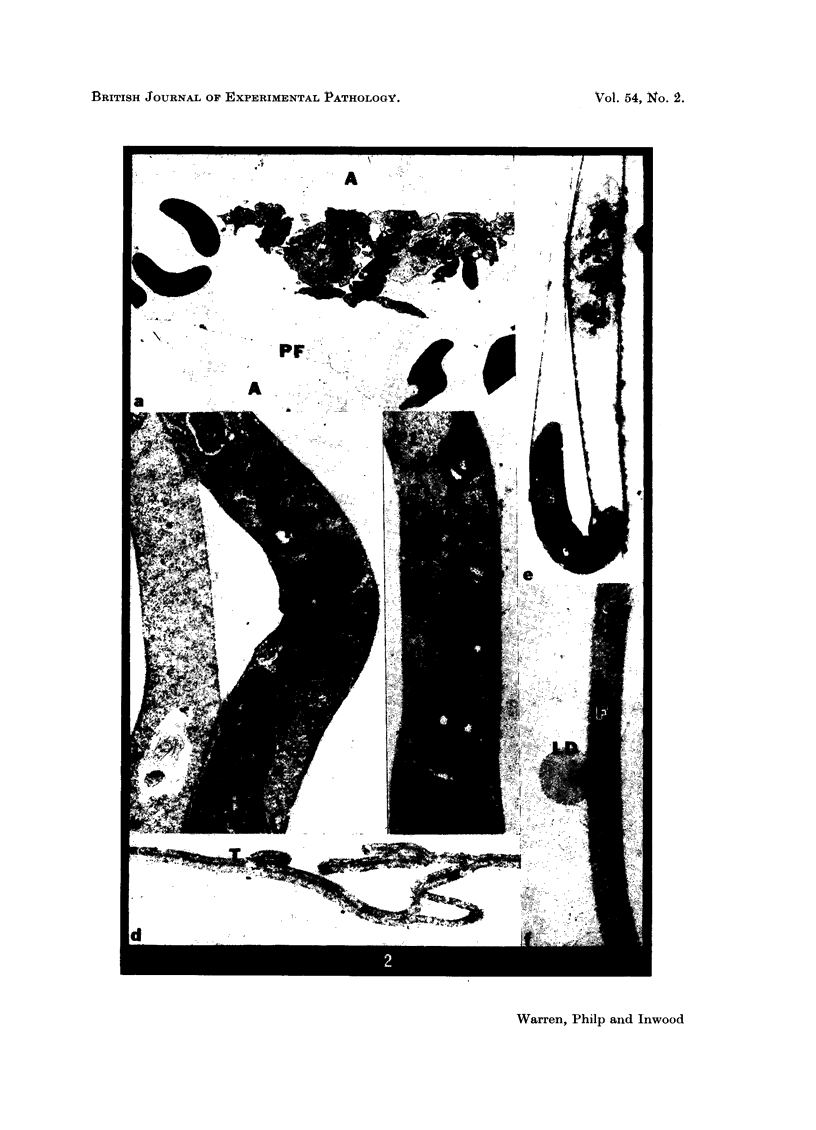
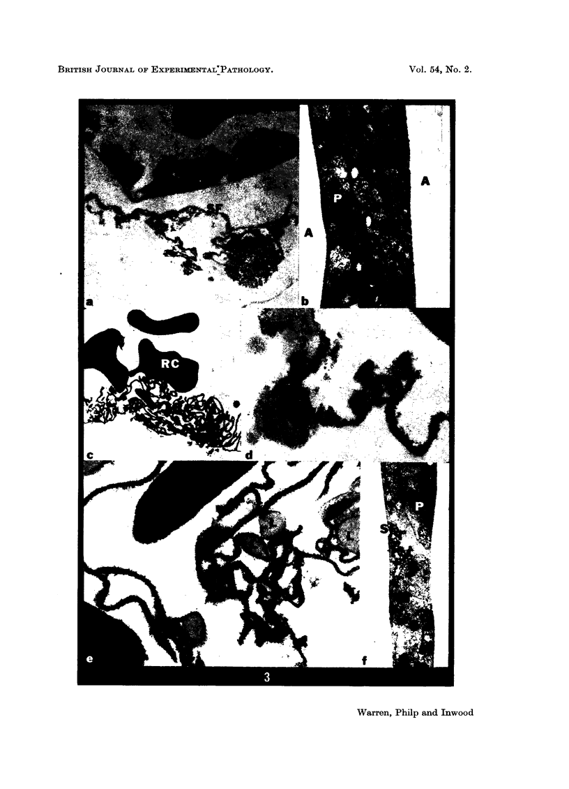
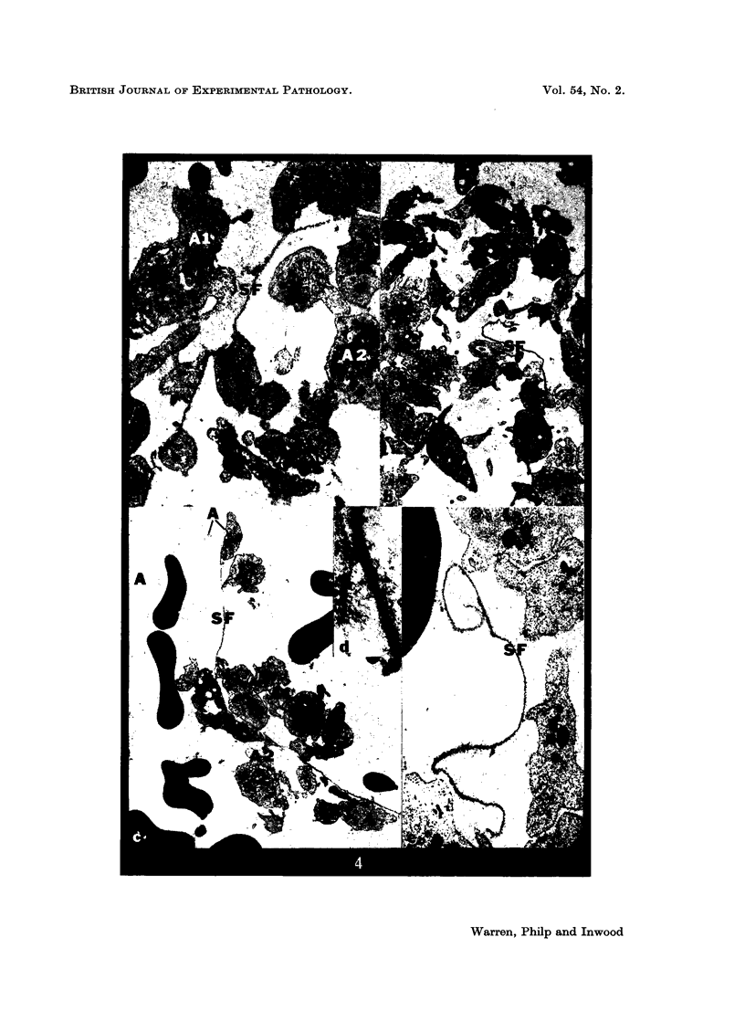
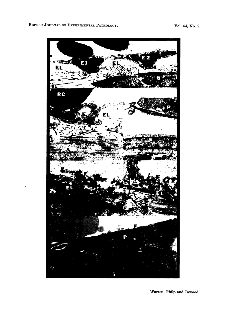
The air/blood interface had the following characteristics: (1) A surface associated protein layer measuring 20 nm which coated the air bubbles and which could slide off the bubble of origin and float freely in the blood. (2) Material morphologically similar to the surface layer was found away from the surface and included small lipid droplets between its layers, and platelets adhered to this to form small aggregates suspended from the interface. (3) The surface layer fused with like laminae and was found within the fluid blood in the vessel, sometimes with adherent platelet aggregates. (4) Platelet adhesion to the bubble interface with the formation of platelet aggregates of an early type i.e. without gross fibrin formation within the aggregates. (5) Pressure damage to underlying endothelial cells by the passage of air bubbles under pressure resulted in herniation of the endothelial cells through fenestrations in the more rigid structures of the vessel wall. (6) Deposits of fibrin on the walls of the vessels were noted after endothelial damage. (7) Lipid droplets were found attached to the surface associated protein on the air side of the air/blood interface and were also found incorporated within it, i.e. covered by this layer on both sides, in which case they took on an ellipsoidal shape.
These alterations would account for certain features of decompression sickness, such as the drop in platelets, even when the mechanical blockage by air bubbles was not of great significance.
Full text
PDF
Images in this article
Selected References
These references are in PubMed. This may not be the complete list of references from this article.
- Clark M. L., Philip R. B., Gowdey C. W. Changes in platelets and lipids in experimental aeroembolism and bends. Aerosp Med. 1969 Oct;40(10):1094–1098. [PubMed] [Google Scholar]
- Niewiarowski S., Regoeczi E., Stewart G. J., Senyl A. F., Mustard J. F. Platelet interaction with polymerizing fibrin. J Clin Invest. 1972 Mar;51(3):685–699. doi: 10.1172/JCI106857. [DOI] [PMC free article] [PubMed] [Google Scholar]
- Philp R. B., Gowdey C. W., Prasad M. Changes in blood lipid concentration and cell counts following decompression sickness in rats and the influence of dietary lipid. Can J Physiol Pharmacol. 1967 Nov;45(6):1047–1059. doi: 10.1139/y67-122. [DOI] [PubMed] [Google Scholar]
- Philp R. B., Inwood M. J., Warren B. A. Interactions between gas bubbles and components of the blood: implications in decompression sickness. Aerosp Med. 1972 Sep;43(9):946–953. [PubMed] [Google Scholar]
- Philp R. B., Schacham P., Gowdey C. W. Involvement of platelets and microthrombi in experimental decompression sickness: similarities with disseminated intravascular coagulation. Aerosp Med. 1971 May;42(5):494–502. [PubMed] [Google Scholar]
- Scarborough D. E., Mason R. G., Dalldorf F. G., Brinkhous K. M. Morphologic manifestations of blood-solid interfacial reactions. A scanning and transmission electron microscopic study. Lab Invest. 1969 Feb;20(2):164–169. [PubMed] [Google Scholar]


