

Abstract
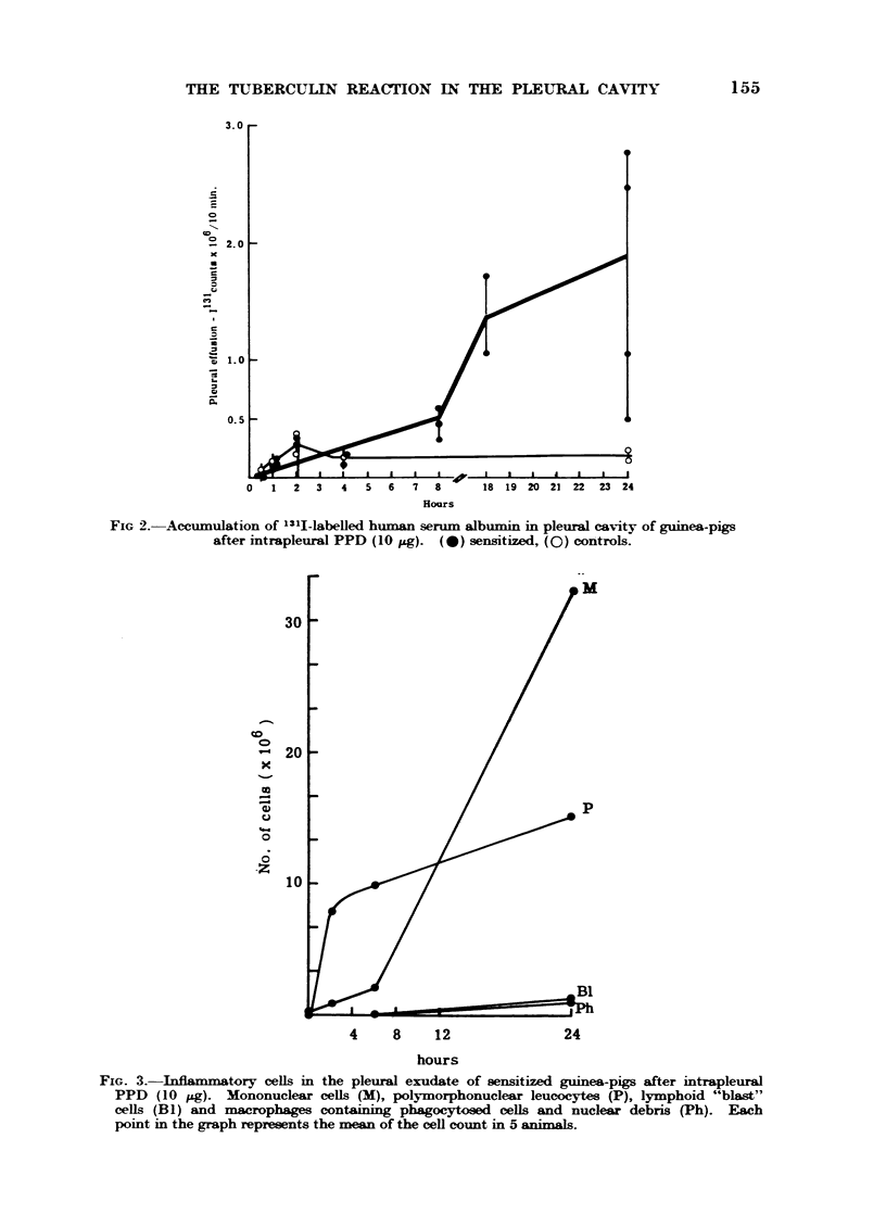
Tuberculin (PPD) injected into the pleural cavity of sensitized guinea-pigs results in the development of a large protein-rich effusion over a period of 24 hours. In the early stages the effusion contains large numbers of polymorphonuclear leucocytes, but later mononuclear cells (lymphocytes and monocytes) predominate. At 24 hours a small percentage of the lymphocytes have the character of lymphoid blast cells, while some of the monocytes contain phagocytosed polymorphonuclear cells or nuclear debris.
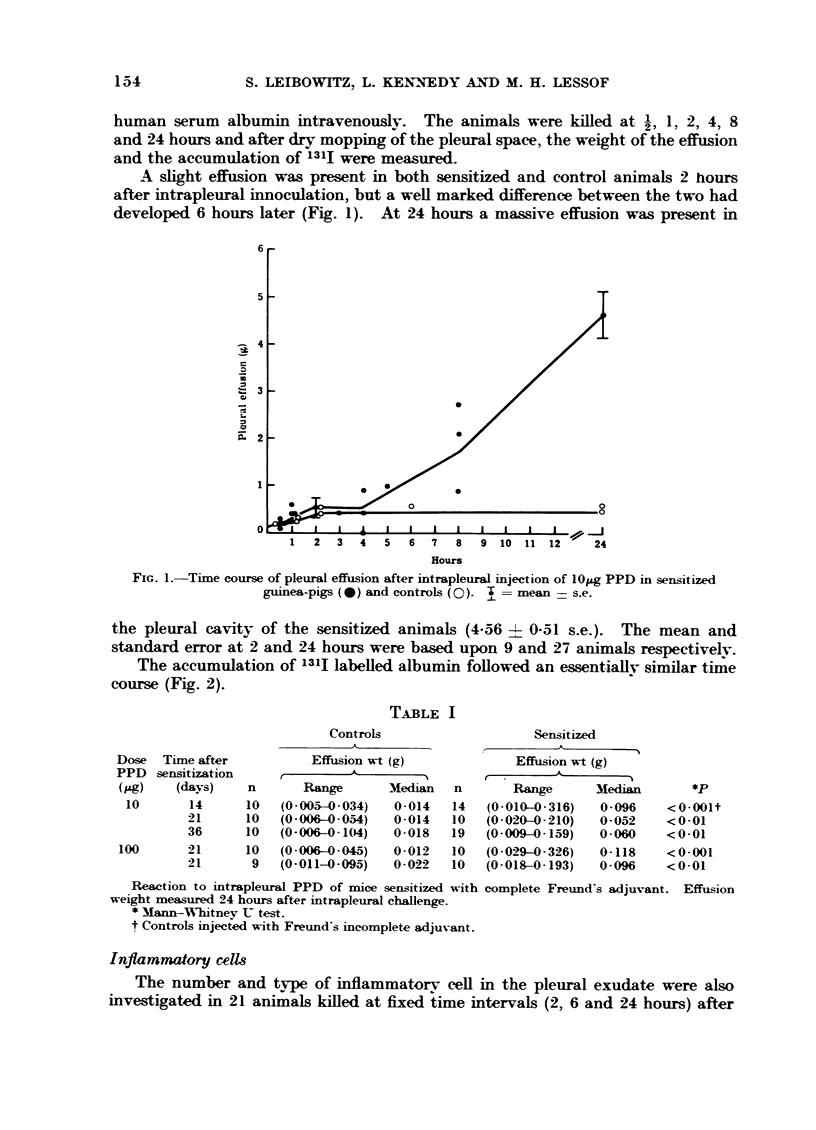
A similar phenomenon can be elicited in mice sensitized with complete Freund's adjuvant. However, in these animals the volume of the effusion is small and difficult to measure accurately.
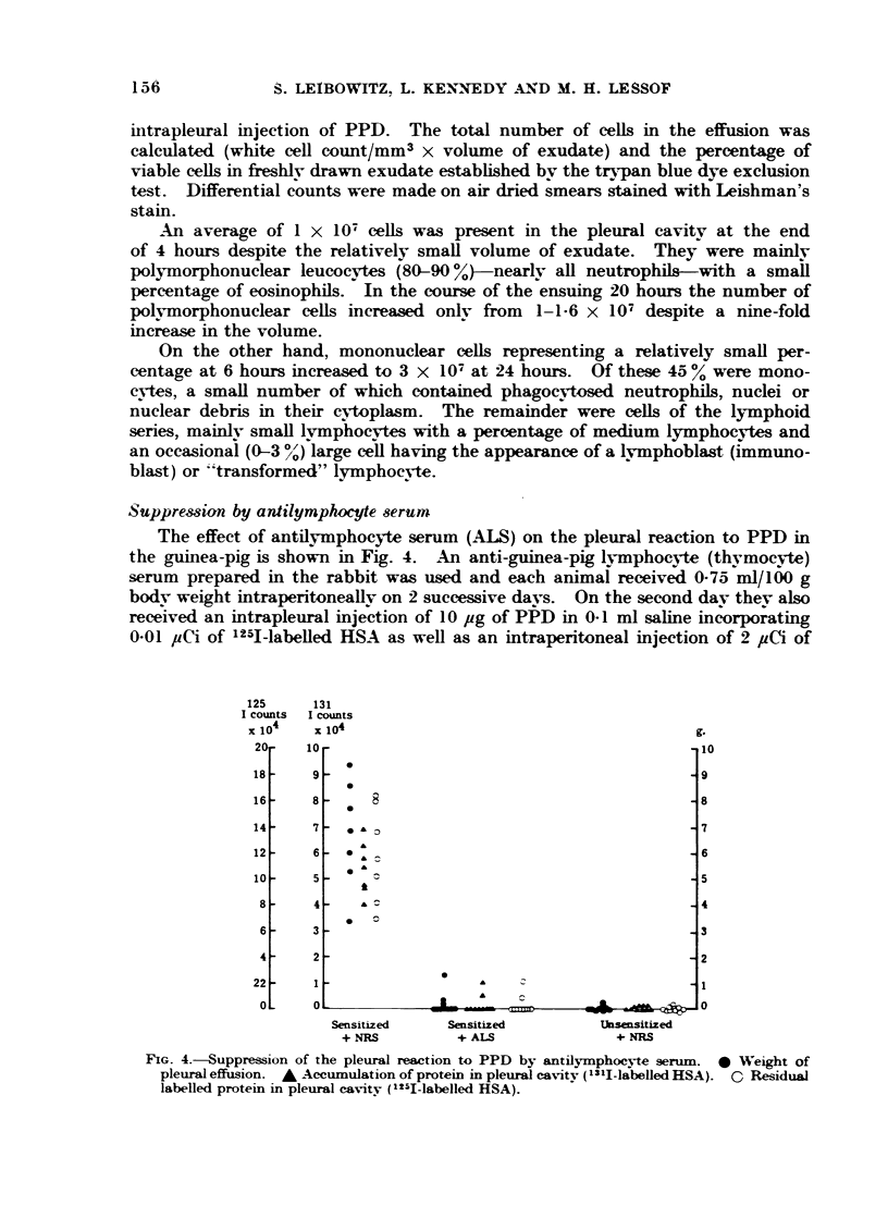
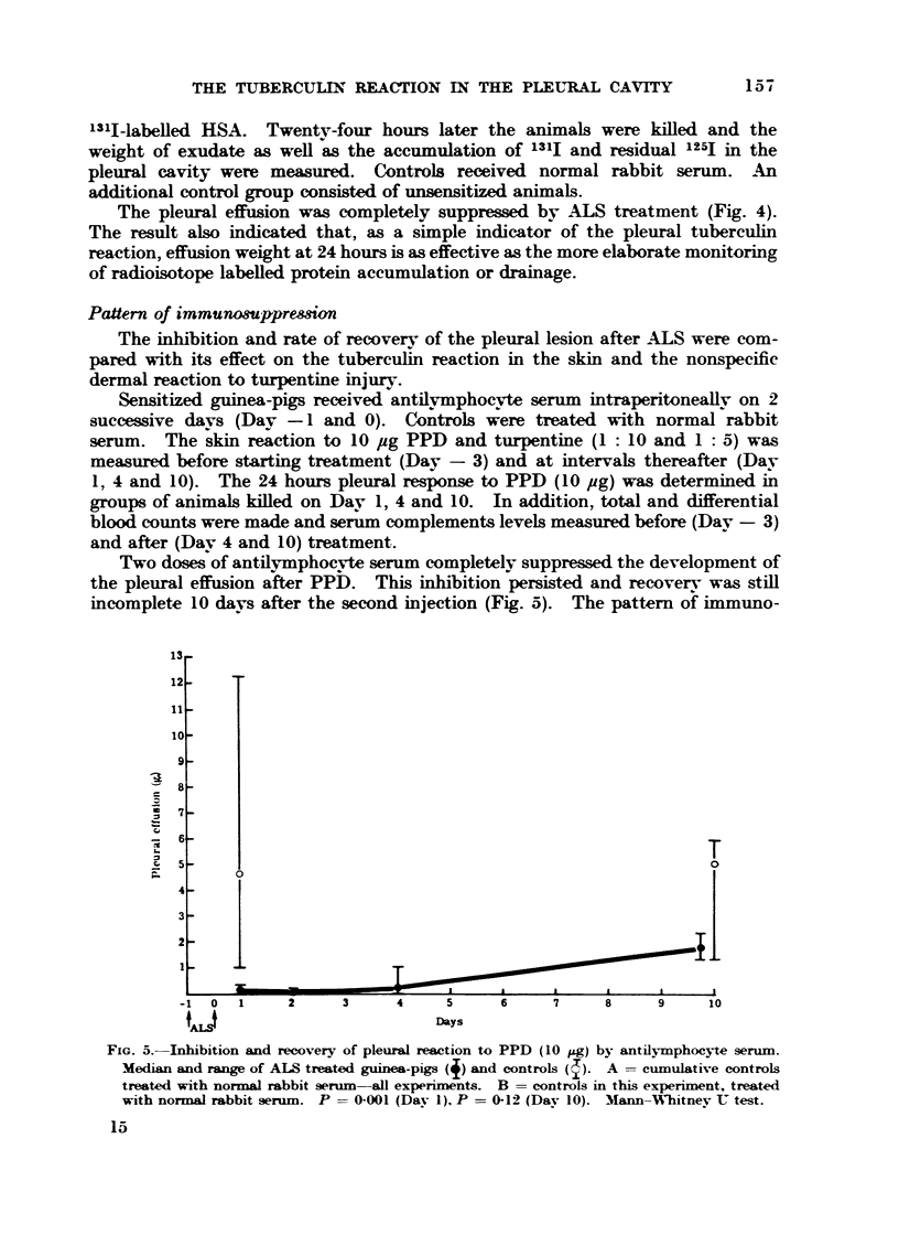
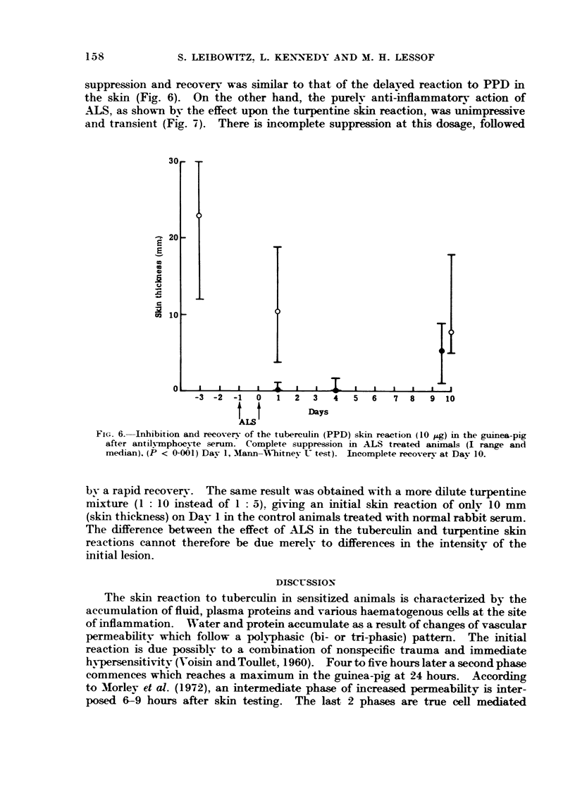
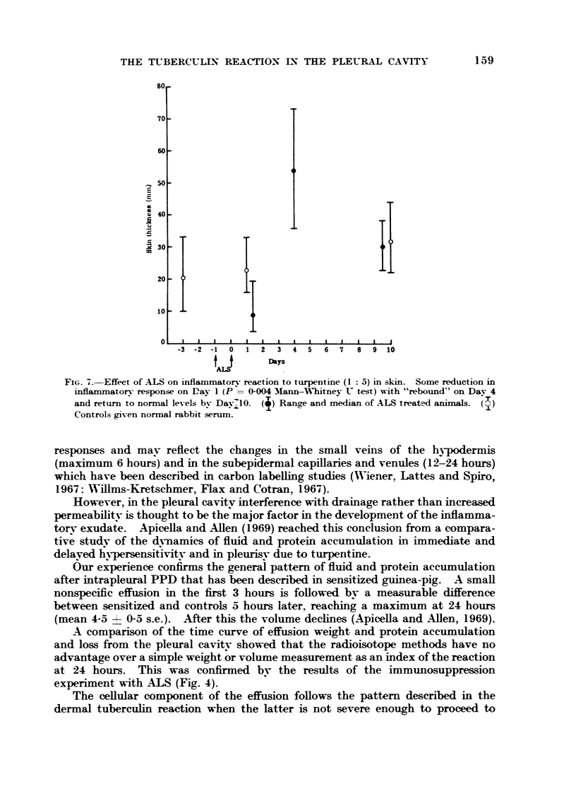
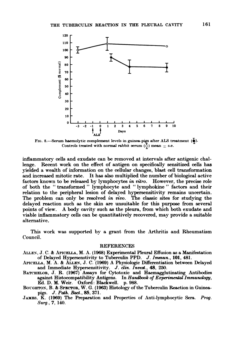
The effusion in the guinea-pig is completely suppressed by antilymphocyte serum (ALS). This effect is relatively long lasting (compared with its transient action on a nonspecific inflammatory lesion due to turpentine) and recovery parallels the return of the tuberculin reaction in the skin—providing further confirmation that the effusion is due to delayed hypersensitivity.
The tuberculin PPD reaction in the pleural cavity provides a useful model of delayed hypersensitivity in which exudate and viable inflammatory cells can be collected for study during the development of the lesion.
Full text
PDF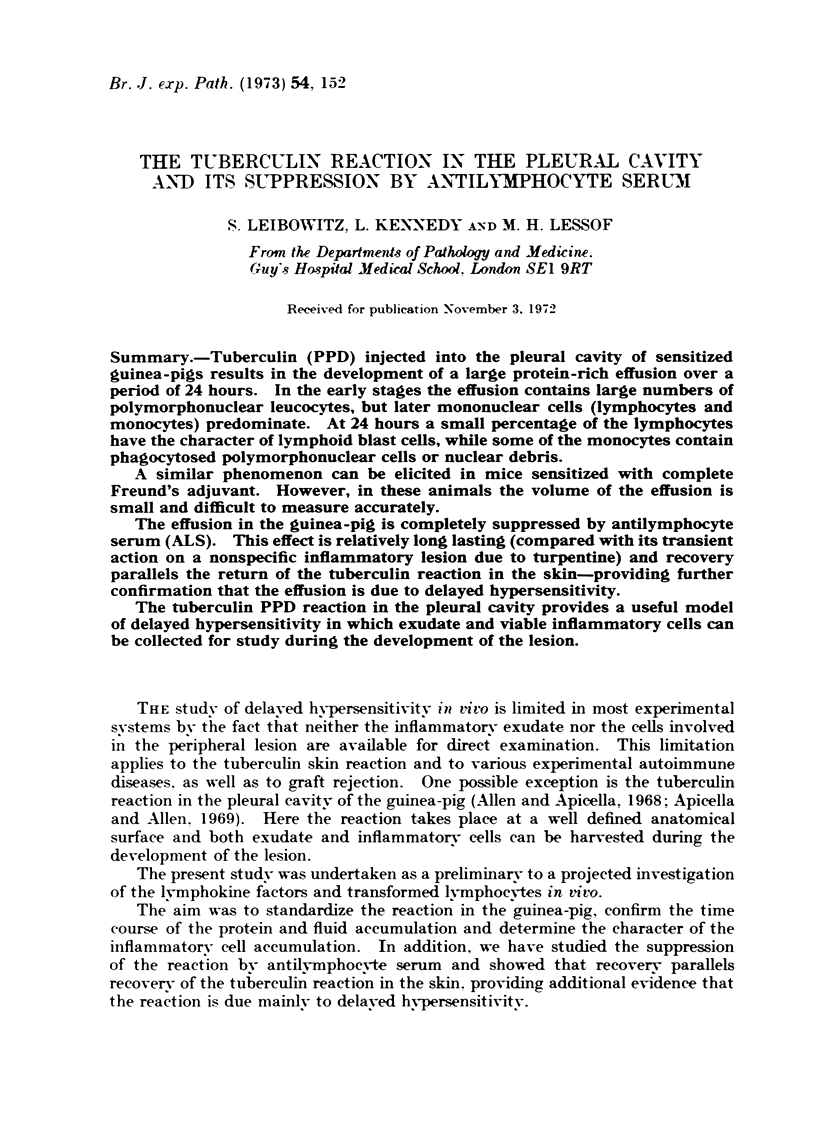

Selected References
These references are in PubMed. This may not be the complete list of references from this article.
- Allen J. C., Apicella M. A. Experimental pleural effusion as a manifestation of delayed hypersensitivity to tuberculin PPD. J Immunol. 1968 Sep;101(3):481–487. [PubMed] [Google Scholar]
- Leibowitz S., Lessof M. H., Kennedy L. A. The effect of anti-lymphocyte serum on experimental allergic encephalomyelitis in the guinea-pig. Clin Exp Immunol. 1968 Oct;3(8):753–760. [PMC free article] [PubMed] [Google Scholar]
- Morley J., Williams T. J., Slater A. J., Cubitt D., Dumonde D. C. Simultaneous measurements of the accumulation of isotope-labelled protein and erythrocytes in skin reactions of allergic inflammation in the guinea-pig. Immunology. 1972 Aug;23(2):113–135. [PMC free article] [PubMed] [Google Scholar]
- Perper R. J., Monovich R. E., Van Gorder T. J. Analysis of the biological activity of antilymphocyte serum. 3. Concentration of immunosuppressive activity in one IgG subfraction and its inhibition by another. Immunology. 1971 Dec;21(6):883–902. [PMC free article] [PubMed] [Google Scholar]
- WASSERMAN E., LEVINE L. Quantitative micro-complement fixation and its use in the study of antigenic structure by specific antigen-antibody inhibition. J Immunol. 1961 Sep;87:290–295. [PubMed] [Google Scholar]
- Wiener J., Lattes R. G., Spiro D. An electron microscopic study of leukocyte emigration and vascular permeability in tuberculin sensitivity. Am J Pathol. 1967 Mar;50(3):485–521. [PMC free article] [PubMed] [Google Scholar]
- Willms-Kretschmer K., Flax M. H., Cotran R. S. The fine structure of the vascular response in hapten-specific delayed hypersensitivity and contact dermatitis. Lab Invest. 1967 Sep;17(3):334–349. [PubMed] [Google Scholar]
- Willoughby D. A., Coote E., Turk J. L. Complement in acute inflammation. J Pathol. 1969 Feb;97(2):295–305. doi: 10.1002/path.1710970215. [DOI] [PubMed] [Google Scholar]