Abstract
A series of cases of rheumatoid arthritis, osteo-arthritis, gout and fibrositis have been investigated by means of the sedimentation rate, Arneth count, saline absorption test, glucose tolerance test, and direct observation of the capillaries, in order to correlate any abnormality of the peripheral circulation with the type and activity of the disease.
The sedimentation rate test, in which a wide bore tube was used, was of great value as an estimation of activity in the rheumatoid arthritic cases, but a small group showed a normal reading. In the gouty subjects it was usually low but was liable to vary greatly within a short time. In the osteo-arthritic and fibrositic groups it was normal.
The Arneth count did not parallel the sedimentation rate; it was usually normal in the gouty subjects but showed abnormalities in the rheumatoid arthritics and sometimes in the osteo-arthritics (a shift usually to the left, but sometimes to the right).
The saline absorption test was valueless except in cases of gross circulatory disturbance.
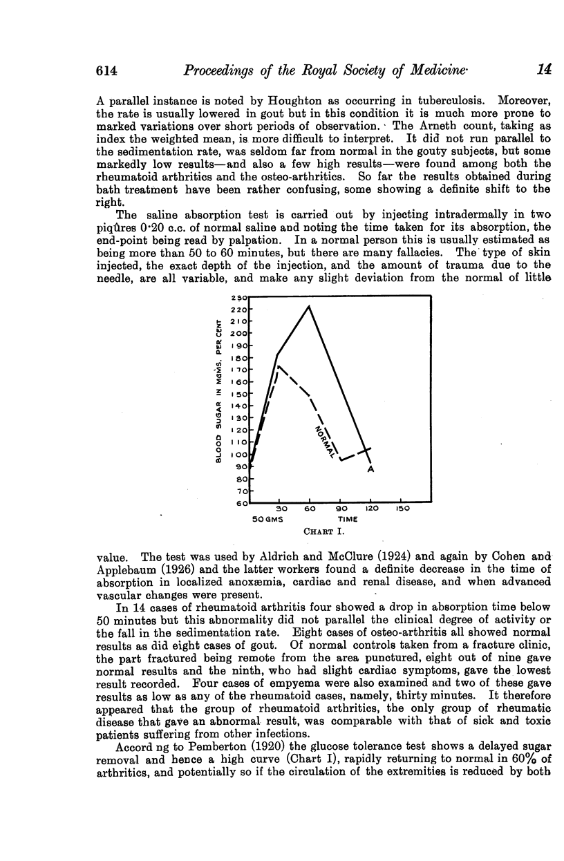
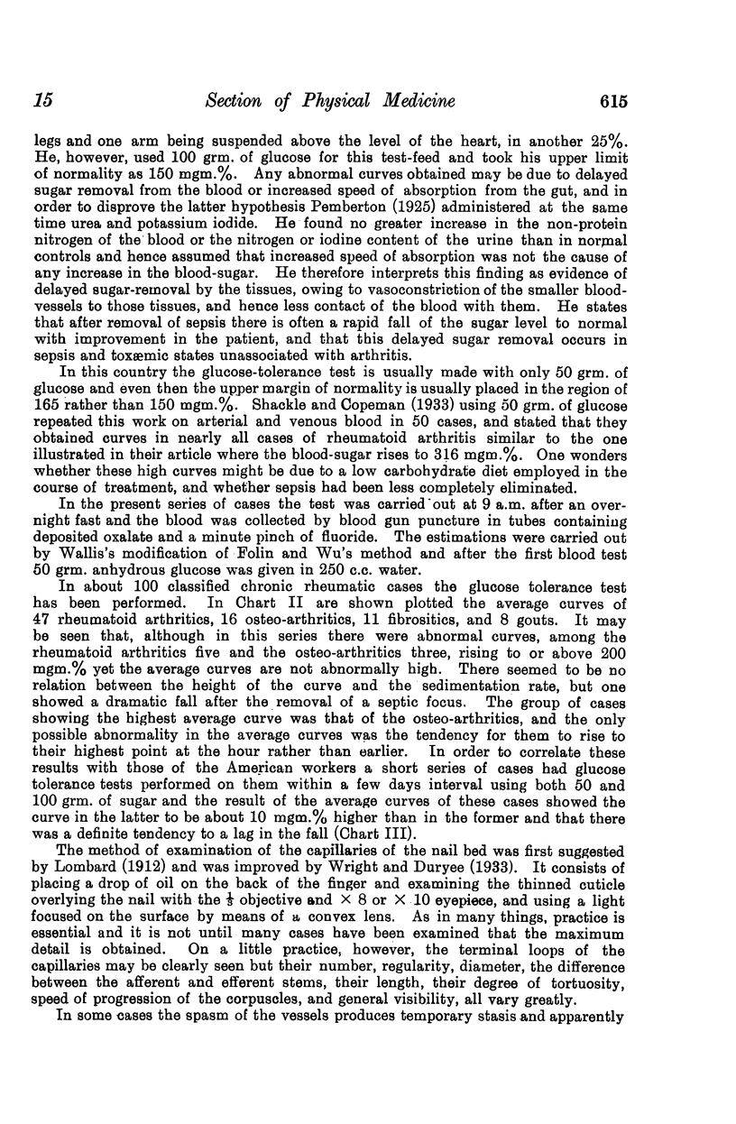
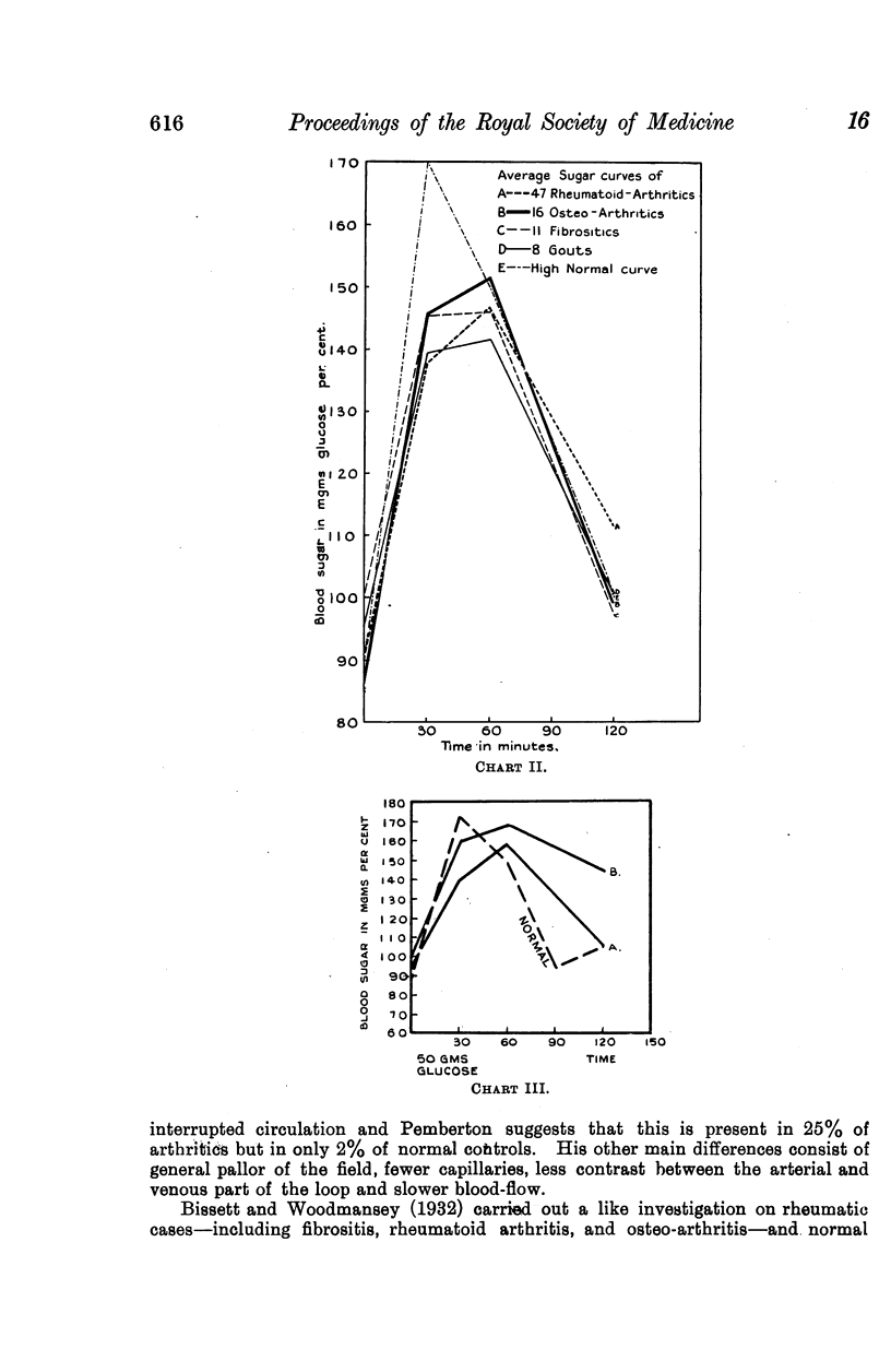
The glucose tolerance test, although producing some abnormal curves, in both the osteo-arthritic and the rheumatoid arthritic groups gave normal curves, taking an average of a large number of cases.
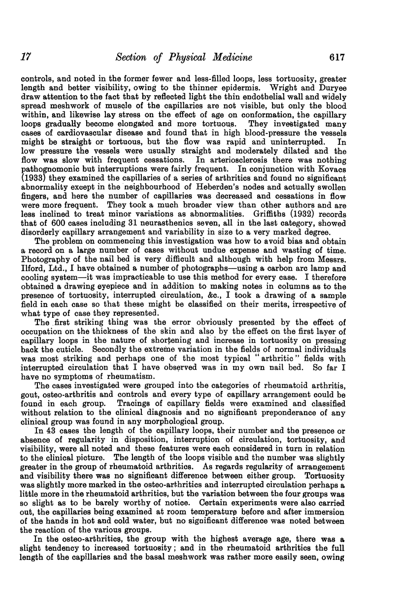
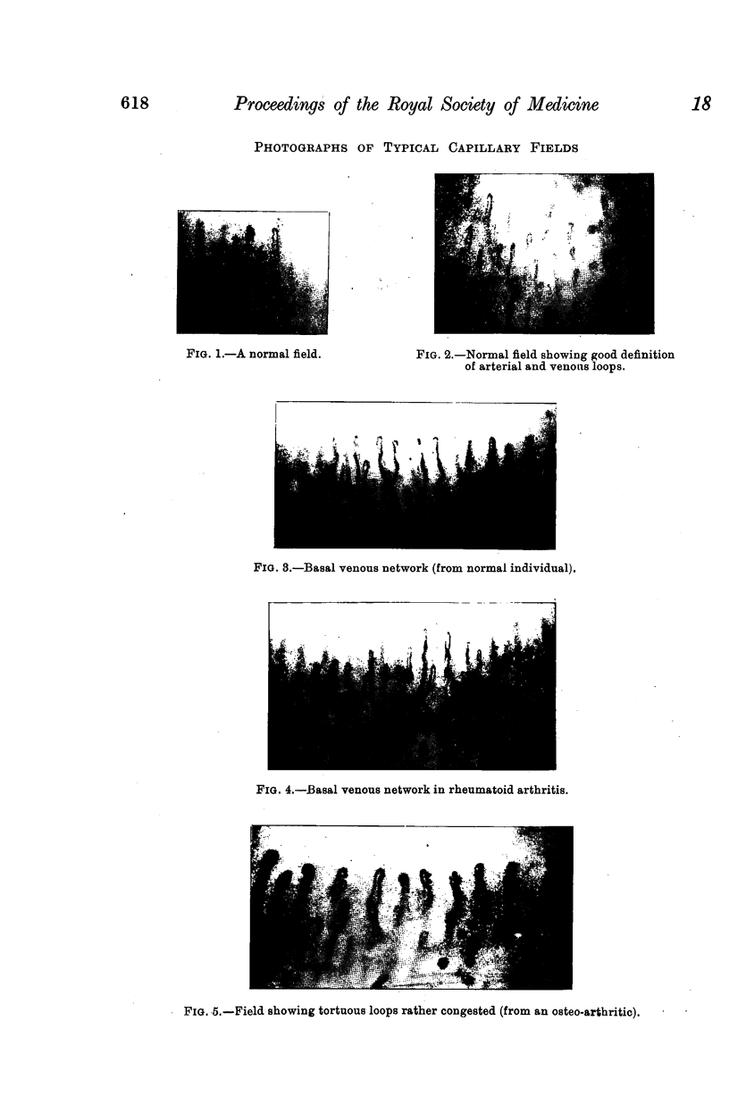
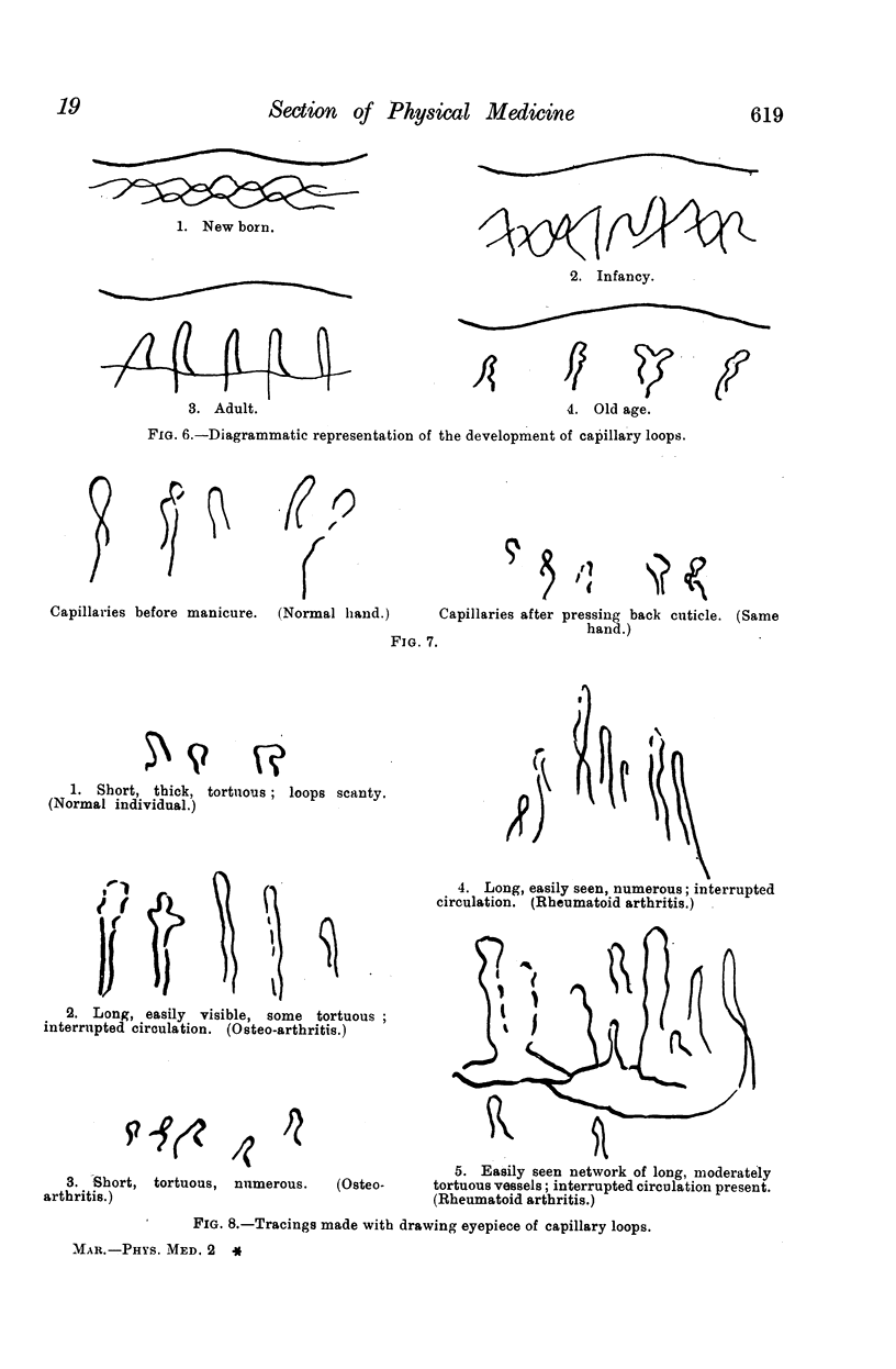
Observation of the capillaries—notes being taken of their number, shape, &c., with a drawing eyepiece and photographs—showed a wide variation in normal individuals. This was so remarkable that the only clear-cut findings were a slight increase in tortuosity in the osteo-arthritic group—probably due to the increase in the average age of this group—and an increase in the visibility of the full-length of the capillary loop and of the basal meshwork in the rheumatoid arthritics, probably due to trophic thinning of the epidermis.
Full text
PDF