

Abstract
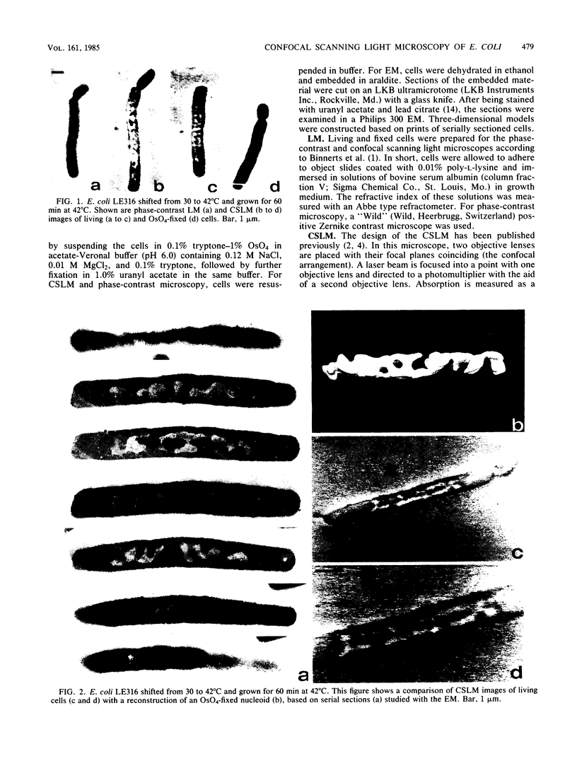
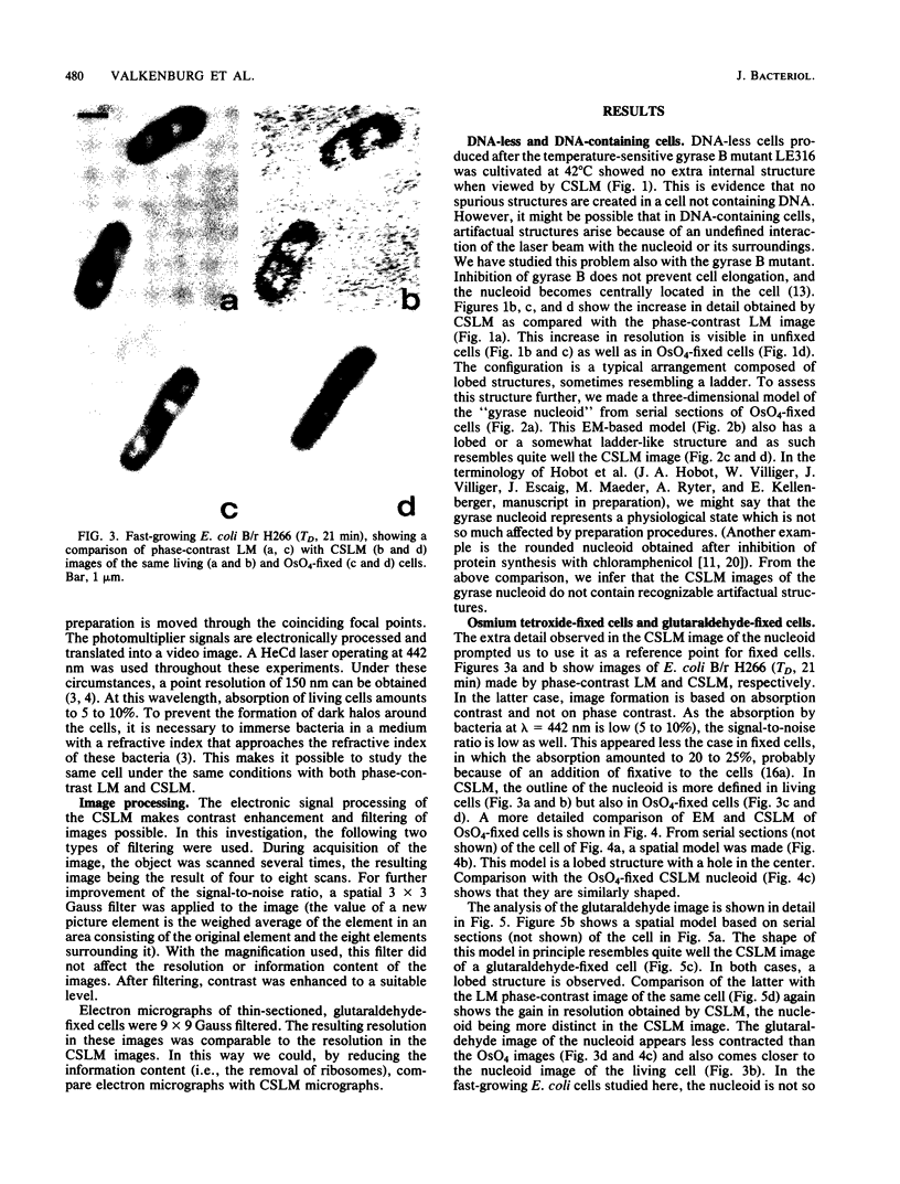
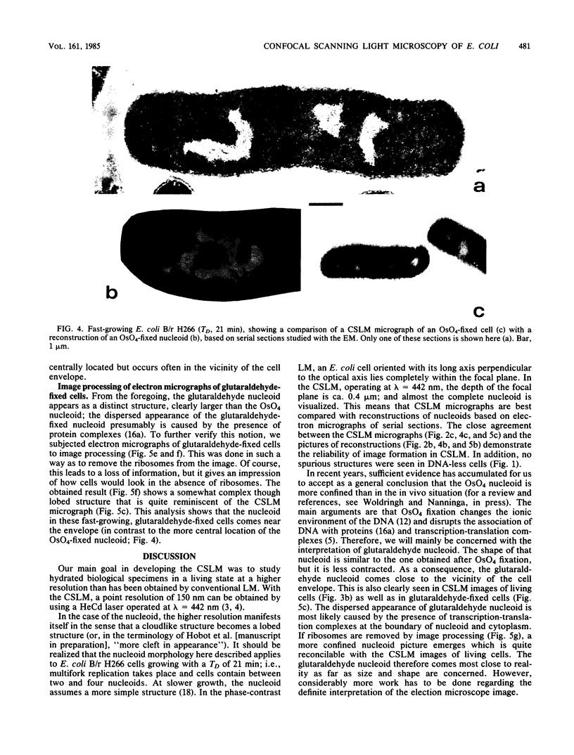
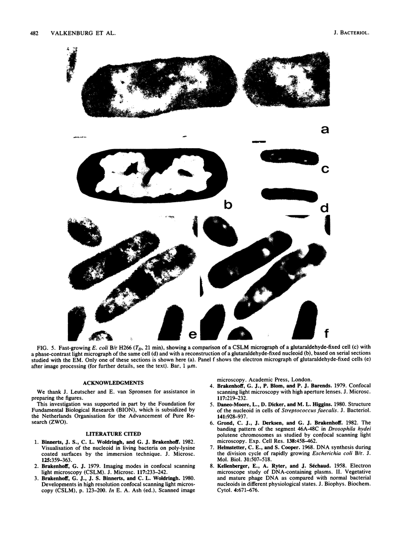
The nucleoid of living and OsO4- or glutaraldehyde-fixed cells of Escherichia coli strains was studied with a phase-contrast microscope, a confocal scanning light microscope, and an electron microscope. The trustworthiness of the images obtained with the confocal scanning light microscope was investigated by comparison with phase-contrast micrographs and reconstructions based on serially sectioned material of DNA-containing and DNA-less cells. This comparison showed higher resolution of the confocal scanning light microscope as compared with the phase-contrast microscope, and agreement with results obtained with the electron microscope. The effects of fixation on the structure of the nucleoid were studied in E. coli B/r H266. Confocal scanning light micrographs and electron microscopic reconstructions showed that the shape of the nucleoid remained similar after OsO4 or glutaraldehyde fixation; however, the OsO4 nucleoid appeared to be somewhat smaller and more centralized within the cell.
Full text
PDF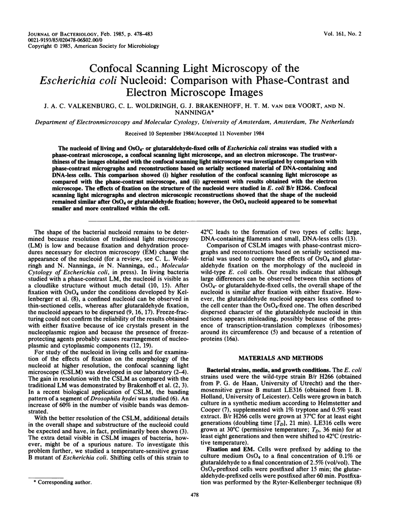
Images in this article
Selected References
These references are in PubMed. This may not be the complete list of references from this article.
- Binnerts J. S., Woldringh C. L., Brakenhoff G. J. Visualization of the nucleoid in living bacteria on poly-lysine coated surfaces by the immersion technique. J Microsc. 1982 Mar;125(Pt 3):359–363. doi: 10.1111/j.1365-2818.1982.tb00351.x. [DOI] [PubMed] [Google Scholar]
- Daneo-Moore L., Dicker D., Higgins M. L. Structure of the nucleoid in cells of Streptococcus faecalis. J Bacteriol. 1980 Feb;141(2):928–937. doi: 10.1128/jb.141.2.928-937.1980. [DOI] [PMC free article] [PubMed] [Google Scholar]
- Grond C. J., Derksen J., Brakenhoff G. J. The banding pattern of the segment 46A-48C in Drosophila hydeï polytene chromosomes as studied by confocal scanning light microscopy (CSLM). Exp Cell Res. 1982 Apr;138(2):458–462. doi: 10.1016/0014-4827(82)90199-9. [DOI] [PubMed] [Google Scholar]
- Helmstetter C. E. DNA synthesis during the division cycle of rapidly growing Escherichia coli B/r. J Mol Biol. 1968 Feb 14;31(3):507–518. doi: 10.1016/0022-2836(68)90424-5. [DOI] [PubMed] [Google Scholar]
- KELLENBERGER E., RYTER A., SECHAUD J. Electron microscope study of DNA-containing plasms. II. Vegetative and mature phage DNA as compared with normal bacterial nucleoids in different physiological states. J Biophys Biochem Cytol. 1958 Nov 25;4(6):671–678. doi: 10.1083/jcb.4.6.671. [DOI] [PMC free article] [PubMed] [Google Scholar]
- MASON D. J., POWELSON D. M. Nuclear division as observed in live bacteria by a new technique. J Bacteriol. 1956 Apr;71(4):474–479. doi: 10.1128/jb.71.4.474-479.1956. [DOI] [PMC free article] [PubMed] [Google Scholar]
- Margaretten W., Morgan C., Rosenkranz H. S., Rose H. M. Effect of hydroxyurea on virus development. I. Electron microscopic study of the effect on the development of bacteriophage T4. J Bacteriol. 1966 Feb;91(2):823–833. doi: 10.1128/jb.91.2.823-833.1966. [DOI] [PMC free article] [PubMed] [Google Scholar]
- Morgan C., Rosenkranz H. S., Carr H. S., Rose H. M. Electron microscopy of chloramphenicol-treated Escherichia coli. J Bacteriol. 1967 Jun;93(6):1987–2002. doi: 10.1128/jb.93.6.1987-2002.1967. [DOI] [PMC free article] [PubMed] [Google Scholar]
- Nanninga N., Woldringh C. L. The interpretation of chemically fixed and freeze-fractured bacterial nucleoplasm. Acta Histochem Suppl. 1981;23:39–53. [PubMed] [Google Scholar]
- Orr E., Fairweather N. F., Holland I. B., Pritchard R. H. Isolation and characterisation of a strain carrying a conditional lethal mutation in the cou gene of Escherichia coli K12. Mol Gen Genet. 1979;177(1):103–112. doi: 10.1007/BF00267259. [DOI] [PubMed] [Google Scholar]
- REYNOLDS E. S. The use of lead citrate at high pH as an electron-opaque stain in electron microscopy. J Cell Biol. 1963 Apr;17:208–212. doi: 10.1083/jcb.17.1.208. [DOI] [PMC free article] [PubMed] [Google Scholar]
- SCHAECHTER M., WILLIAMSON J. P., HOOD J. R., Jr, KOCH A. L. Growth, cell and nuclear divisions in some bacteria. J Gen Microbiol. 1962 Nov;29:421–434. doi: 10.1099/00221287-29-3-421. [DOI] [PubMed] [Google Scholar]
- Séchaud J., Kellenberger E. Electron microscopy of DNA-containing plasms. IV. Glutaraldehyde-uranyl acetate fixation of virus-infected bacteria for thin sectioning. J Ultrastruct Res. 1972 Jun;39(5):598–607. doi: 10.1016/s0022-5320(72)90124-4. [DOI] [PubMed] [Google Scholar]
- Valkenburg J. A., Woldringh C. L. Phase separation between nucleoid and cytoplasm in Escherichia coli as defined by immersive refractometry. J Bacteriol. 1984 Dec;160(3):1151–1157. doi: 10.1128/jb.160.3.1151-1157.1984. [DOI] [PMC free article] [PubMed] [Google Scholar]
- Woldringh C. L. Morphological analysis of nuclear separation and cell division during the life cycle of Escherichia coli. J Bacteriol. 1976 Jan;125(1):248–257. doi: 10.1128/jb.125.1.248-257.1976. [DOI] [PMC free article] [PubMed] [Google Scholar]
- Woldringh C. L., Nanninga N. Organization of the nucleoplasm in Escherichia coli visualized by phase-contrast light microscopy, freeze fracturing, and thin sectioning. J Bacteriol. 1976 Sep;127(3):1455–1464. doi: 10.1128/jb.127.3.1455-1464.1976. [DOI] [PMC free article] [PubMed] [Google Scholar]
- Zusman D. R., Carbonell A., Haga J. Y. Nucleoid condensation and cell division in Escherichia coli MX74T2 ts52 after inhibition of protein synthesis. J Bacteriol. 1973 Sep;115(3):1167–1178. doi: 10.1128/jb.115.3.1167-1178.1973. [DOI] [PMC free article] [PubMed] [Google Scholar]