Abstract
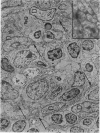
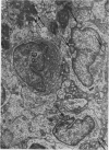
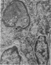
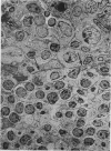
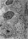
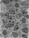
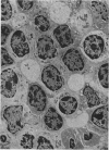
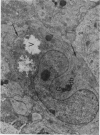
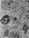
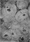
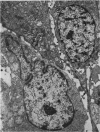
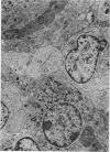
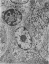
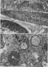
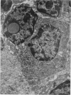
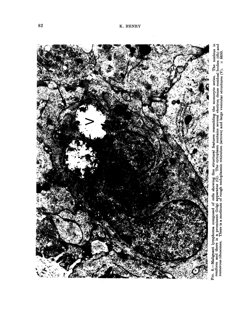
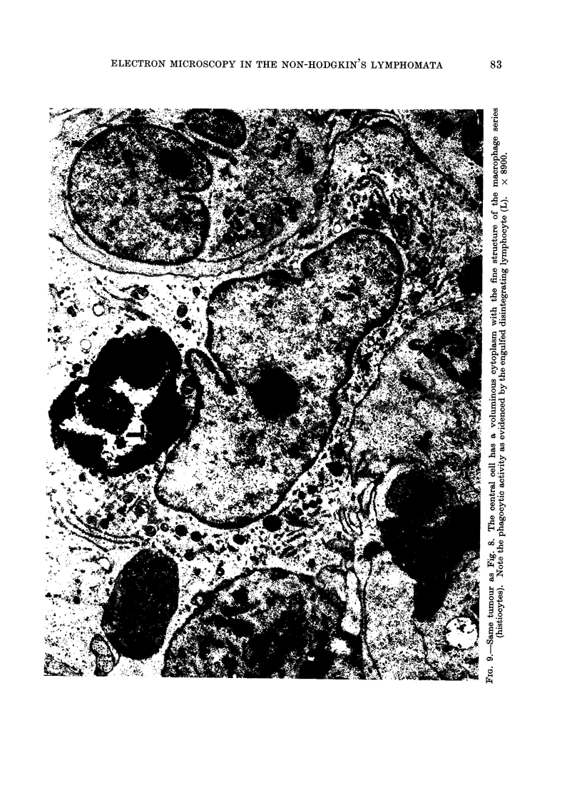
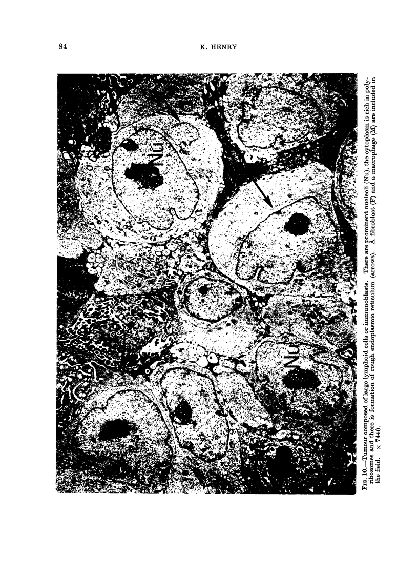
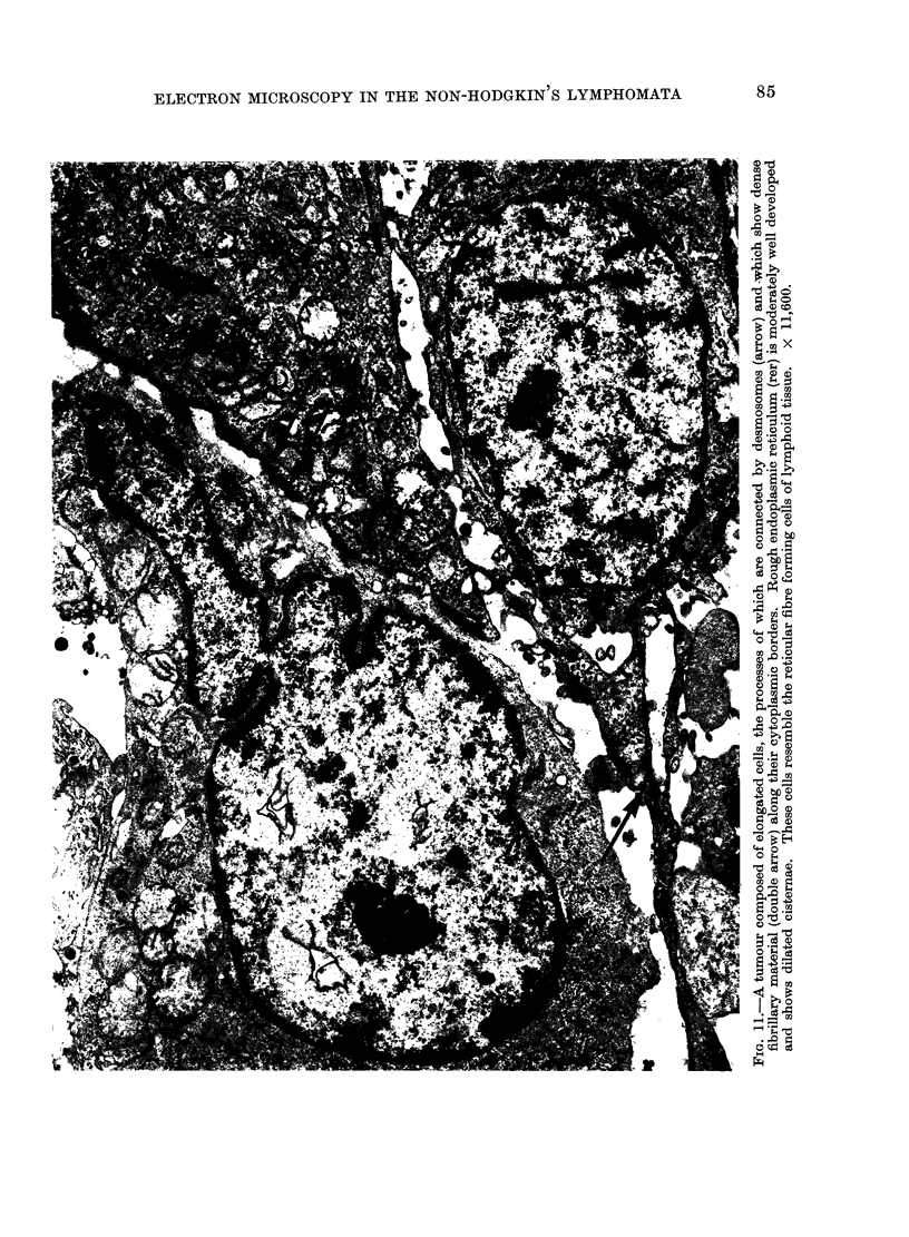
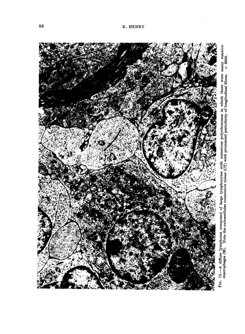
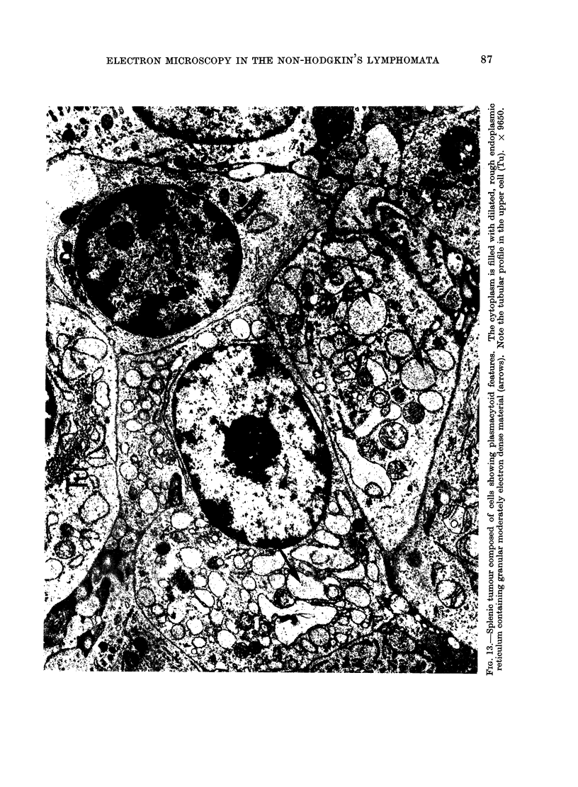
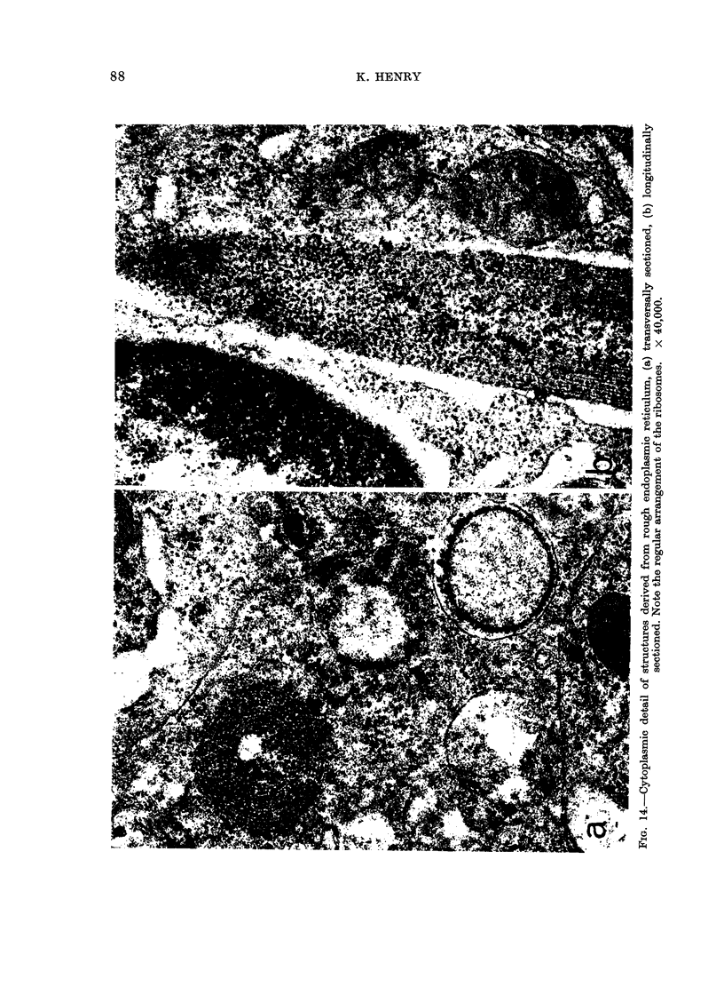
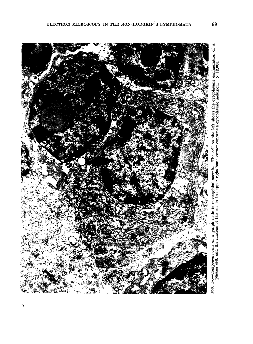
The component cells of peripheral lymphoid tissue have been divided into the lymphocyte and plasma cell lines, mononuclear phagocytic cells, dendritic "reticular cells", the reticular (supporting) cells and endothelial cells, and it is suggested that this system of cells should collectively be referred to as the lymphoreticular monoclear phagocyte system or LRMPS. Seventeen tumours of the LRMPS (excluding Hodgkin's disease) have been studied at ultrastructural level. Of these 17 non-Hodgkin lymphomata 5 were follicular lymphomata and 12 diffuse. It is concluded that electron microscopy plays a valuable role in the diagnosis of this group of tumours. Not only does it allow rejection of a diagnosis of lymphoma in certain anaplastic tumours, but it also enables a more precise identification of the cellular components of a lymphoma as well as indicating the degree of differentiation of the cell line involved. Additional advantages are the visualization of subcellular structures useful as markers, and by means of specialized immunoelectron microscopic techniques the identification of antigens and antibody formation within a given tumour. Two other results of this ultrastructural study are the indication that the dendritic cells of lymphoid follicles are derived from capillary endothelium, and the identification of certain anomalous formations derived from rough endoplasmic reticulum in the case of tumours showing plasmacytoid differentiation.
Full text
PDF
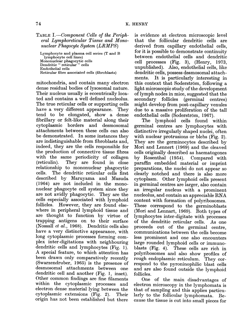
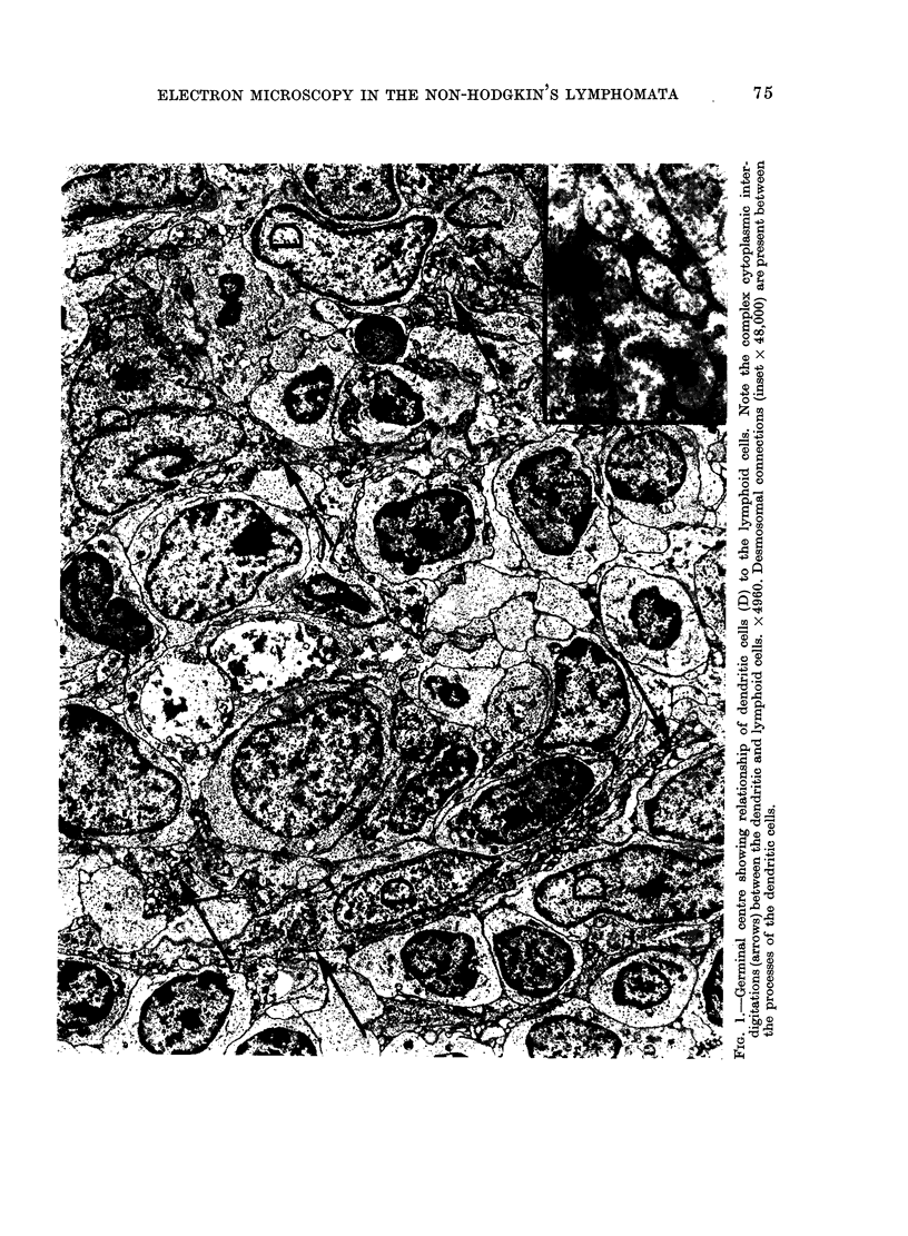
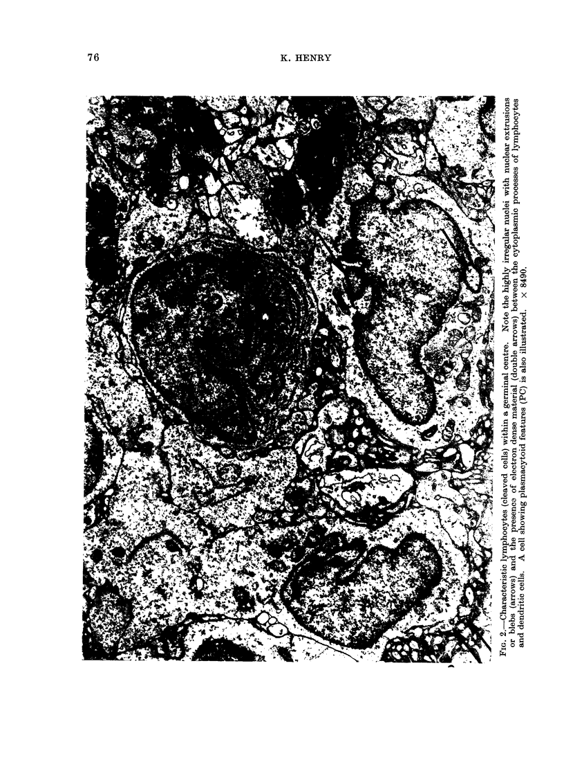
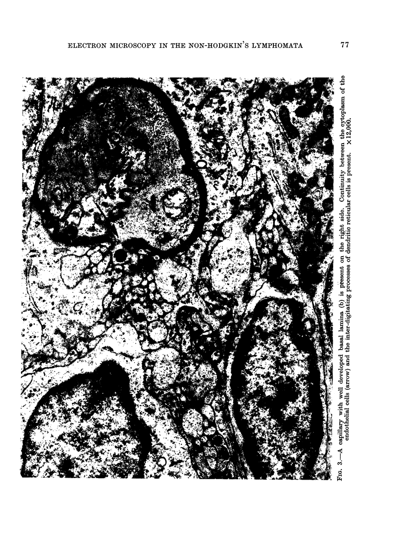
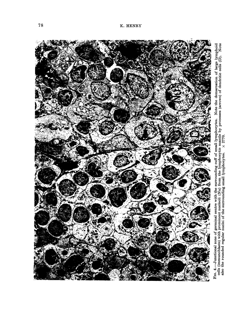
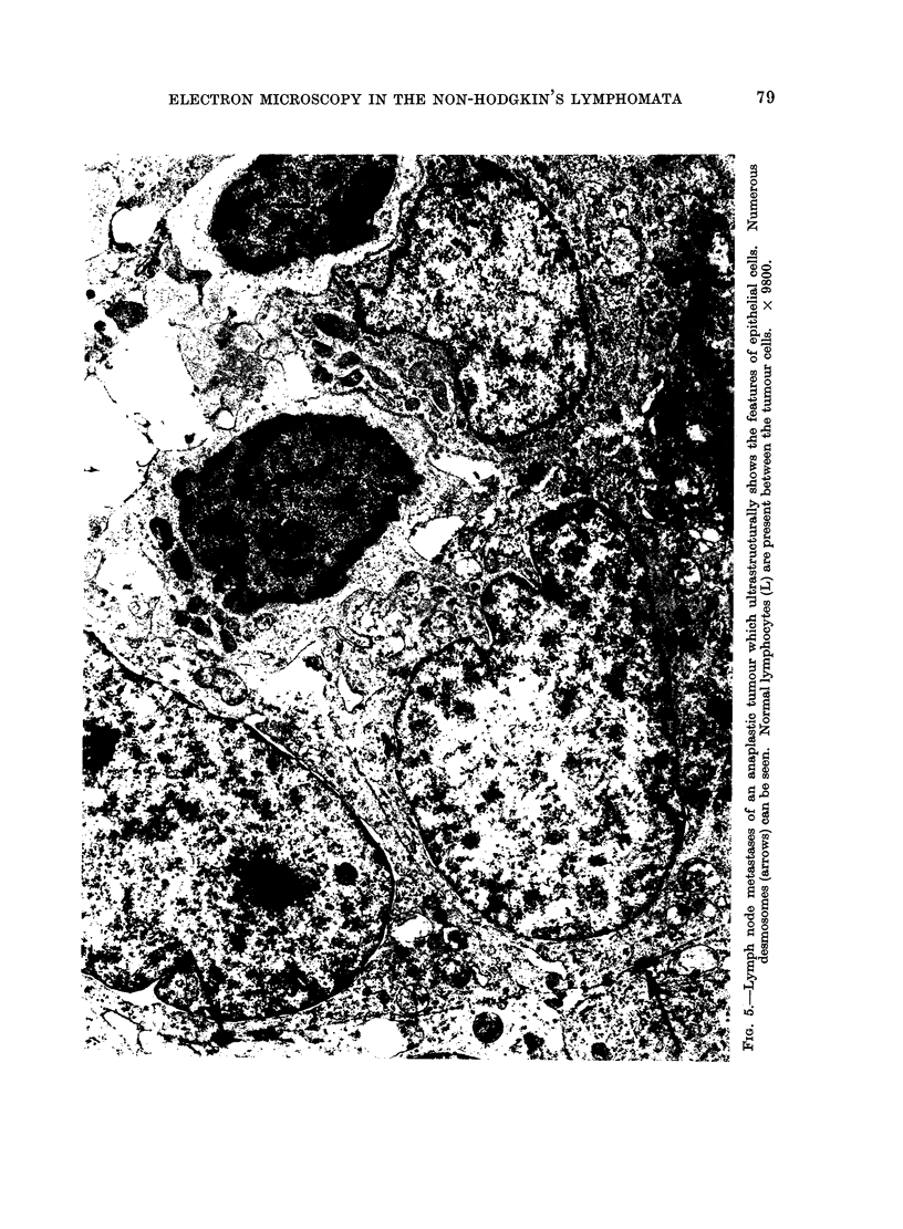
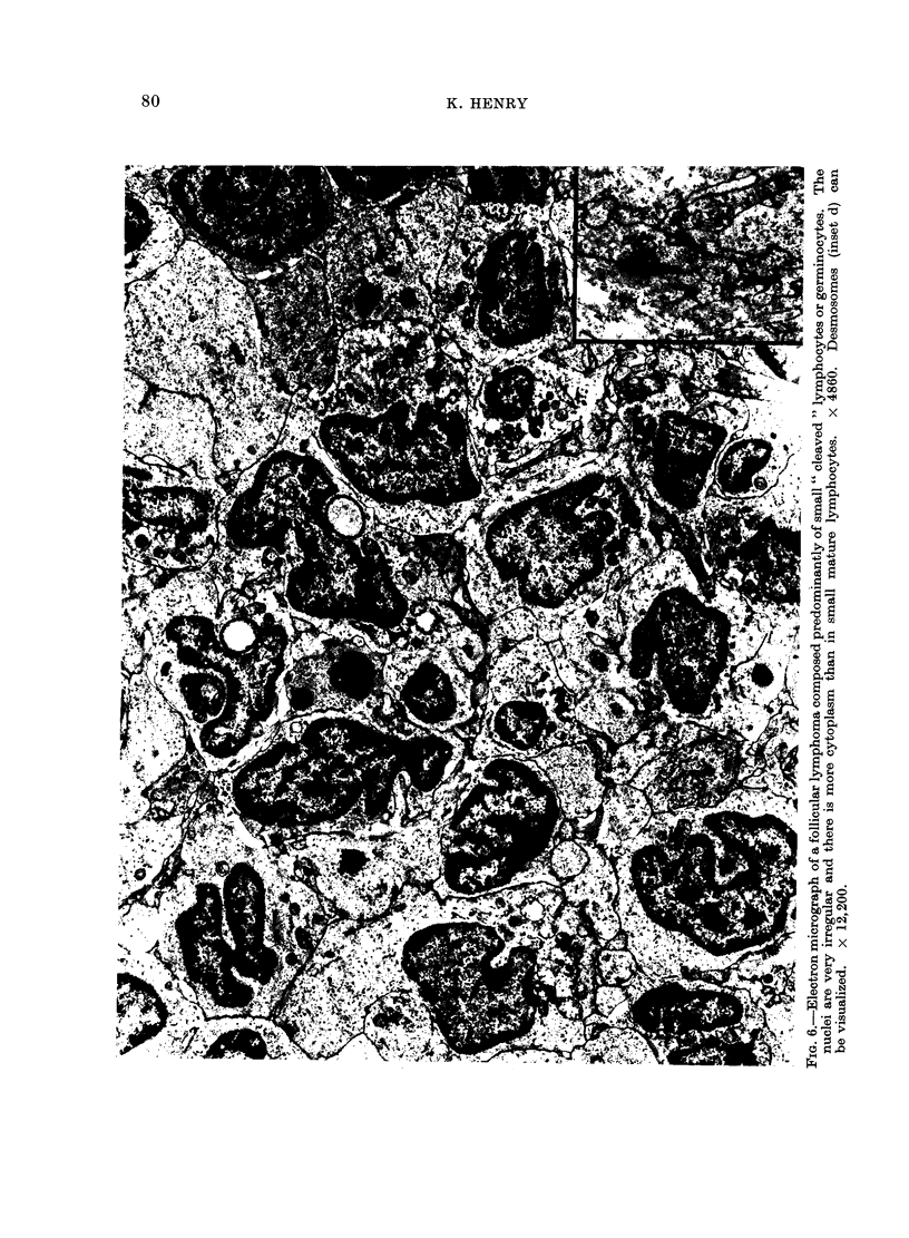
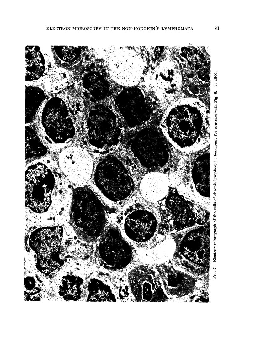
Images in this article
Selected References
These references are in PubMed. This may not be the complete list of references from this article.
- DUTCHER T. F., FAHEY J. L. The histopathology of the macroglobulinemia of Waldenström. J Natl Cancer Inst. 1959 May;22(5):887–917. doi: 10.1093/jnci/22.5.887. [DOI] [PubMed] [Google Scholar]
- Henry K. The lymphoreticular system: morphological organization. J Clin Pathol. 1972 Nov;25(11):1003–1003. doi: 10.1136/jcp.25.11.1003-b. [DOI] [PMC free article] [PubMed] [Google Scholar]
- Lennert K., Niedorf H. R. Nachweis von desmosomal verknüpften Reticulumzellen im follikul-aren Lymphom (Brill Symmers) Virchows Arch B Cell Pathol. 1969;4(2):148–150. [PubMed] [Google Scholar]
- Nossal G. J., Abbot A., Mitchell J., Lummus Z. Antigens in immunity. XV. Ultrastructural features of antigen capture in primary and secondary lymphoid follicles. J Exp Med. 1968 Feb 1;127(2):277–290. doi: 10.1084/jem.127.2.277. [DOI] [PMC free article] [PubMed] [Google Scholar]
- ROSENTHAL N. The lymphomas and leukemias. Bull N Y Acad Med. 1954 Aug;30(8):583–600. [PMC free article] [PubMed] [Google Scholar]
- Roitt I. M., Greaves M. F., Torrigiani G., Brostoff J., Playfair J. H. The cellular basis of immunological responses. A synthesis of some current views. Lancet. 1969 Aug 16;2(7616):367–371. doi: 10.1016/s0140-6736(69)92712-3. [DOI] [PubMed] [Google Scholar]
- Söderström N. Post-capillary venules as basic structural units in the development of lymphoglandular tissue. Scand J Haematol. 1967 Dec;4(6):411–429. doi: 10.1111/j.1600-0609.1967.tb01644.x. [DOI] [PubMed] [Google Scholar]


