Abstract
The breast cancer susceptibility gene BRCA1 encodes a protein that has been implicated in multiple nuclear functions, including transcription and DNA repair. The multifunctional nature of BRCA1 has raised the possibility that the polypeptide may regulate various nuclear processes via a common underlying mechanism such as chromatin remodeling. However, to date, no direct evidence exists in mammalian cells for BRCA1-mediated changes in either local or large-scale chromatin structure. Here we show that targeting BRCA1 to an amplified, lac operator–containing chromosome region in the mammalian genome results in large-scale chromatin decondensation. This unfolding activity is independently conferred by three subdomains within the transactivation domain of BRCA1, namely activation domain 1, and the two BRCA1 COOH terminus (BRCT) repeats. In addition, we demonstrate a similar chromatin unfolding activity associated with the transactivation domains of E2F1 and tumor suppressor p53. However, unlike E2F1 and p53, BRCT-mediated chromatin unfolding is not accompanied by histone hyperacetylation. Cancer-predisposing mutations of BRCA1 display an allele-specific effect on chromatin unfolding: 5′ mutations that result in gross truncation of the protein abolish the chromatin unfolding activity, whereas those in the 3′ region of the gene markedly enhance this activity. A novel cofactor of BRCA1 (COBRA1) is recruited to the chromosome site by the first BRCT repeat of BRCA1, and is itself sufficient to induce chromatin unfolding. BRCA1 mutations that enhance chromatin unfolding also increase its affinity for, and recruitment of, COBRA1. These results indicate that reorganization of higher levels of chromatin structure is an important regulated step in BRCA1-mediated nuclear functions.
Keywords: BRCA1; BRCT; chromatin unfolding; breast cancer; COBRA1
Introduction
Germ line mutations in breast cancer susceptibility gene 1 (BRCA1)* confer elevated risks in the development of familial breast and ovarian cancers (Rahman and Stratton, 1998). BRCA1 encodes a 1,863–amino acid protein with a highly conserved ring finger motif (RING) at the NH2 terminus, and two BRCA1 COOH terminus (BRCT) repeats at the extreme COOH terminus. Whereas most disease-associated mutations of BRCA1 are predicted to result in gross truncation of the protein, 5–10% of the cancer-predisposing mutations cause single amino acid substitutions, many of which are located in the RING domain or BRCT repeats.
Intense research in the past several years has implicated BRCA1 in the regulation of multiple nuclear processes, including DNA repair and transcription (Zhang et al., 1998b; Scully and Livingston, 2000). For example, BRCA1-deficient mouse and human cells are hypersensitive to ionizing radiation due to defects in transcription-coupled repair of oxidative DNA damage, as well as double-strand break-induced homologous recombination (Gowen et al., 1998; Abbott et al., 1999; Moynahan et al., 1999; Scully et al., 1999; Xu et al., 1999). In addition, BRCA1 associates with several repair and recombination proteins such as RAD51 (Scully et al., 1997b), RAD50/MRE11/NBS1 (Zhong et al., 1999; Wang et al., 2000), and MSH2/MSH6 (Wang et al., 2000). BRCA1 also interacts with and is phosphorylated by protein kinases that are key players in the damage checkpoint control, including ATM, ATR, and CHK2 (Cortez et al., 1999; Lee et al., 2000; Tibbetts et al., 2000). Lastly, it has been shown recently that BRCA1 preferentially binds to branched DNA structures (Paull et al., 2001).
In addition to its potential role in DNA repair, BRCA1 has also been implicated in regulation of transcription (Monteiro, 2000; Scully and Livingston, 2000). When tethered to a transcriptional promoter via a heterologous DNA binding domain, the COOH-terminal 304–amino acid (aa) region including the BRCT repeats (Fig. 1 B, AD2, amino acids 1560–1863) can act as a transactivation domain (Chapman and Verma, 1996; Monteiro et al., 1996). More recent work has revealed a second transactivation domain of BRCA1 that resides upstream of the BRCT repeats (Hu et al., 2000) (Fig. 1 B, AD1, aa 1293–1559). The two activation domains (ADs), AD1 and AD2, can cooperatively activate transcription in many cell lines tested (Hu et al., 2000). Consistent with its potential role in transcriptional regulation, the BRCA1 polypeptide is associated with the RNA polymerase II holoenzyme via RNA helicase A (Scully et al., 1997a; Neish et al., 1998). Furthermore, BRCA1 interacts with a number of site-specific transcription factors and modulates their actions in gene activation (Somasundaram et al., 1997; Ouchi et al., 1998, 2000; Zhang et al., 1998a; Fan et al., 1999; Houvras et al., 2000; Zheng et al., 2000).
Figure 1.
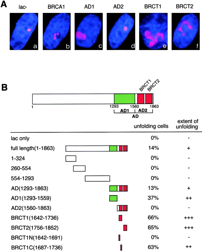
BRCA1 induces large-scale chromatin decondensation. (A) The AO3_1 CHO cell line was transiently transfected with expression vectors for the following proteins: lac repressor (a), lac–BRCA1(b), lac–AD1(c), lac– AD2(d), lac–BRCT1(e), and lac–BRCT2(f). A polyclonal anti–lac repressor antibody and a Cy3-conjugated secondary anti–rabbit IgG were used for immunostaining. Nuclei were visualized by DNA staining with DAPI. (B) The ability of various BRCA1 fragments to unfold chromatin was measured by the percentage of transfected cells that displayed enlarged lac staining and the degree of unfolding. Over 100 transfected cells were surveyed for each construct. Single, double, and triple plus signs indicate various degrees of chromatin unfolding, as exemplified by images for lac–BRCA1 (+), lac–AD1 (++), and lac–BRCT1 (+++). Also shown are schematic diagrams and amino acid coordinates for various BRCA1 fragments.
The multifunctional nature of BRCA1 has raised the possibility that the protein may employ a common mechanism, such as chromatin remodeling, to regulate various chromosomal events. Indeed, the COOH-terminal region of BRCA1 (AD2), which is required for BRCA1 functions in both DNA repair and transcription (Monteiro, 2000; Scully and Livingston, 2000), can induce changes in nucleosome structure when tethered to chromosomal DNA in Saccharomyces cerevisiae (Hu et al., 1999). Furthermore, BRCA1 is associated with histone modifying enzymes (p300 and HDAC) (Neish et al., 1998; Yarden and Brody, 1999; Pao et al., 2000) and an ATP-dependent chromatin remodeling machine (hSNF/SWI) (Bochar et al., 2000). The fact that many cancer-predisposing mutations reduce BRCA1's affinity for these chromatin-modifying proteins suggests that chromatin remodeling may be an important aspect of BRCA1-mediated tumor suppression. However, currently there is no direct evidence in mammalian cells for BRCA1-mediated changes in chromatin structure. This is in part due to the lack of convenient assays for directly monitoring chromatin remodeling at different levels of chromatin structure in mammalian cells.
A lac repressor–based system has allowed direct visualization of large-scale chromatin dynamics in mammalian cells (Belmont, 2001). In this system, multiple copies of the lac operator were engineered into the genome of CHO cells, and together with the surrounding genomic sequences, were amplified to produce a 90-Mb heterochromatic region. By fusing lac repressor with the acidic AD (AAD) of the strong viral transcription factor VP16 and tethering the fusion protein to the heterochromatic chromosome region, this system was used to demonstrate AAD-induced large-scale chromatin decondensation (Tumbar et al., 1999). This large-scale chromatin uncoiling occurred even when RNA pol II–dependent transcription was blocked, suggesting that it was induced through transacting factors recruited by the VP16 AAD, rather than the result of transcription per se. Conceptually, the transacting factors producing this higher order chromatin decondensation could be one of the known chromatin-modifying complexes that modify local nucleosome structure (Peterson and Logie, 2000). Alternatively, AAD-induced chromatin unfolding could involve novel factors acting primarily at the higher levels of chromatin organization.
Although artificial, this lac repressor–tethering system provides a very quick, and therefore powerful, assay to test the possible role of specific proteins in chromatin remodeling and to dissect the protein domains required for the observed large-scale chromatin decondensation. Using this lac repressor–tethering assay, we demonstrate here that BRCA1 induces large-scale chromatin decondensation. We also identify three small subdomains within the transactivation domain of BRCA1 that are capable of independently conferring chromatin unfolding. In addition, cancer-predisposing mutations of BRCA1 display allele-specific effects on the chromatin unfolding activity. Finally, we isolate a novel cofactor of BRCA1 (COBRA1) that binds to one of the chromatin-unfolding domains of BRCA1, and by itself induces large-scale chromatin decondensation. Our results suggest that BRCA1-mediated decondensation of higher levels of chromatin structure may represent a new physiological regulatory pathway related to BRCA1 function. The approach used in the current study also provides a new methodology for identifying novel BRCA1-interacting proteins involved in this regulatory pathway.
Results
BRCA1-mediated large-scale chromatin decondensation in mammalian cells
To assess the impact of BRCA1 on large-scale chromatin structure in mammalian cells, we made use of a CHO cell line, AO3_1, in which multiple copies of the lac operator were engineered to produce a 90-Mb heterochromatic region of the genome (Robinett et al., 1996; Li et al., 1998; Tumbar et al., 1999). The molecular organization of this region consists of ∼400-kb repeats of the 14-kb vector transgene that contains the lac operator repeat and dihydrofolate reductase selectable marker. The repeats are separated on average by ∼1,000 kb of unknown coamplified genomic DNA. Because other cell clones derived from the same selection procedure contain more open, gene-amplified chromosome regions with comparable or greater content of the vector DNA, the heterochromatic appearance of the A03_1 chromosome region is assumed to be due to properties of the coamplified genomic DNA. In vivo binding of lac repressor or its GFP derivatives to this chromosomal site allows direct visualization of large-scale chromatin dynamics without altering the original chromosome structure.
Consistent with previous findings, lac repressor–expressing cells stained with the corresponding antibody exhibited a compact nuclear dot (Fig. 1 A, a). In contrast, expression of lac repressor fused with the full-length BRCA1 induced an irregularly shaped subnuclear structure in 14% of transfected cells (Fig. 1 A, b). Such a staining pattern was not present in any of the cells expressing lac repressor alone. These results suggest that BRCA1, or a BRCA1-associated protein, can induce large-scale chromatin restructuring. The magnitude of this opening was lower than observed for the VP16 AAD, and was present in a lower percentage of cells (14 vs. 60% for VP16 AAD) (Tumbar et al., 1999 and see Fig. 2) . The lack of a response in 100% of cells, even for the VP16 AAD, is not yet understood. It may represent a combination of several factors, including cell cycle–dependent expression as well as the nature of the qualitative assay employed. Transgene arrays have been shown to display coordinated gene silencing effects that are accompanied by cooperative changes in chromatin structure across the entire array (Pikaart et al., 1998). These changes in gene expression and chromatin structure show a variegating phenotype that is clonally inherited. Therefore, it is possible that the large-scale chromatin decondensation induced by a transcriptional activator in the lac system may also display cooperative and variegating responses.
Figure 2.
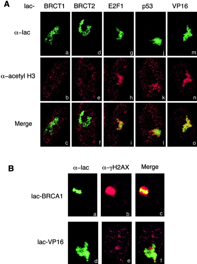
Comparison of chromatin unfolding by various lac fusion proteins. (A) Absence of histone hyperacetylation associated with BRCT-mediated chromatin unfolding. AO3_1 cells were transfected with the expression vectors for lac fused with BRCT1 (a–c), BRCT2 (d–f), E2F1 (g–i), p53 (j–l), or VP16 (m–o). The lac (green), acetylated histone H3 (red), and the merged images were captured by confocal immunofluorescence microscopy. (B) Association of lac–BRCA1 with phosphorylated H2AX. AO3_1 cells were transfected with the lac–BRCA1 expression vector. Cells were double stained with the mouse anti-lac antibody and a rabbit anti–γ-H2AX antibody (1:100 dilution; Upstate Biotechnology).
Deletion analysis showed that chromatin-unfolding activity was conferred by the last 570 aa of BRCA1 (Fig. 1 B, aa 1293–1863). This region of BRCA1, previously designated AD (Hu et al., 1999), consists of two subdomains that act synergistically to stimulate transcription (Fig. 1 B, AD1, aa 1293–1559, and AD2, aa 1560–1863). As illustrated in Fig. 1 B, AD2 contains the two BRCT repeats, BRCT1 and BRCT2. Further domain mapping indicated that AD1, BRCT1, and BRCT2 could independently induce large-scale chromatin unfolding (Fig. 1 A, c, e, and f, and B). It is of note that AD1 often leads to a ball-shaped structure with smooth edges, whereas BRCT1 and BRCT2 tend to give rise to more extended, fiber-like structures with irregular shapes (Fig. 1 A, compare c with e and f). The degree of unfolding by BRCT1 and BRCT2 approached that observed with VP16, with >60% of cells showing this response, whereas the AD1 subdomain showed intermediate unfolding. Interestingly, both the magnitude of this unfolding and the percentage of cells showing unfolding using these subdomains was significantly higher than observed using the full-length BRCA1 fusion protein (Fig. 1 B). Furthermore, AD2, which includes both the BRCT1 and BRCT2 repeats, failed to cause obvious decondensation of high-order chromatin structure (Fig. 1 A, compare d with e and f). As explained below, we interpret this as an indication of a negatively regulated chromatin unfolding activity associated with the full-length BRCA1 and AD2 region.
Further dissection of the BRCT1 domain shows that the 50-aa COOH-terminal half of BRCT1 is sufficient for inducing maximal chromatin unfolding (Fig. 1 B, BRCT1C). In contrast, the NH2-terminal half of BRCT1 (BRCT1N) with a comparable size to BRCT1C, fails to mediate any chromatin decondensation. Furthermore, none of the BRCA1 fragments upstream of AD displayed any activity in chromatin unfolding (Fig. 1 B, 1–324, 260–554, and 554–1293), although they were expressed at similar levels as the chromatin-unfolding domains (unpublished data and see Fig. 5 B). Previous studies have shown that these regions upstream of AD are responsible for BRCA1 interactions with various proteins or protein complexes. For example, the NH2-terminal region of BRCA1 binds BARD1 (Wu et al., 1996), whereas the central region of the protein mediates BRCA1 interactions with hSNF/SWI (Bochar et al., 2000), RAD50/MRE11/NBS1 (Zhong et al., 1999), and RAD51 (Scully et al., 1997b). The inability of these regions to induce large-scale chromatin decondensation argues that chromatin unfolding is not simply due to recruitment of any large proteins or protein complexes to the lac binding sites. Rather, the chromatin unfolding activity is conferred by three specific subdomains in the transactivation domain of BRCA1, suggesting that chromatin decondensation is related to BRCA1 functions in transcriptional regulation and DNA repair.
Figure 5.
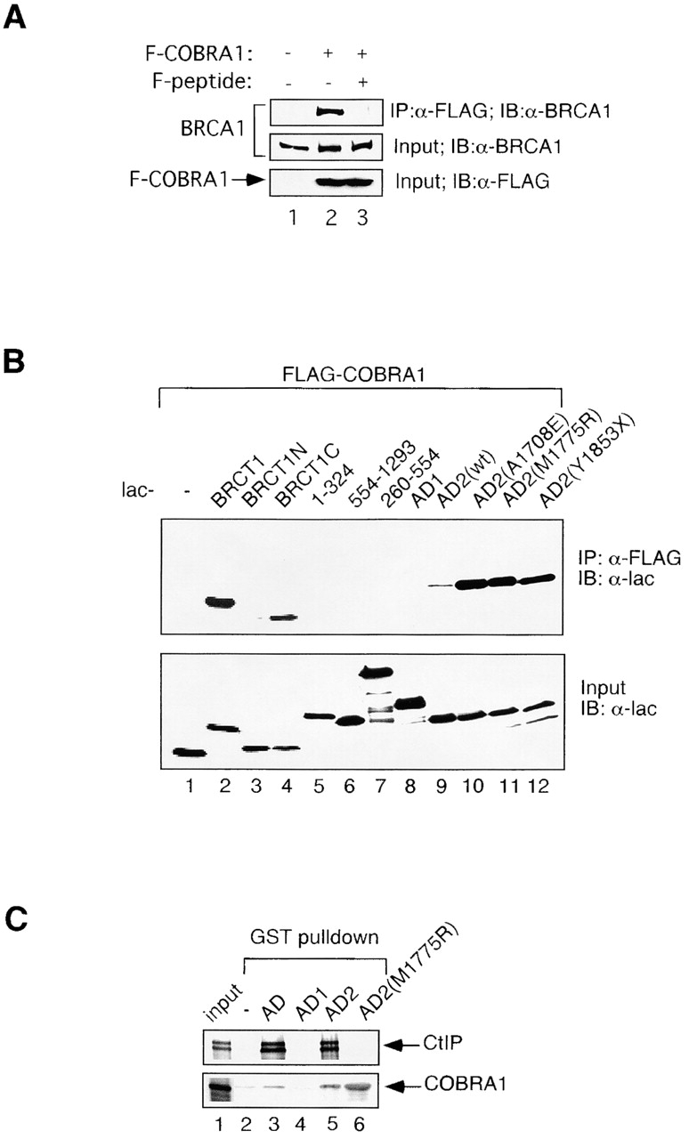
Identification of COBRA1 as a novel BRCA1-interacting protein. (A). COBRA1 interacts with endogenous full-length BRCA1. Human HEK293T cells were transfected with either an empty vector (lane 1) or expression vector for the FLAG-tagged COBRA1 (F-COBRA1; lanes 2 and 3). Cell lysates were immunoprecipitated (IP) with an anti-FLAG antibody conjugated to Protein A agarose beads (Sigma-Aldrich), in the absence (lane2) or presence (lane 3) of the FLAG peptide at a final concentration of 0.8 μg/ml. The immunoprecipitates were immunoblotted (IB) with a monoclonal anti-BRCA1 antibody (AB1 from Oncogene), the results of which are shown in the top panel. As controls, the crude lysates were immunoblotted for the endogenous BRCA1 (middle) and the ectopically expressed FLAG-COBRA1 (bottom). (B) Further characterization of the interaction between BRCA1 and COBRA1. Various lac–BRCA1 fusion constructs and the FLAG-COBRA1 expression vector were cotransfected into HEK293T cells. Cell lysates were immunoprecipitated with an anti-FLAG antibody and subsequently immunoblotted with an anti-lac antibody, the results of which are shown in the top panel. Expression of the lac fusion proteins was determined by immunoblotting of the crude lysates with the anti-lac antibody (bottom). (C) In vitro GST pull-down assay to characterize the BRCA1-COBRA1 interaction. Various GST fusion proteins were expressed in bacteria and coupled to glutathione agarose beads (unpublished data). An equal amount of the GST fusion proteins was used to pull down the 35S labeled, in vitro translated CtIP (top) or COBRA1 (bottom).
Distinction between BRCT and other well-characterized transactivation domains in large-scale chromatin unfolding
A previous study has shown that VP16-induced chromatin unfolding is accompanied by recruitment of histone acetyltransferases and local histone hyperacetylation, a property frequently observed for transcriptionally active or competent chromatin (Tumbar et al., 1999) (Fig. 2 A, m–o). Here we extended the previous work by examining the transactivation domains of two cellular transcription factors, E2F1 and p53. Like lac-VP16, lac-E2F1 and lac-p53 also induced significant chromatin unfolding in 60 and 45% of transfected cells, respectively (Fig. 2 A, g and j). Furthermore, the lac-E2F1– and lac-p53–unfolded chromatin regions were enriched with hyperacetylated histone H3 and H4 (Fig. 2 A, g–i and j–l, and unpublished data). Thus, all three well-characterized transactivation domains (VP16, E2F1, and p53) can simultaneously induce large-scale chromatin unfolding and histone hyperacetylation. However, it remains unknown whether the observed histone hyperacetylation is causally related to chromatin unfolding.
The extent of chromatin decondensation induced by a single BRCT repeat is comparable to that exhibited by these potent transcriptional ADs (Fig. 2 A, compare a and d with g, j, and m). However, no obvious histone H3 or H4 hyperacetylation was detected in the BRCT1- or BRCT2-unfolded chromatin regions (Fig. 2 A, a–c and d–f), suggesting that BRCT-mediated chromatin unfolding is a separable event from histone acetylation. Although both BRCT repeats are required for AD2-mediated transcriptional activation, a single repeat does not serve as a strong AD (Chapman and Verma, 1996; Monteiro et al., 1996) (unpublished data). Therefore, chromatin unfolding by BRCT may be a necessary, but not sufficient step, in transcriptional activation.
In addition to acetylation, histones are subject to other posttranslational modifications under various physiological conditions. Of particular interest, phosphorylation of H2AX, a histone H2A variant, at serine 139 (γ-H2AX) is rapidly stimulated following ionizing radiation (Rogakou et al., 1999). Before irradiation, a subset of γ-H2AX nuclear foci colocalize with BRCA1 foci (Paull et al., 2000). After DNA damage, the number of both γ-H2AX and BRCA1 nuclear foci increases significantly; furthermore, the majority of BRCA1 foci overlapped γ-H2AX foci (Paull et al., 2000). These observations most likely reflect localized recruitment of the putative H2AX kinase and phosphorylation of H2AX-containing nucleosomes that are already present at these sites, rather than recruitment of new H2AX protein.
Using an antibody that specifically recognizes the phosphorylated form of H2AX (γH2AX), we detected colocalization of the endogenous γ-H2AX with the full-length BRCA1 fusion protein in a sub-population (15%) of the lac–BRCA1-transfected cells (Fig. 2 B, a–c). In contrast, lac-VP16 did not display any colocalization with γ-H2AX (Fig. 2 B, d–f), nor did lac-BRCT1 or lac-BRCT2 (unpublished data). It is not clear whether phosphorylation of H2AX is causally linked to BRCA1-mediated chromatin unfolding, as γ-H2AX colocalization is also observed in lac–BRCA1-expressing cells that do not display chromatin decondensation. Consistent with previous reports (Rogakou et al., 1999; Paull et al., 2000), ionizing radiation significantly increased the number and overall intensity of γ-H2AX foci (unpublished data). However, the strong γ-H2AX signal over the entire nucleus made it difficult to examine the effect of DNA damage on the colocalization between γ-H2AX and lac-BRCA1 at the lac binding sites.
Work by Paull et al. (2000) has shown that H2AX at the damaged sites is rapidly phosphorylated after ionizing radiation, which is followed later by colocalization of BRCA1 and other repair proteins (Paull et al., 2000). It is possible that the putative kinase(s) responsible for H2AX phosphorylation directly binds to the full-length BRCA1. In such an event, tethering lac–BRCA1 may simply bring the kinase(s) to the tandem array of the lac binding sites, thus causing hyperphosphorylation of H2AX present in the surrounding chromosomal region. Whereas the functional significance of H2AX phosphorylation in chromatin unfolding remains to be explored, our finding is consistent with the previous suggestion of a physical link between γ-H2AX and BRCA1.
Allele-specific effects of cancer-predisposing mutations of BRCA1 on chromatin unfolding
To determine the effect of cancer-associated mutations on the BRCA1-dependent chromatin unfolding, we introduced a series of common cancer-predisposing mutations into either full-length BRCA1 or AD2. Based on their behaviors in the chromatin-unfolding assay, mutations were classified into three phenotypic categories. The first includes nonsense mutations resulting in truncation of the entire COOH terminus (Fig. 3 B, a). According to previous studies, BRCA1 mutants that lack the COOH terminus of the protein are defective in stimulating transcription and DNA repair (Somasundaram et al., 1997; Abbott et al., 1999; Scully et al., 1999; Jin et al., 2000). As shown in group a of Fig. 3 B, these COOH-terminal truncation mutants also failed to induce chromatin unfolding. The second group of mutants include missense mutations that are located upstream of AD2 (group b, i.e., C61G, S1040N, and R1347G). None of the mutants in this group significantly affects BRCA1-mediated chromatin unfolding.
Figure 3.
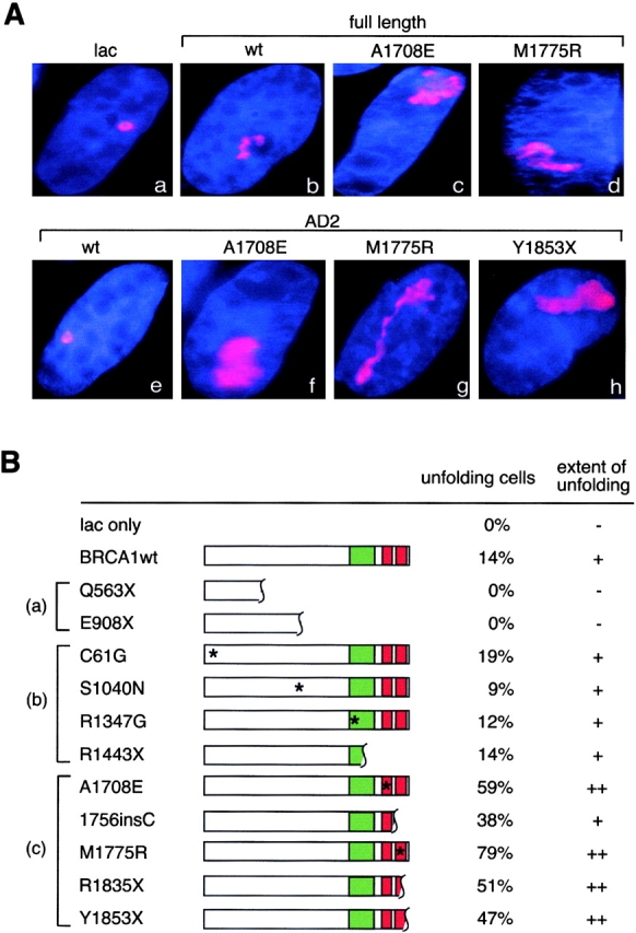
A subset of cancer-predisposing mutations in the COOH-terminal domain of BRCA1 cause increased chromatin unfolding. (A) Cancer-predisposing mutations were introduced into either the full-length BRCA1 (a–d) or AD2 (e–h). The corresponding expression vectors were transfected into AO3_1 cells, and immunostaining was performed as described in Fig. 1. (B) Summary of the effects of different cancer-associated mutations on chromatin unfolding. All mutants shown in this table were tested in the context of full-length BRCA1. Locations of missense mutations are indicated by asterisks, whereas those of nonsense and frameshift mutations are indicated by wavy lines. All mutations are grouped into three (a–c) as discussed in the text.
Contrary to the behaviors of first two groups, mutations in group c markedly enhanced the ability of lac-BRCA1 to induce chromatin unfolding (Fig. 3 B). For example, A1708E, M1775R, and Y1853X led to a pronounced enlargement of the unfolded chromatin structure (Fig. 3 A, compare b with c and d, and e with f–h). The same mutations also significantly increased the percentage of transfected cells that showed chromatin unfolding (Fig. 3 B). For instance, 79% of the cells that expressed the M1775R mutant displayed significant chromatin unfolding, compared with 14% for the wild-type full-length protein. This is an even higher percentage than that previously observed for the VP16 activator (Tumbar et al., 1999).
Interestingly, all mutations in group c result in single aa substitutions or small deletions within the AD2 region. Many of the mutations in this group have been shown previously to abolish AD2 interactions with other transcription-related proteins, including the RNA pol II holoenzyme (Scully et al., 1997a; Neish et al., 1998; Yarden and Brody, 1999). As discussed below, by retaining the chromatin unfolding activity of BRCA1 but blocking its role in other steps of transcriptional activation, these mutations in group c may lead to accumulation of the highly decondensed chromatin structure as observed in the unfolding assay.
Identification of a novel BRCA1-interacting protein
Application of the chromatin unfolding assay allowed us to identify a large-scale chromatin unfolding activity associated with BRCA1, and to narrow down the chromatin-unfolding region of BRCA1 to small subdomains in the COOH terminus of the protein. To identify cofactors recruited by the BRCT repeats to mediate chromatin unfolding, we used BRCT1 as the bait in a yeast two-hybrid screen. One candidate gene, cofactor COBRA1, was isolated from a human ovary cDNA library. It encodes a novel 580-aa protein rich in leucine residues (17%) (Fig. 4) . COBRA1 also contains three repeats of the LXXLL motif, often present in many transcription coactivators and responsible for mediating their ligand-dependent interactions with steroid hormone receptors (Heery et al., 1997). Database searches revealed COBRA1-related hypothetical proteins in mice and flies that share 96 and 51% aa identity with the human protein, respectively (Fig. 4).
Figure 4.
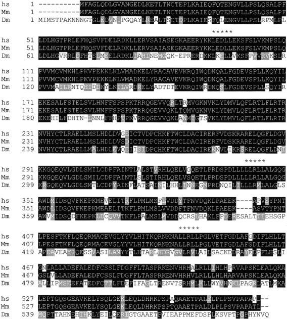
Sequence alignment of human COBRA1 and its homologues from mice and flies. The conserved aa residues are highlighted in black, and the similar residues in gray. The locations of the LXXLL motif are indicated by asterisks.
To confirm the interaction between BRCA1 and COBRA1, a lysate of human HEK293T cells that ectopically expressed FLAG-tagged COBRA1 was immunoprecipitated with an anti-FLAG antibody, followed by immunoblotting with an anti-BRCA1 antibody. As shown in Fig. 5 A, the endogenous human BRCA1 was coprecipitated in a FLAG-COBRA1–dependent manner (lanes 1 and 2). As a control, addition of an excess of FLAG peptide to the immunoprecipitation reaction abolished the BRCA1 signal in the immunoprecipitate (Fig. 5 A, lane 3).
To further assess the binding specificity of COBRA1 to the BRCT1 region of BRCA1, we cotransfected HEK293T cells with FLAG-COBRA1 and lac repressor fused with various fragments of BRCA1. The cell lysates were then immunoprecipitated with the anti-FLAG antibody and subsequently immunoblotted with the anti-lac antibody. As shown in Fig. 5 B, lac-BRCT1 was capable of binding to the FLAG-COBRA1 (lane 2). Consistent with their activity in chromatin unfolding, the COOH-, but not the NH2-terminal half of BRCT1 (Fig. 1 B), interacted with COBRA1 (lanes 3 and 4). None of the BRCA1 fragments upstream of the BRCT repeat, including AD1, displayed any significant affinity for COBRA1 (lanes 5–8). Taken together, our data show that the COBRA1 binding correlates with the BRCT1-mediated large-scale chromatin unfolding.
As shown in Fig. 3, cancer-predisposing mutations in the 3′ region of BRCA1 caused significant enhancement of the chromatin unfolding activity. Intriguingly, the same mutations (A1708E, M1775R, and Y1853X) also increased the affinity for COBRA1 in the coimmunoprecipitation assay (Fig. 5 B, compare lane 9 with lanes 10–12). A similar result was also observed in an in vitro glutathione S-transferase (GST) pull-down assay (Fig. 5 C). In this case, 35S-labeled, in vitro–translated COBRA1 was pulled down by both GST–AD and GST–AD2, but not by GST–AD1 (Fig. 5 C, bottom panel, lanes 3–5). Furthermore, COBRA1 displayed a higher affinity for the mutant (M1775R) than the wild-type GST–AD2 fusion (Fig. 5 C, bottom panel, lanes 5 and 6). As a control, we also used 35S-labeled CtIP, a transcriptional corepressor that binds to the COOH terminus of BRCA1 (Wong et al., 1998; Yu et al., 1998; Li et al., 1999). Consistent with previous findings, CtIP binds specifically to AD2 but, unlike COBRA1, its association with AD2 is abolished by the M1775R mutation (Fig. 5 C, top panel, lanes 5 and 6). Thus, the same cancer-predisposing mutations exert opposite effects on BRCA1 binding to two different partners.
Involvement of COBRA1 in BRCT1-mediated chromatin unfolding
To explore the role of COBRA1 in the BRCT1-mediated chromatin unfolding, we cotransfected FLAG-COBRA1 with various lac–BRCA1 fusion constructs into AO3_1 cells. As detected by confocal immunofluorescent microscopy, FLAG-COBRA1 and lac–BRCT1 colocalized in 96% of the cells that expressed both proteins (Fig. 6 A, a–c). In contrast, we did not detect any enrichment of the FLAG-COBRA1 signal at either the BRCT2- or AD1-unfolded chromatin regions (Fig. 6 A, BRCT2, d–f, and AD1, g–i). Thus, whereas all three subdomains are capable of inducing large-scale chromatin unfolding, they appear to recruit distinct cofactors to mediate this process.
Figure 6.
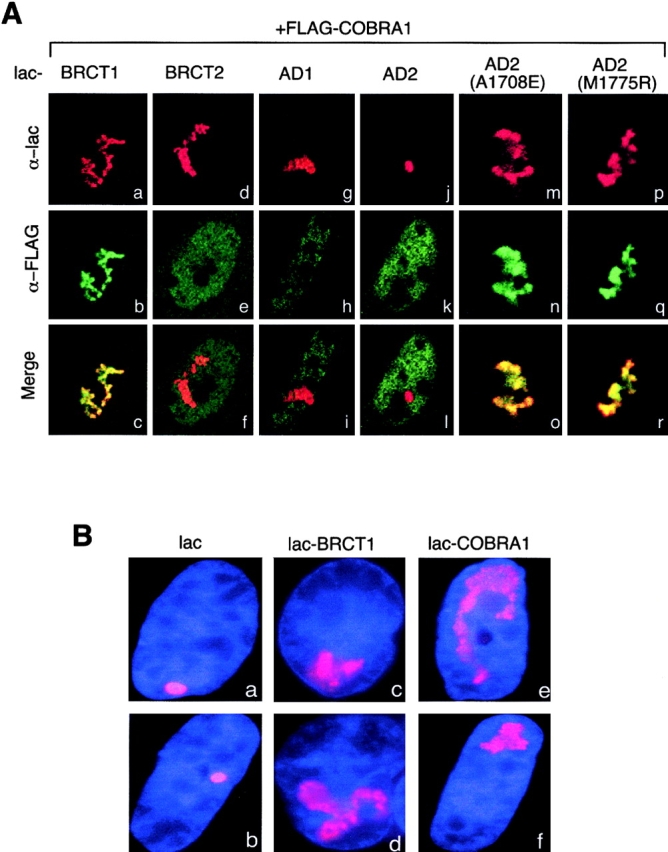
COBRA1 colocalizes with lac–BRCT1 and can induce large-scale chromatin unfolding. (A) Colocalization of lac fusion proteins (red) and FLAG-COBRA1 (green) at the unfolded chromatin region. AO3_1 cells were cotransfected with the expression vectors for FLAG-COBRA1 and lac fused with various fragments of BRCA1. The images were captured by confocal immunofluorescence microscopy. (B) COBRA1 induces chromatin unfolding when directly targeted to the chromosome. AO3_1 cells were transfected with the expression vectors for lac repressor alone (a and b), lac–BRCT1 (c and d), or lac–COBRA1 (e and f). Chromatin unfolding was detected as described in the previous figures.
Wild-type AD2, which failed to induce chromatin unfolding (Fig. 1), did not display any obvious colocalization with COBRA1 (Fig. 6 A, panels j–l). However, two 3′ cancer-predisposing mutations in the same context led to pronounced recruitment of COBRA1 to the unfolded chromatin regions (Fig. 6 A, A1708E, m–o, and M1775R, p–r). Colocalization of COBRA1 and the mutant lac–AD2 fusion proteins was observed in >90% cells that expressed both proteins. Thus, the effect of the 3′ mutations on COBRA1 recruitment correlates with their stimulatory effects on chromatin unfolding.
To directly assess the impact of COBRA1 on large-scale chromatin structure, we used lac repressor to target COBRA1 to the lac binding sites in AO3_1 cells. As shown in Fig. 6 B, 61% of the cells that expressed lac–COBRA1 showed a comparable extent of chromatin unfolding as did lac–BRCT1 (compare c and d with e and f). This finding strongly implicates COBRA1 in BRCT1-mediated chromatin restructuring.
Discussion
Eukaryotic genomes are packaged through multiple steps into higher levels of chromatin structure. It is now well established that remodeling of local chromatin structure is a key step common to the initiation of multiple chromosomal functional events, including transcription, DNA replication, repair, and recombination (Elgin and Workman, 2000; Fyodorov and Kadonaga, 2001). Whereas intense research in the past decade has provided a wealth of information regarding the biochemical basis for chromatin remodeling at the nucleosome level, much less is known about reorganization of higher levels of chromatin structure. It remains unclear whether the known modifications of nucleosome organization are sufficient for changes in large-scale chromatin organization, or whether novel mechanisms acting at higher levels of chromatin structure are responsible for changes in large-scale chromatin organization.
A major difficulty in distinguishing these two possibilities is that most assays for identifying transcriptional activators or coactivators have used transcriptional activity as a final readout. Direct assays for changes in higher order chromatin structure have not been used previously. Because BRCA1 had been functionally implicated in a range of nuclear processes, it was reasonable to postulate that the regulation of these multiple nuclear events might occur through a general chromatin remodeling activity of BRCA1. The lac repressor–tethering system, while artificial in many aspects, provided an excellent assay to pursue this research direction.
Our findings in this current study strongly suggest that BRCA1 recruits COBRA1, a novel factor, to the lac operator–containing chromatin region. Within the constraints of the lac repressor–tethering assay, BRCA1-dependent unfolding of higher levels of chromatin structure appears to be at least partially mediated through recruitment of COBRA1. Notably, BRCA1-mediated chromatin decondensation is distinct from transcriptional activation per se and histone hyperacetylation. It is unclear how unique the histone acetylation–independent chromatin unfolding is. Although the chromatin unfolding produced by VP16, E2F1, and p53 is accompanied by histone hyperacetylation, no causal relationship between histone acetyltransferases recruitment and chromatin unfolding has been demonstrated. Moreover, preliminary data suggests that large-scale decondensation produced by estrogen receptor does not correlate with histone hyperacetylation (A. Nye and A. Belmont, personal communication).
Whereas the lac-based chromatin-unfolding assay provides a new tool for visualizing chromatin dynamics and in vivo protein–protein interactions in mammalian cells, it is important to point out that the molecular and biochemical basis for BRCA1-mediated chromatin decondensation is yet to be understood. Furthermore, utilization of a long tandem array of lac binding sites may raise the concern that the observed chromatin unfolding could simply be due to steric effects of the proteins/protein complexes that are brought to the lac binding sites. However, we believe this possibility is unlikely because our work does not indicate an obvious correlation between the potency of chromatin unfolding and the size or charge of the tethered protein fragments. For example, the minimal chromatin-unfolding domain defined in our study is only 50 aa long (BRCA1C). In contrast, several other BRCA1 fragments that range in size from 324 to 740 aa do not display any chromatin-unfolding activity (Fig. 1 B). In addition, BRCT1 and BRCT2 have a net charge of +5 and –6, respectively, yet both demonstrate strong chromatin-unfolding activity. On the other hand, BRCT1N carries more positive charges (+5) than BRCT1C (+1), but only the latter can induce chromatin decondensation. Finally, in previous work using either lac repressor tetramer, or lac repressor fused to several other protein domains up to ∼350 aa in size (i.e., GFP), no effect on large-scale chromatin structure has been observed (Robinett et al., 1996, and A. Belmont, personal communication).
In our minds, a more serious caveat concerning the lac repressor–tethering system is the question of whether the observed effects produced by BRCA1 and other proteins on large-scale chromatin unfolding are physiologically relevant given the high numbers of lac operator repeats involved. In fact, the exact number of lac repressors binding per lac operator has not been determined and there is reason to believe that lac repressor binding may be significantly limited by steric constraints and phasing of lac operators relative to the nucleosome linker DNA. However, we note that a recent study on a transgene array containing a viral promoter with several glucocorticoid hormone response elements observed a very similar type of large-scale chromatin decondensation produced by glucocorticoid receptor (Muller et al., 2001). Ultimately, validation of the physiological significance of our observations of BRCA1-dependent large-scale chromatin unfolding will depend on the outcome of future experiments exploring the mechanisms of unfolding and identifying the biological functions of other transacting factors involved, such as COBRA1.
With these caveats in mind, we find it particularly intriguing that a subset of cancer-predisposing mutations of BRCA1 lead to increased chromatin unfolding and recruitment of COBRA1. Although the genotype–phenotype relationship in cancer-predisposing mutations of BRCA1 remains to be understood, it is generally assumed that most, if not all, BRCA1 mutations lead to loss of the biological functions of the protein. However, the behaviors of the BRCA1 mutants in the chromatin-unfolding assay clearly demonstrate an allele-specific effect. Consistent with this finding, it has been reported that mutations at different locations along the coding sequence of BRCA1 differentially affect the penetrance of BRCA1-dependent breast and ovarian cancer (Gayther et al., 1995; Risch et al., 2001). It remains to be determined whether the three groups of mutations that cause differential effects on chromatin unfolding (Fig. 3) may indeed lead to distinct clinical consequences in terms of risks, types, or prognosis of BRCA1-associated cancers. In particular, it will be interesting to see whether those 3′ mutations that enhance chromatin unfolding exhibit any dominant or semidominant phenotype in cancer genetics. It is conceivable that constitutive decondensation of large-scale chromatin structure may cause additional deleterious effects on genome stability and thus result in more severe clinical consequences in cancer development.
Our study also indicates that BRCT-mediated chromatin unfolding may be tightly regulated. As shown in Figs. 1 and 3, a single BRCT motif is more potent in chromatin unfolding than the larger fragments of the protein that contain both BRCT repeats. Furthermore, the full-length wild-type BRCA1 only exhibits a moderate chromatin-unfolding activity, whereas the cancer-predisposing mutations in group c (Fig. 3 B) that affect the integrity of the BRCT repeats significantly enhance the chromatin-unfolding activity and COBRA1 binding. These results lead us to the following two models that could explain negative regulation of BRCA1-mediated chromatin unfolding. In a “trans-inhibition” model, we speculate that binding of a putative inhibitor (i.e., CtIP) to AD2 region of BRCA1 may prevent BRCA1 from interacting with its cofactors for chromatin unfolding (i.e., COBRA1). In an alternative, “cis-inhibition” model, the two BRCT tandem repeats may form an intramolecular dimer. This in turn may reduce the affinity of both BRCT repeats for their corresponding cofactors. Conceivably, the “superactivating” mutations in group c may prevent binding of the putative inhibitor or the intra-molecular interaction between the two BRCT motifs, thus rendering the protein constitutively active for binding to the cofactors that mediate chromatin unfolding.
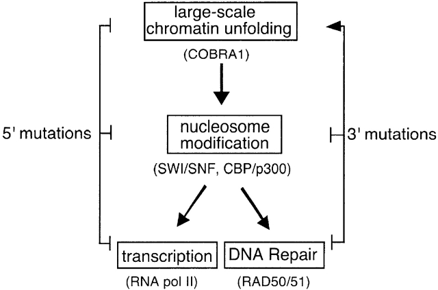
It is plausible that BRCT-mediated chromatin unfolding may lead to a novel nuclear function of BRCA1 in global reorganization of the genome. However, in light of the known function of the COOH-terminal region of BRCA1 in transcription and DNA repair, the observed chromatin decondensation may represent the first step in BRCA1-mediated regulation of these two nuclear processes (Fig. 7) . In such a model, higher order chromatin decondensation may be followed by BRCA1-mediated chromatin modification at the nucleosomal level (i.e., histone hyperacetylation) and recruitment of the transcription or repair machineries. As shown in Fig. 7, nonsense mutations that result in truncation of the entire COOH-terminal region (5′ mutations) may abolish BRCA1 functions in all three steps, resulting in a completely inactive mutant protein. On the other hand, mutations located at the 3′ end of the gene (3′ mutations) may render BRCA1 incompetent at the second and third steps, but still allow constitutive chromatin decondensation at the first step. This could then lead to accumulation of extensively unfolded chromatin structure as seen in our study. Consistent with this model, many 3′ cancer-predisposing mutations abolish BRCA1 interactions with RNA pol II holoenzyme and the histone modifying enzymes (Scully et al., 1997a; Neish et al., 1998; Yarden and Brody, 1999), as well as nucleosome remodeling in yeast (Hu et al., 1999). Thus, chromatin unfolding may be a necessary but not sufficient step for BRCA1-dependent transcriptional activation. Additional steps such as histone modification and recruitment of the basal machinery may also be required for fulfilling BRCA1 function in transcription and DNA repair.
Figure 7.
Model for BRCA1-mediated nuclear functions. Inhibitory and stimulatory effects of the 5′ and 3′ mutations on the three steps are indicated by bars and arrows on the sides, respectively. Factors in parentheses are those that may be targeted or recruited by BRCA1 to facilitate a specific step in activation of transcription or DNA repair. See text for more detail.
Materials and methods
Chromatin unfolding assay
To construct the EGFP-lac-E2F1 and EGFP-lac-p53 fusion expression vectors, the PCR fragments that encode the E2F1 (aa 368–437) and p53 (aa 1–73), respectively, were cloned into the AscI site in the plasmid p3′SS d tb Cl EGFP AscI (NYE4) (A.C. Nye and A.S. Belmont, personal communication). The correct orientation of the inserts was identified by colony hybridization and confirmed by DNA sequencing. To construct the lac-BRCA1 plasmids, the sequence for lac repressor was first amplified by PCR from the plasmid NYE4. The lac sequence was cloned into the HindIII–NotI sites of pRC-CMV (Invitrogen), generating pRC-lac. Various BRCA1 fragments and the COBRA1 sequence were amplified by PCR and inserted into the unique AscI site of pRC-lac.
The chromatin unfolding experiments were performed as previously described (Tumbar et al., 1999). Briefly, AO3_1 cells were transiently transfected with the lac expression vectors using the FuGENE 6 transfection reagent (Roche). The medium was changed 24 h after transfection and cells were immunostained 48 h after transfection. Cells grown on glass coverslips were fixed with 1.6% paraformaldehyde for 30 min in PBS, permeabilized with 0.2% Triton X-100 in PBS for 5 min, and blocked in 1% normal goat serum in PBS for 1 h. The coverslips were then incubated with primary antibodies at room temperature for 1 h, followed by incubation with the appropriate secondary antibodies for 1 h. Unless otherwise specified, a rabbit polyclonal anti–lac repressor antibody (Stratagene) and mouse monoclonal anti-FLAG antibody (Sigma-Aldrich) were applied at 1:20,000 dilution. The anti–acetylated histone H3 antibody was raised against di-acetylated H3 (Lys9 and Lys14) (Boggs et al., 1996) (Lin et al., 1989), a gift from Drs. C. Mizzen and C.D. Allis (University of Virginia, Charlottesville, VA). The secondary antibodies were goat anti–rabbit IgG-conjugated with Cy3 (Amersham), and horse anti–mouse IgG-conjugated with fluorescein isothiocyanate (FITC; Vector Laboratories).
For visualization of the nuclei, cells were stained with 0.2 μg/ml 4,6-diamidino-2-phenylindole (DAPI) for 5 min before mounting. Fluorescent images were acquired by a charged-coupled device camera (Hamamatsu ORCA) that was mounted on a Nikon Microphot-SA microscope and equipped with Improvision Openlab software. Confocal images were collected on a Zeiss LSM410 confocal microscope. Figs. were assembled using Adobe Photoshop (v. 5.5).
Yeast two-hybrid screen
To identify proteins that specifically interact with the BRCT1 repeat of BRCA1, the standard yeast two-hybrid screen was performed in the following manner. First, the bait plasmid was generated by inserting a PCR-amplified cDNA fragment encoding the BRCT1 sequence (aa 1642–1736) into the NdeI–EcoRI restriction sites of pAS2–1 (CLONTECH Laboratories, Inc.), resulting in an in-frame fusion with the GAL4 DNA-binding domain. The resultant plasmid, pAS2-BRCT1, and a human ovary cDNA prey library (CLONTECH Laboratories, Inc.) were sequentially transformed into the S. cerevisiae strain CG1945 according to the manufacturer's instructions (CLONTECH Laboratories, Inc.). Transformants were plated on synthetic medium lacking tryptophan, leucine and histidine but containing 1 mM 3-aminotriazole. Approximately 2.3 million transformants were screened. The candidate clones were retrieved from the yeast cells and reintroduced back to the same yeast strain to verify the interaction between the candidates and the BRCT1 bait. The specificity of the interaction was determined by comparing the interactions between the candidates and various bait constructs.
Coimmunoprecipitation
HEK293T cells were transfected using LipofectAmine 2000 (GIBCO BRL). 24 h after transfection, cells were washed twice with PBS and lysed in 0.5 ml lysis buffer (50 mM Hepes, pH 8, 250 mM NaCl, 0.1% NP-40, and protease inhibitor tablets from Roche). After brief sonication, the lysate was centrifuged at 16,000 g for 12 min at 4°C. The supernatant was used for subsequent coimmunoprecipitation. 20 μl of the supernatant was used as crude extract for detecting protein expression level. 15 μl of a 50% slurry of the anti-FLAG agarose beads (Sigma-Aldrich) was used in each immunoprecipitation. Immunoprecipitation was performed overnight at 4°C. The beads were centrifuged at 3,300 rpm for 2 min, and washed three times with washing buffer (50 mM Hepes, pH8, 500 mM NaCl, 0.5% NP-40) and three times with RIPA buffer (50 mM Tris, pH 8.0, 150 mM NaCl, 1% NP-40, 0.1% SDS, and 0.5% sodium deoxycholate). Each wash was performed for at least 30 min. The precipitates were then eluted in 15 μl 2× SDS-PAGE sample buffer. Gel electrophoresis was followed by immunoblotting according to standard procedures.
GST pulldown assay
The PCR fragments encoding various BRCA1 fragments were cloned into pGEX-2T and the constructs were confirmed by sequencing. The GST-BRCA1 proteins were made and purified, with the induction of protein expression performed at 19°C overnight. pcDNA3 vector containing the COBRA1 gene was used for in vitro transcription and translation in the TnT Reticulocyte Lysate system (Promega). The 35S-labeled COBRA1 was translated in vitro according to the manufacturer's instructions and mixed with 10 μg the GST-bound bead in 0.5 ml binding buffer (50 mM Tris-HCl, pH 7.5, 150 mM NaCl, 1 mM EDTA, 0.3 mM DTT, 0.1% NP-40 and protease inhibitor tablet). The binding reaction was performed at 4°C overnight and the beads were subsequently washed four times with washing buffer (same as binding buffer except 0.5% NP-40 was used), 30 min each time. The beads were eluted in 10 μl 2 × SDS-PAGE sample buffer and the proteins were resolved on 10% denaturing gel. The gel was then dried and exposed to x-ray films for overnight.
Acknowledgments
We thank David Allis, Michael Christman, and Mark Alexandow for critical reading of the manuscript, and the Center for Cell Signaling at the University of Virginia (Charlottesville, VA) for the microscopy facility.
This work was supported by National Institutes of Health grants (GM58460 to A. Belmont and CA83990 to R. Li), the American Cancer Society (RPG99-211-01-MBC to R. Li), and a National Institutes of Health Traineeship (T32-GM07283) and a Howard Hughes Medical Institute Predoctoral Fellowship to A.C. Nye.
Footnotes
Abbreviations used in this paper: aa, amino acid(s); AAD, acidic activation domain; BRCA1, breast cancer susceptibility gene 1; BRCT, BRCA1 COOH terminus; COBRA1, cofactor of BRCA1; EGFP, enhanced green fluorescent protein; GST, glutathione S-transferase; RING, ring finger motif.
References
- Abbott, D.W., M.E. Thompson, C. Robinson-Benion, G. Tomlinson, R.A. Jensen, and J.T. Holt. 1999. BRCA1 expression restores radiation resistance in BRCA1-defective cancer cells through enhancement of transcription-coupled DNA repair. J. Biol. Chem. 274:18808–18812. [DOI] [PubMed] [Google Scholar]
- Belmont, A.S. 2001. Visualizing chromosome dynamics with GFP. Trends Cell Biol. 11:250–257. [DOI] [PubMed] [Google Scholar]
- Bochar, D.A., L. Wang, H. Beniya, A. Kinev, Y. Xue, W.S. Lane, W. Wang, F. Kashanchi, and R. Shiekhattar. 2000. BRCA1 is associated with a human SWI/SNF-related complex: linking chromatin remodeling to breast cancer. Cell. 102:257–265. [DOI] [PubMed] [Google Scholar]
- Boggs, B.A., B. Connors, R.E. Sobel, A.C. Chinault, and C.D. Allis. 1996. Reduced levels of histone H3 acetylation on the inactive X chromosome in human females. Chromosoma. 105:303–309. [DOI] [PubMed] [Google Scholar]
- Chapman, M.S., and I.M. Verma. 1996. Transcriptional activation by BRCA1. Nature. 382:678–679. [DOI] [PubMed] [Google Scholar]
- Cortez, D., Y. Wang, J. Qin, and S.J. Elledge. 1999. Requirement of ATM-dependent phosphorylation of brca1 in the DNA damage response to double-strand breaks. Science. 286:1162–1166. [DOI] [PubMed] [Google Scholar]
- Elgin, S.C.R., and J.L. Workman. 2000. Chromatin structure and gene expression. Frontiers in Molecular Biology. B.D. Hames and D.M. Glover, editors. Oxford University Press, Oxford. 354 pp.
- Fan, S., J. Wang, R. Yuan, Y. Ma, Q. Meng, M.R. Erdos, R.G. Pestell, F. Yuan, K.J. Auborn, I.D. Goldberg, and E.M. Rosen. 1999. BRCA1 inhibition of estrogen receptor signaling in transfected cells. Science. 284:1354–1356. [DOI] [PubMed] [Google Scholar]
- Fyodorov, D.V., and J.T. Kadonaga. 2001. The many faces of chromatin remodeling: switching beyond transcription. Cell. 106:523–525. [DOI] [PubMed] [Google Scholar]
- Gayther, S.A., W. Warren, S. Mazoyer, P.A. Russell, P.A. Harrington, M. Chiano, S. Seal, R. Hamoudi, E.J. van Rensburg, A.M. Dunning, et al. 1995. Germline mutations of the BRCA1 gene in breast and ovarian cancer families provide evidence for a genotype-phenotype correlation. Nat. Genet. 11:428–433. [DOI] [PubMed] [Google Scholar]
- Gowen, L.C., A.V. Avrutskaya, A.M. Latour, B.H. Koller, and S.A. Leadon. 1998. BRCA1 required for transcription-coupled repair of oxidative DNA damage. Science. 281:1009–1012. [DOI] [PubMed] [Google Scholar]
- Heery, D.M., E. Kalkhoven, S. Hoare, and M.G. Parker. 1997. A signature motif in transcriptional co-activators mediates binding to nuclear receptors. Nature. 387:733–736. [DOI] [PubMed] [Google Scholar]
- Houvras, Y., M. Benezra, H. Zhang, J.J. Manfredi, B.L. Weber, and J.D. Licht. 2000. BRCA1 physically and functionally interacts with ATF1. J. Biol. Chem. 275:36230–36237. [DOI] [PubMed] [Google Scholar]
- Hu, Y.-F., T. Miyake, Q. Ye, and R. Li. 2000. Characterization of a novel trans-activation domain of BRCA1 that functions in concert with the BRCA1 C-terminal (BRCT) domain. J. Biol. Chem. 275:40910–40915. [DOI] [PubMed] [Google Scholar]
- Hu, Y.F., Z.L. Hao, and R. Li. 1999. Chromatin remodeling and activation of chromosomal DNA replication by an acidic transcriptional activation domain from BRCA1. Genes Dev. 13:637–642. [DOI] [PMC free article] [PubMed] [Google Scholar]
- Jin, S., H. Zhao, F. Fan, P. Blanck, W. Fan, A.B. Colchagie, A.J. Fornace, Jr., and Q. Zhan. 2000. BRCA1 activation of the GADD45 promoter. Oncogene. 19:4050–4057. [DOI] [PubMed] [Google Scholar]
- Lee, J.-S., K.M. Collins, A.L. Brown, C.-H. Lee, and J.H. Chung. 2000. hCds1-mediated phosphorylation of BRCA1 regulates the DNA damage response. Nature. 404:201–204. [DOI] [PubMed] [Google Scholar]
- Li, G., G. Sudlow, and A.S. Belmont. 1998. Interphase cell cycle dynamics of a late-replicating, heterochromatic homogenously staining region: precise choreography of condensation/decondensation and intranuclear positioning. J. Cell Biol. 140:975–989. [DOI] [PMC free article] [PubMed] [Google Scholar]
- Li, S., P.L. Chen, T. Subramanian, G. Chinnadurai, G. Tomlinson, C.K. Osborne, Z.D. Sharp, and W.H. Lee. 1999. Binding of CtIP to the BRCT repeats of BRCA1 involved in the transcription regulation of p21 is disrupted upon DNA damage. J. Biol. Chem. 274:11334–11338. [DOI] [PubMed] [Google Scholar]
- Lin, R., J.W. Leone, R.G. Cook, and C.D. Allis. 1989. Antibodies specific to acetylated histones document the existence of deposition- and transcription-related histone acetylation in Tetrahymena. J. Cell Biol. 108:1577–1588. [DOI] [PMC free article] [PubMed] [Google Scholar]
- Monteiro, A.N. 2000. BRCA1: exploring the links to transcription. TIBS. 25:469–474. [DOI] [PubMed] [Google Scholar]
- Monteiro, A.N., A. August, and H. Hanafusa. 1996. Evidence for a transcriptional activation function of BRCA1 C-terminal region. Proc. Natl. Acad. Sci. USA. 93:13595–13599. [DOI] [PMC free article] [PubMed] [Google Scholar]
- Moynahan, M.E., J.W. Chiu, B.H. Koller, and M. Jasin. 1999. Brca1 controls homology-directed DNA repair. Mol. Cell. 4:511–518. [DOI] [PubMed] [Google Scholar]
- Muller, W.G., D. Walker, G.L. Hager, and J.G. McNally. 2001. Large-scale chromatin decondensation and recondensation regulated by transcription from a natural promoter. J. Cell Biol. 154:33–48. [DOI] [PMC free article] [PubMed] [Google Scholar]
- Neish, A.S., S.F. Anderson, B.P. Schlegel, W. Wei, and J.D. Parvin. 1998. Factors associated with the mammalian RNA polymerase II holoenzyme. Nucleic Acids Res. 26:847–853. [DOI] [PMC free article] [PubMed] [Google Scholar]
- Ouchi, T., A.N. Monteiro, A. August, S.A. Aaronson, and H. Hanafusa. 1998. BRCA1 regulates p53-dependent gene expression. Proc. Natl. Acad. Sci. USA. 95:2302–2306. [DOI] [PMC free article] [PubMed] [Google Scholar]
- Ouchi, T., S.W. Lee, M. Ouchi, S.A. Aaronson, and C.M. Horvath. 2000. Collaboration of signal transducer and activator of transcription 1 (STAT1) and BRCA1 in differential regulation of IFN-gamma target genes. Proc. Natl. Acad. Sci. USA. 97:5208–5213. [DOI] [PMC free article] [PubMed] [Google Scholar]
- Pao, G.M., R. Janknecht, H. Ruffner, T. Hunter, and I.M. Verma. 2000. CBP/p300 interact with and function as transcriptional coactivators of BRCA1. Proc. Natl. Acad. Sci. USA. 97:1020–1025. [DOI] [PMC free article] [PubMed] [Google Scholar]
- Paull, T.T., E.P. Rogakou, V. Yamazaki, C.U. Kirchgessner, M. Gellet, and W.M. Bonner. 2000. A critical role for histone H2AX in recruitment of repair factors to nuclear foci after DNA damage. Curr. Biol. 10:886–895. [DOI] [PubMed] [Google Scholar]
- Paull, T.T., D. Cortez, B. Bowers, S.J. Elledge, and M. Gellert. 2001. From the cover: direct DNA binding by Brca1. Proc. Natl. Acad. Sci. USA. 98:6086–6091. [DOI] [PMC free article] [PubMed] [Google Scholar]
- Peterson, C.L., and C. Logie. 2000. Recruitment of chromatin remodeling machines. J. Cell. Biochem. 78:179–185. [DOI] [PubMed] [Google Scholar]
- Pikaart, M., F. Recillas-Targa, and G. Felsenfeld. 1998. Loss of transcriptional activity of a transgene is accompanied by DNA methylation and histone deacetylation and is prevented by insulators. Genes Dev. 12:2852–2862. [DOI] [PMC free article] [PubMed] [Google Scholar]
- Rahman, N., and M.R. Stratton. 1998. The genetics of breast cancer susceptibility. Annu. Rev. Genet. 32:95–121. [DOI] [PubMed] [Google Scholar]
- Risch, H.A., J.R. McLaughlin, D.E. Cole, B. Rosen, L. Bradley, E. Kwan, E. Jack, D.J. Vesprini, G. Kuperstein, J.L. Abrahamson, et al. 2001. Prevalence and penetrance of germline BRCA1 and BRCA2 mutations in a population series of 649 women with ovarian cancer. Am. J. Hum. Genet. 68:700–710. [DOI] [PMC free article] [PubMed] [Google Scholar]
- Robinett, C.C., A. Straight, G. Li, C. Willhelm, G. Sudlow, A. Murray, and A.S. Belmont. 1996. In vivo localization of DNA sequences and visualization of large-scale chromatin organization using lac operator/repressor recognition. J. Cell Biol. 135:1685–1700. [DOI] [PMC free article] [PubMed] [Google Scholar]
- Rogakou, E.P., C. Boon, C. Redon, and W.M. Bonner. 1999. Megabase chromatin domains involved in DNA double-strand breaks in vivo. J. Cell Biol. 146:905–915. [DOI] [PMC free article] [PubMed] [Google Scholar]
- Scully, R., and D.M. Livingston. 2000. In search of the tumor-suppressor functions of BRCA1 and BRCA2. Nature. 408:429–432. [DOI] [PMC free article] [PubMed] [Google Scholar]
- Scully, R., S.F. Anderson, D.M. Chao, W. Wei, L. Ye, R.A. Young, D.M. Livingston, and J.D. Parvin. 1997. a. BRCA1 is a component of the RNA polymerase II holoenzyme. Proc. Natl. Acad. Sci. USA. 94:5605–5610. [DOI] [PMC free article] [PubMed] [Google Scholar]
- Scully, R., J. Chen, A. Plug, Y. Xiao, D. Weaver, J. Feunteun, T. Ashley, and D.M. Livingston. 1997. b. Association of BRCA1 with Rad51 in mitotic and meiotic cells. Cell. 88:265–275. [DOI] [PubMed] [Google Scholar]
- Scully, R., S. Ganesan, K. Vlasakova, J. Chen, M. Socolovsky, and D.M. Livingston. 1999. Genetic analysis of BRCA1 function in a defined tumor cell line. Mol. Cell. 4:1093–1099. [DOI] [PubMed] [Google Scholar]
- Somasundaram, K., H. Zhang, Y.X. Zeng, Y. Houvras, Y. Peng, G.S. Wu, J.D. Licht, B.L. Weber, and W.S. El-Deiry. 1997. Arrest of the cell cycle by the tumour-suppressor BRCA1 requires the CDK-inhibitor p21WAF1/CiP1. Nature. 389:187–190. [DOI] [PubMed] [Google Scholar]
- Tibbetts, R.S., D. Cortez, K.M. Brumbaugh, R. Scully, D. Livingston, S.J. Elledge, and R.T. Abraham. 2000. Functional interactions between BRCA1 and the checkpoint kinase ATR during genotoxic stress. Genes Dev. 14:2989–3002. [DOI] [PMC free article] [PubMed] [Google Scholar]
- Tumbar, T., G. Sudlow, and A.S. Belmont. 1999. Large-scale chromatin unfolding and remodeling induced by VP16 acidic activation domain. J. Cell Biol. 145:1341–1354. [DOI] [PMC free article] [PubMed] [Google Scholar]
- Wang, Y., D. Cortez, P. Yazdi, N. Neff, S.J. Elledge, and J. Qin. 2000. BASC, a super complex of BRCA1-associated proteins involved in the recognition and reapir of aberrant DNA structures. Genes Dev. 14:927–939. [PMC free article] [PubMed] [Google Scholar]
- Wong, A.K., P.A. Ormonde, R. Pero, Y. Chen, L. Lian, G. Salada, S. Berry, Q. Lawrence, P. Dayananth, P. Ha, et al. 1998. Characterization of a carboxy-terminal BRCA1 interacting protein. Oncogene. 17:2279–2285. [DOI] [PubMed] [Google Scholar]
- Wu, L.C., Z.W. Wang, J.T. Tsan, M.A. Sillman, A. Phung, X.L. Xu, M.C. Yang, L.Y. Hwang, A.M. Bowcock, and R. Baer. 1996. Identification of a RING protein that can interact in vivo with the BRCA1 gene product. Nat. Genet. 14:430–440. [DOI] [PubMed] [Google Scholar]
- Xu, X., Z. Weaver, S.P. Linke, C. Li, J. Gotay, X.W. Wang, C.C. Harris, T. Ried, and C.X. Deng. 1999. Centrosome amplification and a defective G2-M cell cycle checkpoint induce genetic instability in BRCA1 exon 11 isoform-deficient cells. Mol. Cell. 3:389–395. [DOI] [PubMed] [Google Scholar]
- Yarden, R.I., and L.C. Brody. 1999. BRCA1 interacts with components of the histone deacetylase complex. Proc. Natl. Acad. Sci. USA. 96:4983–4988. [DOI] [PMC free article] [PubMed] [Google Scholar]
- Yu, X., L.C. Wu, A.M. Bowcock, A. Aronheim, and R. Baer. 1998. The C-terminal (BRCT) domains of BRCA1 interact in vivo with CtIP, a protein implicated in the CtBP pathway of transcriptional repression. J. Biol. Chem. 273:25388–25392. [DOI] [PubMed] [Google Scholar]
- Zhang, H., K. Somasundaram, Y. Peng, H. Tian, H. Zhang, D. Bi, B.L. Weber, and W.S. El-Deiry. 1998. a. BRCA1 physically associated with p53 and stimulates its transcriptional activity. Oncogene. 16:1713–1721. [DOI] [PubMed] [Google Scholar]
- Zhang, H., G. Tombline, and B.L. Weber. 1998. b. BRCA1, BRCA2, and DNA damage response: collision or collusion? Cell. 92:433–436. [DOI] [PubMed] [Google Scholar]
- Zheng, L., H. Pan, S. Li, A. Flesken-Nikitin, P.-L. Chen, T.G. Boyer, and W.-H. Lee. 2000. Sequence-specific transcriptional corepressor function for BRCA1 through a novel zinc finger protein, ZBRK1. Mol. Cell. 6:757–768. [DOI] [PubMed] [Google Scholar]
- Zhong, Q., C.-F. Chen, S. Li, Y. Chen, C.-C. Wang, J. Xiao, P.-L. Chen, Z.D. Sharp, and W.-H. Lee. 1999. Association of BRCA1 with the hRad50-hMre11-p95 complex and the DNA damage response. Science. 285:747–750. [DOI] [PubMed] [Google Scholar]


