Abstract
Large conductance (BK-type) calcium-activated potassium channels utilize alternative splicing and association with accessory β subunits to tailor BK channel properties to diverse cell types. Two important modulators of BK channel gating are the neuronal-specific β4 subunit and alternative splicing at the STREX exon. Individually, these modulators affect the gating properties of the BK channel as well as its response to phosphorylation. In this study, the combined functional consequences of STREX and the β4 subunit on BK channel biophysical properties were investigated in transfected HEK293 cells. Surprisingly, we found that the combined effects of STREX and β4 are non-additive and even opposite for some properties. At high calcium, β4 or the STREX exon individually share properties that promote BK channel opening via slowing of deactivation. However, the combined effects are a speeding of deactivation and a decreased open probability. β4 also inhibits BK channel opening by a slowing of activation. This effect occurs across calcium concentrations in the absence of STREX, but predominates only at low calcium for STREX containing channels. BK channel response to phosphorylation status are also altered by the combination of the β4 subunit and the STREX exon. β4/STREX channels show a slowing of activation kinetics following dephosphorylation whereas β4 channels lacking STREX do not. In contrast, β4 confers a speeding of activation in response to cAMP-dependent phosphorylation in channels lacking STREX, but not in channels containing the STREX exon. These results indicate that the combination of the β4 subunit and STREX exon confer non-additive and unique properties to BK channels. Analysis of expression in brain slices suggests that STREX and β4 mRNA overlap expression in the dentate gyrus of the hippocampus and the cerebellar purkinje cells, suggesting that these unique properties of BK channels may underlie BK channel gating in these cells.
Keywords: hippocampus, calcium, potassium channel, ion, voltage clamp, phosphatase
INTRODUCTION
Large-conductance, calcium-activated (so called “BK-type”) potassium channels are activated by intracellular calcium and by depolarization. In the brain BK channels are expressed in a large variety of neurons (Tseng-Crank et al., 1994) and have diverse roles. Such roles include controlling action potential shape, regulating firing frequency (Shao et al., 1999; Faber and Sah, 2002, 2003) and regulating neurotransmitter release (Hu et al., 2001; Raffaelli et al., 2004). Tailoring of BK channel biophysical properties for these diverse functions is partially mediated by alternative splicing of the pore-forming α subunit, Slo gene (Atkinson et al., 1991, Butler et al., 1993, Tseng-Crank et al., 1996, Xie and McCobb, 1998, Fettiplace and Fuchs, 1999, Ramanathan et al., 1999, Yu et al., 2006). In addition a family of tissue-specific accessory (β) subunits modulate BK channel properties (McManus et al., 1995, Xia et al., 1999, Behrens et al., 2000, Brenner et al., 2000, Meera et al., 2000, Weiger et al., 2000, Lippiat et al., 2003).
The brain-specific β4 subunit (Behrens et al., 2000; Brenner et al., 2000) inhibits BK channels in sub-and low micromolar calcium (Brenner et al., 2000; Wang et al., 2006). The β4 subunit confers resistance to peptide blockers, charybdotoxin and iberiotoxin (Behrens et al., 2000; Meera et al., 2000), and slows activation and deactivation kinetics (Brenner et al., 2000, Ha et al., 2004). Finally, β4 subunit effects on BK channels are dependent on phosphorylation status (Jin et al., 2002).
Whereas the β4 subunit inhibits channel activation, alternatively splicing of the BK channel Slo gene to include the so called “STREX exon” dramatically enhances BK channel openings (Saito et al., 1997; Xie and McCobb, 1998). The STREX exon name derives from its splicing regulation by stress-axis hormones (Xie and McCobb, 1998). The STREX exon is the most thoroughly studied site of alternative splicing of the Slo gene due to its numerous effects on BK channels. Such effects include changes in channel biophysical properties (Saito et al., 1997, Fettiplace and Fuchs, 1999, Ramanathan et al., 1999), regulation by hormones (Xie and McCobb, 1998, Mahmoud and McCobb, 2004, Lai and McCobb, 2006), response to cAMP-dependent phosphorylation (Tian et al., 2001, Chen et al., 2005) and response to oxidation status (Erxleben et al., 2002, McCartney et al., 2005) (also reviewed in (Shipston, 2001, Fury et al., 2002)). The STREX exon resides in the C2 site of the carboxyl terminal domain of the channel pore-forming α subunit. Two of four splice isoforms at the C2 site are expressed in the brain (Chen et al., 2005; MacDonald et al., 2006). These are the cysteine-rich STREX exon (Saito et al., 1997; Xie and McCobb, 1998) and a second isoform that lacks an insertion called ZERO (Xie and McCobb, 1998). Relative to ZERO BK channels, STREX BK channels speed activation and slow deactivation kinetics resulting in a dramatic enhancement of BK channel openings (Saito et al., 1997; Xie and McCobb, 1998).
Surprisingly, STREX BK channels were proposed to enhance rather than reduce excitability of adrenal chromaffin cells by sustaining the afterhyperpolarization of action potentials and removing sodium channel inactivation (Solaro et al., 1995, Lovell and McCobb, 2001). More recently it has been shown that a gain-of-function mutation (D434G) of the Slo gene in humans causes epilepsy (Du et al., 2005). Similar to STREX BK channels, the D434G mutation speeds activation and slows deactivation of BK channels (Diez-Sampedro et al., 2006). The concept that BK channel gain-of-function is pro-epileptic underscores the importance of tight regulation of BK channel expression and biophysical properties in the CNS. Indeed deletion of the β4 subunit, which otherwise inhibits BK channel activation at all but high calcium, also enhances excitability in neurons and causes an epileptic phenotype in knockout mice (Brenner et al., 2005).
The finding that the D434G gain-of-function mutations result in pathological effects on neuronal excitability suggests that STREX channel expression and gating properties should be tightly regulated to the needs of individual neuronal types. Though both the STREX exon and the β4 subunit are expressed in the brain, the combinations of these two regulatory effectors of BK channels have not been studied. So called “type II BK channels”, which are now known to be a product of BK channel α/β4 subunits (Brenner et al., 2005), have diverse properties in their gating kinetics and responses to kinase and phosphatase (Reinhart and Levitan, 1991). Such channel properties may be mediated by a combination of alternative splicing of BK channels and association with auxiliary β subunits. Therefore, we investigated where STREX and β4 subunit expression overlap in the CNS, and the consequence these two modulatory elements have on gating properties of the channels.
EXPERIMENTAL PROCEDURES
Expression of cDNAs
We used the mouse ZERO α subunit cDNA expression vector in pcDNA3 (GenBank/EMBL/DDBJ accession no. MMU09383), and mouse β4 in the Invitrogen vector pcDNA3.1Hygro(+). We subcloned the mouse STREX exon from an EST Clone (# EMM1002-4705604, Open Biosystems, Huntsville, AL which is similar to previously published AF295094) into mouse ZERO α subunit cDNA in pcDNA3 vector. The STREX construct was verified by sequencing. Mouse BK channel α alone (ZERO or STREX splice isoform) or α+ β4 (molar ratio 1:10) were transfected into HEK 293T cells using 2–3 μg total DNA and 10 μl Lipofectamine Reagent (Invitrogen, Carlsbad, CA) per 35 mm dish of cells. After 4–5 hours of incubation, the cells were washed and replated on German glass coverslip (Bioindustrial Products, San Antonio, Texas) and analyzed by electrophysiology for the following 1–3 days. GFP expression from cotransfection (0.2 μg) of the EGFP-N1 plasmid (CLONTECH Laboratories, Inc.) was used to identify channel-expressing cells.
Patch-clamp Electrophysiology
Macropatch recordings were made using the patch-clamp technique in the inside-out configuration. Experiments were performed at room temperature. Data were sampled at 10–30-μs intervals and low-pass filtered at 8.4 kHz using the HEKA EPC9 four-pole Bessel filter. Data were analyzed without further filtering. Leak currents were subtracted after the test pulse using P/5 negative pulses from a holding potential of −120 mV (−140 mV for 120 μM calcium). Patch pipettes (borosilicate glass, World Precision Instruments, Sarasota, FL) were coated with Sticky Wax (Kerr Corp., Romulus, MI) and fire polished to 3–5 MΩ resistance. The external recording solution (electrode solution) was composed of 20 mM HEPES, 140 mM KMeSO3, 2 mM KCl, 2 mM MgCl2, pH 7.3. Internal solutions were composed of a pH 7.3 solution of 20 mM HEPES, 140 mM KMeSO3, 2 mM KCl, and buffered with 5 mM HEDTA and added CaCl2 (VWR, West Chester, PA) to the appropriate concentrations to give 1.0 and 5.3 μM estimated free Ca2+ concentrations. Higher Ca2+ solution (estimated as 120 μM) was buffered with 5 mM NTA. Although attempts were made to collect data at different Ca2+ (Figs. 1–3) from the same patch, the patches were not always stable enough to collect all solution changes. The estimated free calcium concentration was confirmed using a calcium-sensitive electrode (Orion, Thermo Electron Corp., Beverly, MA) that was calibrated by defined free-calcium solutions ranging from 0.01 μM to 100 mM (CALBUF1, WPI, Sarasota, FL).
Figure 1.
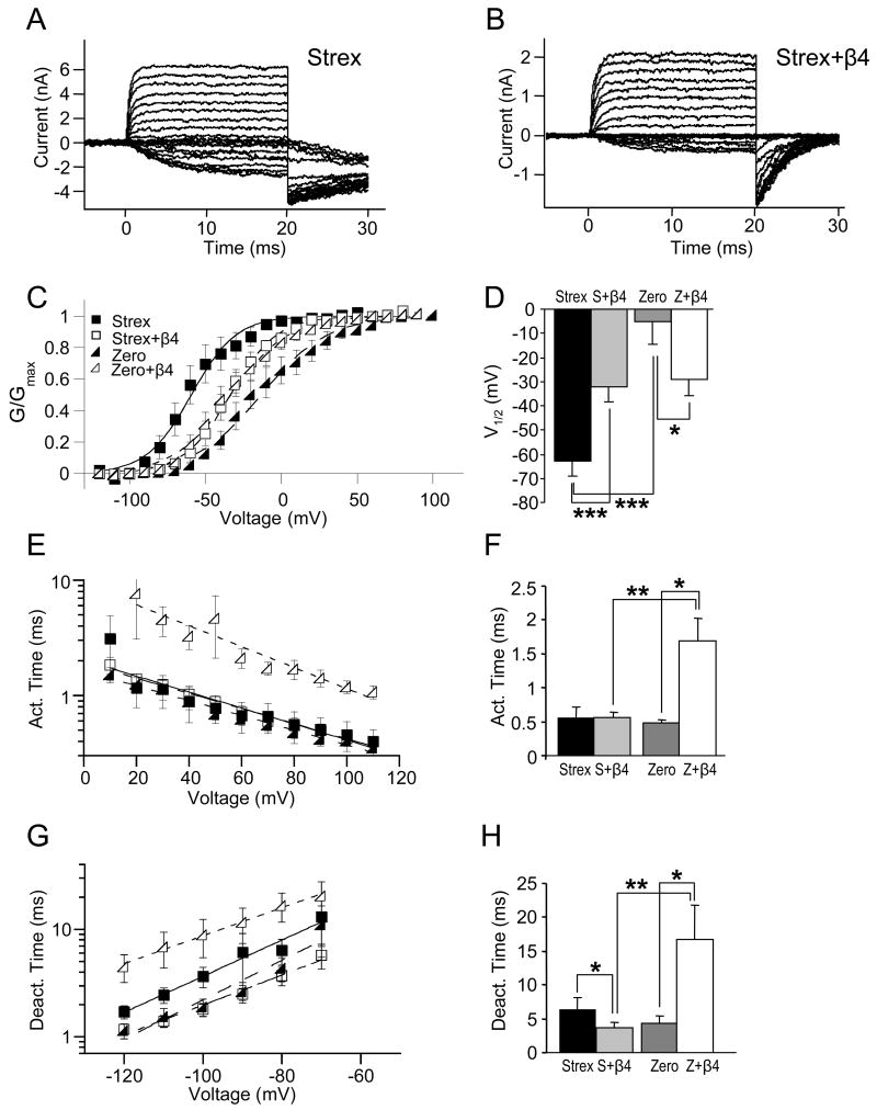
The β4 subunit inhibits STREX and activates ZERO BK channels in 120 μM calcium. Representative current traces for STREX alone (A) and STREX+β4 BK channels (B). C, plot of conductance versus voltage (G-V) relationship for STREX and ZERO BK channels with or without the β4 subunit. D, plot of average V1/2 for STREX, STREX+β4, ZERO, and ZERO+β4 BK channels. E, plot of activation time constants as a function of voltage. F, average activation time constants at +80 mV. G, graph of deactivation time constants as a function of voltage. H, average deactivation time constant at −80 mV. In C, E, F, solid symbol represent BK channels without the β4 subunit, whereas open symbol represent BK channels co-expressed with the β4 subunit. Squares show STREX and triangles ZERO BK channels. In D, F, H, black columns show values for STREX alone, light grey for STREX+β4, dark grey for ZERO alone, and white for ZERO+β4 BK channels. One asterisk represents P < 0.05, two represent P < 0.01, and three represent P < 0.001; unpaired T-Test.
Figure 3.
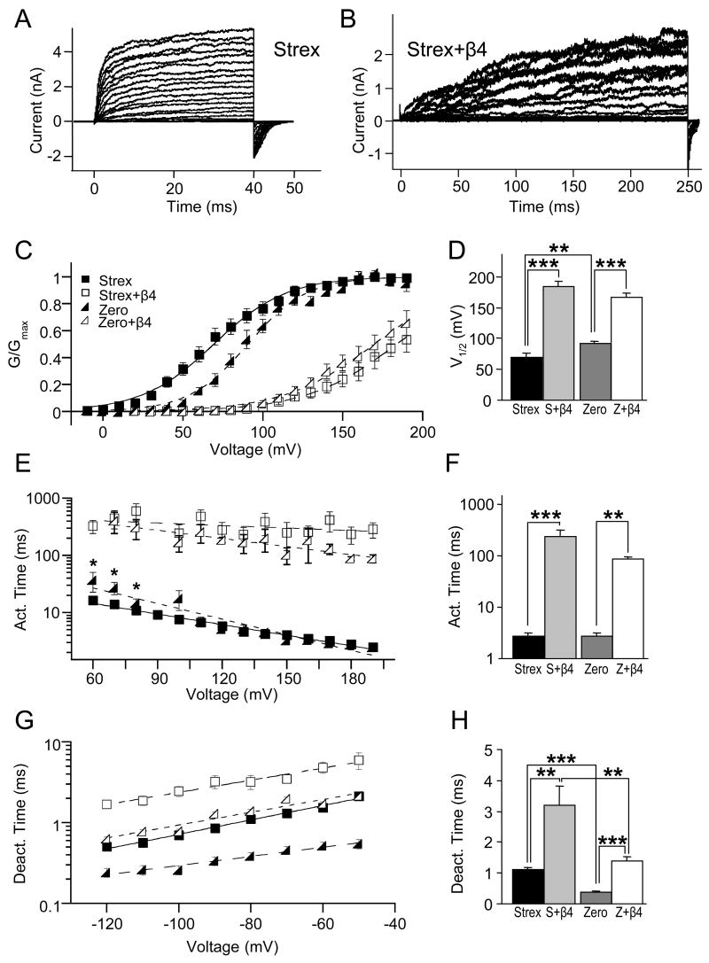
Inhibitory effects of the β4 subunit on STREX BK channels are dramatically increased in low calcium. Representative current traces measured in 1.0 μM calcium for STREX alone (A) and STREX+β4 BK channels (B). C, G-V relationships are plotted as a function of voltage. D, plot of average V1/2 for STREX, STREX+β4, ZERO and ZERO+β4 BK channels. Plot of activation (E) and deactivation (G) time constants as a function of voltage. F, average activation time constants taken at +180 mV. H, average deactivation time constant at −80 mV. In C, E, F, solid symbols represent BK channels without the β4 subunit, whereas open symbols are for BK channels co-expressed with the β4 subunit. Squares show STREX and triangles show ZERO BK channels. In D, F, H black columns show values for STREX alone, light grey for STREX+β4, dark grey for ZERO alone, and white for ZERO+β4 BK channels. One asterisk represents P < 0.05, two represent P < 0.01, and three represent P < 0.001; unpaired T-Test.
Conductance–voltage (G-V) relationships were obtained using a test pulse to positive potentials followed by a step to a negative voltage (−80 mV) and then measuring instantaneous tail current amplitudes 200 μs after the test pulse. V1/2 and Z was determined by fitting G-V curves to a Boltzmann function (G=Gmax{1/(1+e−(V-V1/2)ZF/RT)}) and normalizing to the maximum of the fit. At 1.0 μM Ca2+, where maximum conductance could not be obtained in the presence of β4, conductance was normalized to maximal conductance at higher Ca2+. Measurements of activation rates were obtained from currents elicited from −80 mV to more positive potentials. Activation and deactivation time constant (τ) was obtained by fitting of curves to a single exponential function, I = I0 e(−t/τ). Tail currents were evoked by a 10-ms step to 160 mV (for 120 μM Ca2+) or 200 mV (for 5.3 and 1.0 μM free calcium) and then measured by stepping down to various voltages for durations at least greater than three times the deactivation time constants. KaleidaGraph (Synergy Software, Reading, PA), Excel (Microsoft, Redmont, WA) and Igor Pro (WaveMetrics, Portland, Oregon) were used for calculations and presentation of the data.
Phosphorylation experiments
Macroscopic currents were recorded in buffered 5.3 μM Ca2+ solution for at least 5 minutes before application of alkaline phosphatase (AP, 10 DEA U/ml, Sigma, St. Louis, MO). Following dephosphorylation, the inside-out patches were perfused by buffered 5.3 μM Ca2+ solution with 1 mM ATP, 1 mM cAMP and 10 nM okadaic acid (all from Sigma, St. Louis, MO), pH = 7.3. Data were obtained 5 minutes after application of each solution change and represent mean ± SEM. Statistical analysis used paired T-Test. Un-paired T-Test was used for Δ V1/2 and for Δ activation and Δ deactivation kinetics if the data were compared with or without co-expression of β4 subunit. Δ Activation and Δ deactivation kinetics represent subtraction of individual paired data points (before vs. phosphatase, or phosphatase vs cAMP treatment) of activation or deactivation time constants at a given potential. In the figures, one asterisk represents P < 0.05, two represent P < 0.01, and three represent P < 0.001.
In Situ Hybridizations (ISH) on Free-Floating Brain Sections
Templates for riboprobes were synthesized from BK channel cDNA-specific PCR fragments that were subcloned into the pCR-Blunt Vector (Invitrogen, Carlsbad, CA) according to manufacturer’s instructions. cRNA for sense or anti-sense probes to a non-alternatively spliced region (representing total mRNA) and to the STREX exon were transcribed from linnearized plasmids using the AmpliScribe T7 Transcription Kit (Epicentre Biotechnologies, Madison, Wi) with digoxigenin-UTP (Roche, Mannheim, Germany) in the ratio of 6.5 mM UTP to 3.5 mM digoxigenin-UTP. For Total BK channels, residues encompassing 1659 to 2093 (of cDNA MMU09383) were used. For STREX BK channels, the complete STREX insert was used to generate a probe.
Brain slice preparation for ISH was modified from previous methods (Strassle et al., 2005). BK channel β4 subunit knockout male mice (Brenner et al., 2005) or control C57BL/6J animals from 4–5 months were used. All animal procedures were reviewed and approved by the University of Texas Health Science Center at San Antonio Institutional Animal Care and Use Committee. Animals were deeply anesthetized with isoflurane and transcardially perfused with an initial 50 ml solution of sterile, DEPC-treated 0.9% saline and then with 60 ml ice-cold 4% paraformaldehyde in 0.1 M phosphate buffer saline (pH 7.4, PFA solution). Brains were removed and post-fixed for 3 hours at 4°C in 4% PFA solution. The brains were then placed in 0.1 M DEPC-treated PBS with 20% nuclease-free sucrose (Sigma, St. Louis, MO) overnight in 4°C until they sank. The following day the brains were cryosectioned at 40 micron thickness under nuclease-free conditions. The sections were post-fixed in 4%-PFA for 15 minutes and washed twice for 10 minutes in 0.1 M DEPC-treated PBS.
Brain sections were pre-hybridized for 3 hours at 42°C in hybridization buffer containing 5X sodium citrate sodium chloride buffer, SSC (pH 7.5) in DEPC-treated water, 50% formamide, salmon sperm DNA (ssDNA, 100 μM, Sigma, St. Louis, MO), transfer RNA, type X-SA (100 μM, Sigma, St. Louis, MO) and 0.1% Tween-20 (Fisher Scientific, Houston, TX). The pre-hybridized slices were incubated with the DIG-labeled riboprobes (400 ng/ml hybridization buffer) overnight at 45°C. Following hybridization, the sections were washed twice for 25 minutes in 2X SSC with 0.2% Tween-20 at room temperature, twice for 25 minutes in 2X SSC with 0.2% Tween-20 at 50 °C and twice in 0.2X SSC with RNase A (50 μg/ml, Sigma, St. Louis, MO) at 55°C. The sections were equilibrated for 2 hours at room temperature in blocking solution (pH 7.5) containing 0.9% NaCl, 50 mM Tris Base, TBS (Fisher Scientific, Houston, TX), 0.2% Tween-20 and 2% bovine serum albumin (ICN Biomedicals, Aurora, OH). After blocking, the sections were incubated for 1.5 hours at room temperature with an anti-DIG antibody conjugated to alkaline phosphatase (1:4000, Roche). Post-antibody washes included three washes for 25 minutes of TBS with 2 mM levamisole (Sigma, St. Louis, MO) at room temperature and one 30-minute wash in staining buffer (pH 9.5) containing 0.1 M NaCl, 50 mM Tris, 0.2% Tween-20 and 2 mM levamisole for 30 minutes. The section was then stained in the dark with nitro blue tetrazolium/5-bromo-4-chloro-3-indoxyl phosphate (7.5 μl/ml SB, Roche) for 1–2 hours at room temperature. The reaction was stopped by washing in PBS (pH 7.5, twice for 15 minutes). The sections were post-fixed in 4% PFA for 15 minutes followed by two brief PBS washes. Finally, the sections were dehydrated in a PBS/glycerol series (10%, 20%, 40%, 60% and 80% glycerol) and mounted.
Immunohistology
β4 mRNA expression can be detected utilizing a knockin of the eGFP reporter in the β4 gene locus (Brenner et al., 2005). Briefly, brain sections from eGFP/β4 knockin mice were obtained as described above, stained with rabbit anti-GFP antibody (ab6556, Abcam, 1:5,000 dilution) and detected with biotinylated goat anti-rabbit secondary antibody at 1:200 dilutions. The staining was visualized using the Peroxidase Substrate Kit (Vector Laboratories, Burlingame, CA).
RESULTS
To compare β4 subunit modulation of BK channels, we co-expressed ZERO or STREX BK channels with or without the β4 subunit in HEK 293T cells. To control internal calcium, recordings were made of cell-excised macropatches in the inside-out configuration. Previously, we have shown that the effects of the β4 subunit on BK channels are calcium dependent. The β4 subunit promotes BK channel activation at high intracellular calcium, but inhibits channel activation at low calcium (Brenner et al., 2000; Wang et al., 2006). Therefore, we compared β4 effects on BK channel splice isoforms at relatively high (120 μM), moderate (5.3 μM) and very low (1 μM) calcium concentrations (Fig. 1–Fig. 3 respectively). These data are also summarized in Table I.
Table I.
Summary Data
| V1/2 (mV) | Q (e) | Act. Time (ms) | Deact. Time (ms) | N | |
|---|---|---|---|---|---|
| 120 μM Ca2+ | |||||
| Zero | −5.41 ± 9.19 | 1.95 ± 0.07 | 0.48 ± 0.05 | 4.34 ± 1.07 | 10 |
| Zero+β4 | −29.20 ± 6.70 | 1.56 ± 0.18 | 1.69 ± 0.32 | 16.7 ± 5.07 | 13 |
| Strex | −62.98 ± 6.28 | 4.78 ± 1.23 | 0.55 ± 0.17 | 6.36 ± 1.72 | 8 |
| Strex+β4 | −31.98 ± 6.35 | 2.05 ± 0.21 | 0.56 ± 0.07 | 3.7 ± 0.71 | 9 |
| 5 μM Ca2+ | |||||
| Zero | 31.21 ± 8.45 | 1.35 ± 0.08 | 1.71 ± 0.30 | 1.69 ± 0.23 | 9 |
| Zero+β4 | 61.63 ± 5.43 | 1.09 ± 0.06 | 8.15 ± 1.14 | 5.41 ± 0.63 | 8 |
| Strex | −18.24 ± 3.85 | 1.34 ± 0.06 | 2.07 ± 0.26 | 11.32 ± 3.72 | 8 |
| Strex+β4 | 2.64 ± 5.08 | 1.21 ± 0.05 | 1.13 ± 0.18 | 2.1 ± 0.26 | 9 |
| 1 μM Ca2+ | |||||
| Zero | 91.22 ± 3.40 | 1.59 ± 0.13 | 2.70 ± 0.49 | 0.38 ± 0.04 | 16 |
| Zero+β4 | 166.78 ± 6.61 | 1.07 ± 0.11 | 87.68 ± 6.65 | 1.39 ± 0.13 | 12 |
| Strex | 70.26 ± 5.42 | 1.28 ± 0.09 | 2.79 ± 0.31 | 1.11 ± 0.08 | 11 |
| Strex+β4 | 183.28 ± 9.24 | 1.06 ± 0.11 | 234.71 ± 78.7 | 3.21 ± 0.62 | 13 |
At high calcium, β4 has opposite effects on G-Vs of ZERO and STREX channels
Representative current traces are shown for STREX BK channels alone (Fig. 1A) and for STREX+β4 BK channels (Fig. 1B) in 120 μM Ca2+. Mean steady-state conductance-voltage (G-V) relations are shown in Fig. 1C. The voltage for half-maximal activation (V1/2) was derived from individual G-V curves to quantify the effects of the β4 subunit (Fig. 1D). When co-expressed with ZERO BK channels at high calcium, the β4 subunit significantly shifts V1/2 towards negative potentials (Behrens et al., 2000; Brenner et al., 2000; Wang et al., 2006). The β4 subunit shifts V1/2 from −5.41 ± 9.19 mV (ZERO alone) to −29.20 ± 6.70 mV (ZERO+ β4, Fig. 1D). However, co-expression of the β4 subunit with STREX BK channels has opposite effects. β4 dramatically shifts STREX V1/2 towards positive potentials from −62.98 ± 6.28 mV (STREX alone) to −31.98 ± 6.35 mV (STREX+β4, Fig. 1D). Interestingly, the presence of the β4 subunit eliminates the greater than 50 mV difference in V1/2 of the two splice forms, making their steady-state openings very similar (Fig. 1D).
An additional property of β4 and other β subunits is that they reduce the slope (effective gating charge, Q) of the G-V relations for ZERO channels (Wang and Brenner, 2006, Wang et al., 2006). Similarly, we found here that β4 significantly reduces the Q for both ZERO and STREX channels across all calcium concentrations (Table I). For example, at 5.3 μM calcium β4 reduces Q from 1.35 ± 0.08 e to 1.09 ± 0.06 e for ZERO (P = 0.007), and from 1.34 ± 0.06 e to 1.21 ± 0.05 e for STREX channels (P = 0.04). Similar effects were seen in 1 μM calcium (see Table I). One distinct effect of STREX channels in 120 μM calcium was a significant increase in Q that was not seen in ZERO channels (Fig. 1C, Q is 4.78 ± 1.23e for STREX versus 1.95 ± 0.07e for ZERO channels, P = 0.026) nor in STREX channels alone at lower calcium (1.34 ± 0.06 e for STREX alone in 5.3 μM calcium and 1.28 ± 0.09 e for STREX alone in 1 μM calcium). Nevertheless, coexpression of β4 with STREX channels in 120 μM calcium reduced Q to values similar to ZERO alone or ZERO/β4 channels (STREX alone, 4.78 ± 1.23 e; STREX/β4, 2.05 ± 0.21 e; ZERO alone, 1.56 ± 0.18 e; ZERO/β4, 1.95 ± 0.07 e).
At high calcium, effects on deactivation underlie differences between ZERO and STREX channels with β4
Fig. 1E shows average activation kinetics plotted as a function of voltage for both ZERO and STREX BK channels with or without the β4 subunit. The data are summarized in Fig 1F for activation time constants taken at +80 mV. We did not observe a significant difference in activation kinetics between STREX alone (0.55 ± 0.17 ms) and STREX+β4 (0.56 ± 0.07 ms) BK channels in 120 μM Ca2+. However activation kinetics of ZERO BK channels were significantly slowed by the presence of the β4 subunit (from 0.48 ± 0.05 ms to 1.69 ± 0.32 ms).
Both STREX and BK channel β subunits slow deactivation kinetics (Ramanathan et al., 1999, Brenner, et al., 2000). This is shown at various voltages in Fig. 1G and 2G, and summarized for −80 mV in Fig. 1H and 2H. Interestingly, whereas both the β4 subunit and STREX individually slow deactivation kinetics to promote channel openings in high calcium, their combination speeds deactivation of BK channels (Fig. 1H and 2H). With STREX BK channels, the β4 subunit significantly speeds the deactivation time constant from 6.36 ± 1.72 ms (STREX alone) to 3.7 ± 0.71 (STREX+β4) whereas with ZERO BK channels it slows the deactivation time constant (p < 0.05) from 4.34 ± 1.07 ms (ZERO alone) to 16.7 ± 5.07 ms (ZERO+β4). Thus, the opposing effects of β4 subunit on STREX and ZERO BK channel deactivation kinetics underlie opposing effects on steady-state conductance.
Figure 2.
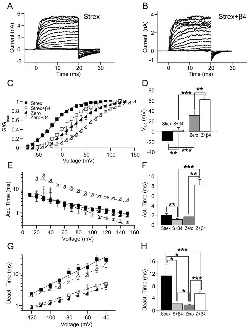
In 5.3 μM calcium, the β4 subunit inhibits both STREX and ZERO BK channels by different effects on kinetics. Representative current traces for STREX alone (A) and STREX+β4 BK channels (B). C, plot of conductance versus voltage (G-V) relationship for STREX and ZERO BK channels with or without the β4 subunit. D, plot of average V1/2 for STREX, STREX+β4, ZERO and ZERO+β4 BK channels. Activation (E) and deactivation (G) time constants are plotted as a function of voltage. F, activation time constant at +80 mV. H, deactivation time constant at −80 mV. In C, E, F, solid symbols represent BK channels without the β4 subunit, whereas open symbols are for BK channels co-expressed with the β4 subunit. Squares show STREX and triangles show ZERO BK channels. In D, F, H, black columns show values for STREX alone, light grey for STREX+β4, dark grey for ZERO alone, and white for ZERO+β4 BK channels. One asterisk represents P < 0.05, two represent P < 0.01, and three represent P < 0.001; unpaired T-Test.
At intermediate and low calcium, β4 dramatically inhibits both ZERO and STREX conductance
While the β4 subunit conferred opposite effects on V1/2 with ZERO versus STREX BK channels in 120 μM Ca2+, in 5.3 μM Ca2+ β4 inhibits both ZERO and STREX isoforms (Fig. 2A–D). Summary data in Fig. 2D shows that the β4 subunit significantly shifts V1/2 of STREX and ZERO more positive by +20 mV (−18.24 ± 3.85 mV, STREX alone; to +2.64 ± 5.08 mV, STREX+β4) and +30 mV, respectively (+31.21 ± 8.45 mV, ZERO alone; +61.63 ± 5.43 mV, ZERO+β4). Although the positive shifts of STREX and ZERO V1/2 are similar, they are mediated by different β4 effects on channel gating kinetics. At this Ca2+ concentration, the β4 subunit inhibits STREX BK channels by speeding deactivation, but inhibits ZERO BK channels by slowing activation. Activation time constant at +80 mV for STREX is reduced slightly (2.07 ± 0.26 ms, STREX alone; to 1.13 ± 0.18 ms, STREX+β4) but is increased dramatically for ZERO (1.71 ± 0.3 ms, ZERO alone; 8.15 ± 1.14 ms, ZERO+β4). Opposite effects of the β4 subunit on ZERO versus STREX BK channels are also observed in deactivation kinetics (Fig. 2G). Deactivation time is dramatically decreased by the β4 subunit for STREX (Fig. 2H, 11.32 ± 3.72 ms for STREX alone; 2.1 ± 0.26 ms for STREX+β4) but increased for ZERO BK channels (1.69 ± 0.23 ms, ZERO alone; 5.41 ± 0.63 ms, ZERO+β4).
An interesting effect unique to STREX channels is that deactivation times is actually slower at 5 μM calcium than 120 μM (Table I). A similar effect is seen for STREX+β4 channels at 1 μM calcium as compared to 5 μM calcium. These findings were further confirmed by paired recordings of STREX channels during solution changes from 120 to 5 μM calcium. We indeed found that STREX patches (7/8) showed a speeding of deactivation with an average 5.5 ± 3.9 ms (P = 0.09) increase in time constant at −80 mV and a 0.75 ± 0.28 ms (P = 0.015) increase in time time constant at −120 mV as calcium was reduced. In contrast, reducing calcium always speeds deactivation times for ZERO and ZERO+β4 (Table I).
To investigate the effects of the β4 subunit in calcium concentrations closer to resting cytoplasmic levels, we compared STREX BK channels alone to STREX+β4 BK channels in buffered 1.0 μM Ca2+ solution (Fig. 3). β4 in low calcium shifts BK channel G-V curves to very positive potentials where it is not feasible to achieve the maximum steady-state conductance. Therefore, recordings from each patch in low calcium were subsequently exposed to high Ca2+ (120 μM) to normalize to the maximal conductance. Representative current traces are shown for STREX alone (Fig. 3A) and STREX+β4 BK channels (Fig. 3B). Fig. 3C shows G-V curves for all four variants as a function of voltage. Plots of V1/2 values (Fig. 3D) show that β4 dramatically shifts both STREX and ZERO BK channels to positive potentials, with a greater effect on STREX BK channels. The β4 subunit shifts V1/2 of STREX BK channels from 70.26 ± 5.42 mV to 183.28 ± 9.24 mV. V1/2 of ZERO BK channels are also significantly shifted by the β4 subunit to more positive values from 91.22 ± 3.40 mV to 166.78 ± 6.61 mV. Taken together, these results suggest that the β4 subunit reduces the difference in conductance that arises by alternatively splicing to include the STREX exon. In addition, β4 dramatically inhibits BK channel activation at low Ca2+ (1.0 μM), regardless of the splice isoform.
At low calcium, β4 inhibits both STREX and ZERO BK channels by slowing activation
At low calcium β4 effects on activation kinetics underlies the large positive shift of the V1/2. Fig. 3E and 3F summarizes activation time constants as a function of voltage. The β4 subunit slows activation by orders of magnitude, from 2.79 ± 0.31 ms to 234.71 ± 78.7 ms for STREX or from 2.70 ± 0.49 ms to 87.68 ± 6.65 ms for ZERO (at +180 mV). The β4 subunit also slows deactivation kinetics, albeit to lesser extents at this Ca2+ concentration (Fig. 3G). Deactivation time of BK channels is significantly slowed by the β4 subunit; from 1.11 ± 0.08 ms to 3.21 ± 0.62 ms (STREX) or from 0.38 ± 0.04 ms to 1.39 ± 0.13 ms (ZERO) (Fig. 3H). Interestingly, although β4 slows ZERO BK channel deactivation across calcium concentrations, only at low calcium concentrations does it slow STREX BK channel deactivation.
The β4 subunit confers sensitivity to dephosphorylation of STREX but not ZERO BK channels
It has been reported that the β4 subunit (Jin et al., 2002) and the STREX exon (Tian et al., 2001) each alter the response of BK channels to dephosphorylation and cAMP-dependent phosphorylation status. Here we measured the combined effect of both STREX and the β4 subunit. The experimental approach was to measure responses to dephosphorylation by alkaline phosphatase followed by cAMP + ATP application to measure effects of pKA-dependent phosphorylation (BK channels in HEK cells have pKA pre-tethered to the channel and therefore do not require application of the catalytic enzyme (Tian et al., 2001, Tian et al., 2003)). Because all data was paired, we could measure both effects on steady-state values (Fig. 4 and Fig. 5, left panels) and relative changes from control values (Fig. 4 and Fig. 5, right panels). Results are shown for effects on V1/2 and gating kinetics in 5.3 μM Ca2+ when STREX and ZERO BK channels, respectively, are expressed with and without the β4 subunit.
Figure 4.
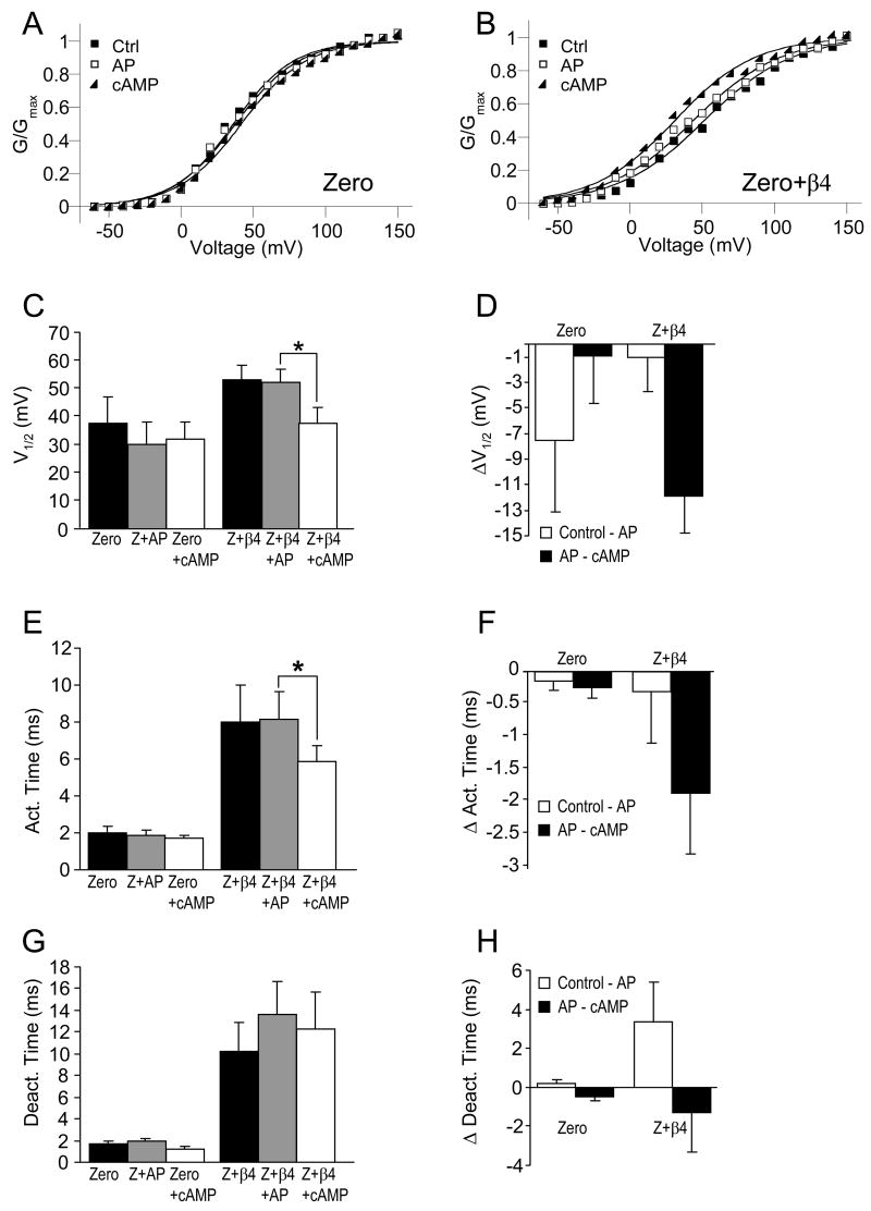
STREX+β4 but not STREX BK channels are inhibited by dephosphorylation. Recordings were made in 5.3 μM internal calcium. Representative G-V relationships for STREX (A) and STREX+β4 BK channels (B) for pre-treatment control (filled squares), dephosphorylation by alkaline phosphatase (AP, empty squares) and cAMP-dependent phosphorylation (cAMP, triangles). C, summarized V1/2. D, ΔV1/2 of control versus dephosphorylation (white bars), and dephosphorylated versus cAMP (dark bars). E, mean of activation time constant taken at +80 mV. G, mean of deactivation time constant taken at −80 mV. Δ Activation (F) and Δ deactivation (H) show means of paired subtraction of activation and deactivation time constants at +80 and −80 mV respectively. In C, E, G, control values (black) are compared with AP treatment (grey) and subsequent cAMP application (white). In D, F, H subtraction of control versus dephosphorylated values (white) is compared with subtraction of dephosphorylated versus cAMP-treated values (black). Paired T-test is used for statistical analysis (STREX n = 8; STREX+β4 n = 11). Un-paired T-test is used only for comparison of ΔV1/2. One asterisk represents P < 0.05, two represent P < 0.01, and three represent P < 0.001.
Figure 5.
cAMP-dependent phosphorylation activates ZERO+β4 BK channels. Recordings were made in 5.3 μM internal calcium. Representative G-V relationships for ZERO (A) and ZERO+β4 BK channels (B) for pre-treatment control (filled squares), dephosphorylation by alkaline phosphatase (AP, empty squares) and cAMP-dependent phosphorylation (cAMP, triangles). C, Summarized V1/2. D, ΔV1/2 of control versus dephosphorylation (white bars), and dephosphorylated versus cAMP (dark bars). E, mean of activation time constant taken at +80 mV. G, mean of deactivation time constant taken at −80 mV (G). Δ activation (F) and Δ deactivation (H) show means of paired subtraction of activation and deactivation time constants at +80 mV and −80 mV, respectively. In C, E, G, control values (black) are compared with AP treatment (grey) and subsequent cAMP application (white). In D, F, H subtraction of control versus dephosphorylated values (white) is compared with subtraction of dephosphorylated versus cAMP-treated values (black). Paired T-test is used for statistical analysis (ZERO n = 7; ZERO+β4 n = 11). Un-paired T-test is used only for comparison of ΔV1/2. One asterisk represents P < 0.05, two represent P < 0.01, and three represent P < 0.001.
Representative G-V relations for STREX illustrate that application of alkaline phosphatase to STREX channels tends to shift V1/2 to positive potentials (Figs. 4A, C–D, from −18.24 ± 3.85 mV to −9.38 ± 6.17 mV), although this shift is not statistically significant (P = 0.086). In contrast, STREX+β4 BK channels show a greater response to dephosphorylation (Fig. 4B–D). Dephosphorylation causes a significant positive shift of V1/2 from −1.6 ± 6.22 mV of control to 19.14 ± 4.89 mV (Figs. 4C–D). This is mediated predominantly by a slowing of activation kinetics (Fig. 4E–F, STREX+β4 is 1.1 ± 0.17 versus STREX+β4 phosphatase 2.1 ± 0.11 ms, P = 0.002).
Activation of pKA has been shown to inhibit STREX BK channels in HEK293 cells (Tian et al., 2001). Consistent with this, we found that activation of pKA with 1 mM cAMP and 1 mM ATP caused a small but significant speeding of deactivation kinetics (Fig. 4G and H) for both STREX (from 7.16 ± 1.22 ms to 5.53 ± 0.92 ms, P = 0.03) and STREX+β4 BK channels (from 2.30 ± 0.21 ms to 1.66 ± 0.19 ms, P = 0.02). However, the small change in kinetics conferred a relatively small positive shift of the V1/2 that did not reach statistical significance (Figs. 4C–D from −9.38 ± 6.17 mV to 5.07 ± 7.73 mV, P = 0.09 for STREX and from 19.14 ± 4.89 mV to 22.49 ± 6.08 mV, P = 0.33 for STREX+β4). Interestingly, the combined effects of dephosphorylation and cAMP-dependent phosphorylation showed a significant inhibition of STREX BK channels (Fig. 4C, P = 0.011 for net difference between control and cAMP).
ZERO BK channels are activated by cAMP-dependent phosphorylation when co-expressed with the β4 subunit
Contrary to STREX+β4 BK channels that are inhibited by dephosphorylation (Fig. 4), ZERO and ZERO+β4 BK channels are not significantly affected by dephosphorylation (Fig. 5). The application of cAMP after dephosphorylation, however, produces a small but statistically significant shift of the V1/2 to more negative potentials for ZERO+β4 BK channels (from 52.27 ± 5.02 mV to 38.07 ± 5.54 mV). This negative V1/2 shift is mediated by faster activation (Fig. 5E, from 8.11 ± 1.5 ms for dephosphorylation to 5.88 ± 0.81 mV for cAMP application). We did not observe a significant V1/2 shift for ZERO BK channels (Fig. 5C–D). In summary, the β4 subunit confers a significantly greater sensitivity of STREX BK channels to dephosphorylation, and ZERO BK channels to pKA-dependent phosphorylation.
The β4 subunit and STREX BK channels are coexpressed in the hippocampus and cerebellum
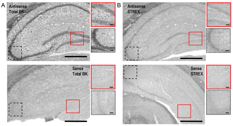
Our findings using an in vitro expression system suggest that the β4 subunit differentially affects alternative splice products of the BK channel and their response to phosphorylation status. Using quantitative PCR, it has been previously shown that the STREX exon is relatively abundant in pituitary mRNA but weakly expressed in whole brain mRNA (Chen et al., 2005). Utilizing an eGFP knockin reporter for β4 gene expression, it was found that the β4 subunit also has abundant expression in the anterior and posterior pituitary (Brenner et al., 2005). To better understand the physiological relevance of β4 subunit/STREX BK channel interactions in other central brain regions, we used in situ mRNA hybridization (ISH) combined with eGFP immunohistochemistry to reveal if STREX BK channels overlap in their mRNA expression with the β4 subunit in the brain. We made two specific cRNA riboprobes labeled with digoxigenin-UTP for STREX and total BK channels. The antisense strand riboprobes were tested for specific cRNA-mRNA hybridization towards mRNA of STREX and total BK channels when compared to negative controls of corresponding sense riboprobes (Fig. 6). Coronal sections of the hippocampus show that signals for total BK channels (Fig. 6A upper) and for STREX BK channels (Fig 6B upper) are absent when control sense riboprobes are used (lower panel of Fig. 6A and 6B).
Figure 6.
Riboprobes for STREX only and Total BK channels are specific. Antisense strand cRNA probe against Total BK channels independently of a splice isoform (A, top panel) or antisense strand cRNA against STREX exon only (B, top panel) were tested on coronal sections of the hippocampus from 4 month old male C57BL/6J mice. As negative controls, sense strands of cRNA of the same cDNA templates that were used for antisense probes were transcribed. A, bottom panel shows sense strand riboprobe for Total BK channels. B, bottom panel shows sense strand for STREX BK channels. Insets show the dental gyrus area (solid line) and the CA3 area (dashed line) of the hippocampus. The scale bar in the panels corresponds to 100 μm and to 10 μm in the insets. Incubation of the brain sections with the sense strand riboprobes results in almost total loss of signal when compared with the antisense strand riboprobes suggesting very low background hybridization.
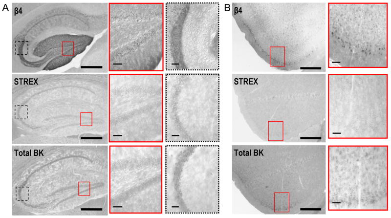
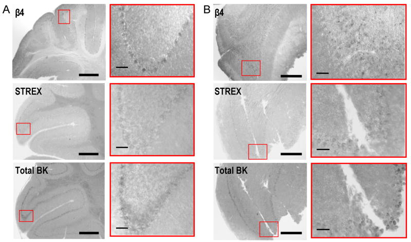
Figs. 7 and 8 compare the expression pattern of the β4 subunit (top panels) with STREX (middle panels) and total BK channels (lower panels) in sagittal sections of the hippocampus (Fig. 7A), the frontal lobe neocortex (Fig. 7B), the cerebellum (Fig. 8A), and in the olfactory bulb (Fig. 8B). In general, the expression of STREX BK channels represented a smaller fraction of total BK channels. In the hippocampus, we observed expression of both the β4 subunit and STREX in the granule cells of the dentate gyrus and in the CA3 pyramidal neurons (insets in Fig. 7A). Because eGFP is able to diffuse throughout neuronal processes, the β4 reporter is observed in both the dendritic layers of the granule cells (lined inset) and the mossy fiber projections (dashed inset) (Fig. 7A upper panel). The in situ hybrization for STREX and total BK channels is instead localized in the cell body layers of the dentate gyrus and CA3 regions (Fig. 7A middle and lower panels). The expression of the β4 subunit in the CA1 region was much weaker although both STREX and total BK channels were homogeneously expressed in the whole hippocampus including CA1 pyramids. In the neocortex (frontal lobe) expression of both β4 and total BK channels predominate, whereas STREX channels are very weakly expressed (Fig 7B). On the other hand, STREX and β4 show overlapping expression in the Purkinje neurons of the cerebellum (insets of Fig. 8A). Interestingly, β4 subunit immunohistochemistry showed more widespread expression in the ventral olfactory bulb (insets of Fig. 8B) than BK channels, which were more confined to a very narrow layer of the granule neurons.
Figure 7.
STREX BK channels are expressed in the hippocampus and very weakly in the neocortex. Expression of STREX versus Total BK channels is compared to the expression of the β4 subunit. Specific antisense strand riboprobes were used for detecting STREX and Total BK channels in wild-type male C57BL/6J mice. Anti-GFP immunohistochemistrry was employed to detect the β4 expression pattern in the male β4 −/− C57BL/6J mice since the β4-specific promoter drives expression of GFP as a transgenic marker for the β4 subunit. Panels show serial sections of the hippocampus (A) and the frontal lobe (B) stained for the β4 subunit (top panels), STREX (middle panels), and Total BK channels (bottom panels). In the hippocampus, insets show the granule neurons of the dental gyrus (solid line frame) and the pyramidal neurons of the CA3 region (dashed line frame). In the neocortex of the frontal lobe, insets show pyramidal neurons of the layer II/III. We detected distinguishable signal for STREX exon overlapping with the β4 subunit expression in the hippocampus but fairly weak signal for STREX exon in the neocortex of the frontal lobe. Scale bars of the panels correspond to 100 μm. In the insets, scale bars represent 10 μm.
Figure 8.
STREX BK channels are expressed in the cerebellum and weakly in the olfactory bulb. As in Fig. 7, expression of STREX versus Total BK channels is compared to the expression of the β4 subunit. Panels show serial sections of the cerebellum (A) and the olfactory bulb (B) stained for the β4 subunit (top panels), STREX (middle panels), and Total BK channels (bottom panels). In the cerebellum, insets show signal for STREX exon and the β4 subunit in the Purkinje pyramidal neurons. In the olfactory bulb, the expression of both Total BK channels and STREX exon is limited to a restricted neuronal population whereas the β4 subunit displays widespread distribution. Scale bars of the panels correspond to 100 μm. In the insets, scale bars represent 10 μm.
DISCUSSION
The STREX exon shows relatively weak CNS expression as measured by PCR of whole brains (Chen et al., 2005). In contrast, STREX appears to show greatest neuronal expression in early CNS development and in neurosecretory tissues such as adrenal gland and pituitary (MacDonald et al., 2006). Consistent with this, we found that the STREX shows significant expression only in few brain regions. Despite relatively weak expression of STREX in some brain regions, the finding that a single STREX containing subunit dominates the properties of a BK channel tetramer (Tian et al., 2004) suggests that even weak expression may be functionally important. It has been recently reported that gain-of-function of BK channels in humans and mice results in epilepsy phenotypes (Du, et. al, 2005; Brenner, et al, 2005). Thus, the ability of the β4 subunit to inhibit STREX BK channels in a wide range of calcium concentrations may have protective effects in some central neurons where STREX has limited expression. These may include the hippocampus dentate gyrus and the cerebellum purkinje cells where STREX and β4 expression overlap.
The mechanism used by β4 to inhibit STREX BK channels appears to be calcium dependent. At low calcium β4 reduces channel openings by slowing activation. At intermediate and high calcium (> 5.3 μM Ca2+), β4 reduces BK channel opening by opposing the slow deactivation conferred by the STREX exon. Effects on deactivation are somewhat surprising given that β4, for channels lacking STREX, promotes BK channel opening by slowing deactivation. Yet the combined effects of STREX and β4 at high Ca2+ speed deactivation relative to either alone. This is in contrast to the avian beta subunit that creates a further slowing of deactivation kinetics when the STREX exon and β subunits are combined (Ramanathan et al., 1999). The mechanisms that underlie differences between the avian β subunit and the mouse β4 are not understood. Given that the avian β subunit has higher similarity to the related β1 and β2 subunit family members (Xia, et al., 1999), these differences may be related to functional divergence between β1/β2 and β4 paralogues. In contrast to STREX BK channels, β4 slows both activation and deactivation of ZERO channels. In high Ca2+, this causes a net effect that shifts the G-V relationship to negative potentials. As calcium is reduced (1 μM Ca2+) the slow activation conferred by β4 predominates to strongly inhibit both STREX and ZERO BK channels. This is from millisecond times, in the absence of β4, to hundreds of milliseconds for BK channels with β4. Thus, at low calcium, the β4 subunit slows activation kinetics to well outside normal physiological times-scales for processes such as action potential repolarization or regulation of neurotransmitter release.
Intracellular calcium concentrations in microdomains can indeed approach 20–30 μM during calcium sparks (ZhuGe et al., 2000) and up to several hundred μM in the presynaptic sites near calcium channels (Oheim et al., 2006). In neurons, BK channels have been found in close vicinity of microdomain calcium (Franciolini et al., 2001; Parsons et al., 2002) or in presynaptic sites (Hu et al., 2001). We can hypothesize that BK channels may experience brief but very robust elevations of calcium. Therefore, the local calcium environment in combination with ZERO/STREX splicing may determine whether BK/β4 channels can contribute to shaping of action potentials or regulation of neurotransmitter release. For example, given the significantly faster activation rates at intermediate calcium concentrations (such as 5 μM, Fig. 2F) the β4 subunit may permit STREX channels but not ZERO BK channels to contribute to action potential repolarizations. In addition, the speeding of deactivation of STREX channels by β4 would limit BK channel contribution to the afterhyperpolarization. As calcium concentrations rise to higher levels, activation rates are much faster and deactivation much slower (independent of STREX or ZERO splicing, Fig. 1) so that either BK channel isoform may contribute to action potential repolarization and dampen firing rates.
Modulation of BK channels involves many mechanisms (Weiger et al., 2002; Lu et al., 2006) including reversible phosphorylation. An additional distinction between STREX and ZERO BK channels is their response to cAMP-dependent phosphorylation. STREX containing BK channels are frequently inhibited by cAMP dependent phosphorylation in HEK293 cells whereas ZERO isoforms are activated in the majority of channel patches (Tian et al., 2001). Our results indicate that the ZERO/β4 channels have a greater sensitivity to cAMP-dependent activation than ZERO channels alone. The fact that ZERO BK channel were not significantly activated by cAMP may be due to pretreatment of the channels with alkaline phosphatase. BK channel response to phosphorylation has been shown to be quite variable in neurons (Reinhart et al., 1989, Reinhart et al., 1991, Reinhart and Levitan, 1995, Widmer et al., 2003) and even in transfected HEK293 cells (Tian et al., 2001). For example, application of cAMP to ZERO BK channels expressed in HEK293 cells has been shown to activate 10 out of 16 BK channel patches (Tian et al., 2001). In part, this may arise from cell-to-cell variation in basal levels of phosphorylation of channels. Therefore, to produce a more reproducible response to pKA (protein kinase A)-dependent phosphorylation, we pretreated all patches with phosphatase. Although phosphatase may homogenize the channel’s response to cAMP-dependent phosphorylation, it may affect other phosphorylation sites that alter the response of ZERO channels but not ZERO/β4 channels to cAMP-dependent phosphorylation. For example, in cerebellar purkinje cells cAMP-dependent channel activation of BK channels appears to be conditional on the previous phosphorylation state. Protein kinase C and protein phosphatase-2A occlude cAMP-dependent activation, whereas protein phosphatase-1 promote pKA activation (Widmer et al., 2003).
Whereas our results indicate that BK channels alone are not significantly affected by dephosphorylation, dephosphorylation of β4 containing channels confer a slower activation of BK channels along with a positive shift of the G-V relations. Surprisingly, this effect was dependent on the presence of STREX. In HEK cells β4 subunits are tonicly phosphorylated (Jin et al., 2002). These results suggest that the inhibition of BK STREX/β4 channels by dephosphorylation may be attributed to the phosphorylation state of the β4 subunit. It would also indicate that tonic phosphorylation of the β4 subunit reduces its full ability to inhibit STREX BK channels.
The non-additive effects of STREX and the β4 subunit suggest that β4 somehow perturbs interactions of STREX within BK channels. The physical basis of this is only a matter of speculation. Many effects of β1 are mediated by their intracellular domains (Orio and Latorre, 2005, Orio et al., 2006, Wang and Brenner, 2006). To date, the α subunit domains found to interact with β subunits are the N-terminal, transmembrane S0 domain (Wallner et al., 1996) and the carboxyl terminal tail region (Qian et al., 2002). The STREX coding region lies within a variable region (Schreiber and Salkoff, 1997) of the carboxyl terminus that links two conserved functional domains of the channel. Amino terminal to STREX is the so called “RCK1” (regulator of potassium conductance) calcium and magnesium sensing domain (Shi et al., 2002, Xia et al., 2002). Carboxyl terminal to STREX is a putative second RCK-like domain (RCK2). Although the RCK2 sequence shows weak evidence of RCK homology (Fodor and Aldrich, 2006), it is required for channel function and may provide a hydrophobic interface for calcium-dependent gating (Kim et al., 2006). Positioned between the two domains, it is indeed possible that STREX may modulate RCK domain interactions. Whether or not the β4 intracellular domains directly interact with this region is yet to be determined.
Early characterization of BK channel subtypes in neuronal synaptosomal membrane preparations identified two classes of BK channels. These were the fast-gated, iberiotoxin sensitive (type I) BK channels and the slow gated, iberiotoxin resistant (type II) BK channels (Reinhart and Levitan, 1991). Type II BK channels are now realized to encode BK/β4 subunit channels due to the iberiotoxin resistance they confer (Behrens et al., 2000; Meera et al., 2000). However, type I and type II BK channels show diverse properties in response to kinase and phosphatase-dependent modulation (Reinhart and Levitan, 1991). Our findings suggest that the line between type I and type II channels may not be so distinct. STREX/β4 BK channels, which may be classified as type II BK channels based on iberiotoxin resistance, could nevertheless appear as fast-gated type I BK channels due to their fast deactivation kinetics at high calcium and activation by phosphatase treatment (Reinhart and Levitan, 1991). Thus, the combination of alternative splicing and association with accessory β subunits may create a greater diversity in BK channel properties than previously expected.
Acknowledgments
We thank Bin Wang and Rachel J. Brenner for critical reading of the manuscript. This work was supported by a National Institutes of Health Grant NS052574, a National American Heart Association Grant 0335007N and an Epilepsy Foundation of America Grant.
List of Abbreviations
- BK Channel
Large conductance, calcium-activated potassium channel
- β4
beta4 accessory subunit
- Ca2+
calcium
- cAMP
Cyclic Adenosine Monophosphate
- eGFP
enhanced green fluorescent protein
- G-V
conductance-voltage relationship
- ISH
in situ hybridization
- PBS
phosphate buffered saline
- PCR
polymerase chain reaction
- PFA
paraformaldehyde
- pKA
protein kinase A
- Q
effective gating charge of conductance-voltage relations
- STREX
stress axis hormone-regulated exon
- V1/2
voltage for half-maximal activation
Footnotes
Publisher's Disclaimer: This is a PDF file of an unedited manuscript that has been accepted for publication. As a service to our customers we are providing this early version of the manuscript. The manuscript will undergo copyediting, typesetting, and review of the resulting proof before it is published in its final citable form. Please note that during the production process errors may be discovered which could affect the content, and all legal disclaimers that apply to the journal pertain.
References
- Adelman JP, Shen KZ, Kavanaugh MP, Warren RA, Wu YN, Lagrutta A, Bond CT, North RA. Calcium-activated potassium channels expressed from cloned complementary DNAs. Neuron. 1992;9:209–216. doi: 10.1016/0896-6273(92)90160-f. [DOI] [PubMed] [Google Scholar]
- Atkinson NS, Robertson GA, Ganetzky B. A component of calcium-activated potassium channels encoded by the Drosophila slo locus. Science. 1991;253:551–555. doi: 10.1126/science.1857984. [DOI] [PubMed] [Google Scholar]
- Behrens R, Nolting A, Reimann F, Schwarz M, Waldschutz R, Pongs O. hKCNMB3 and hKCNMB4, cloning and characterization of two members of the large-conductance calcium-activated potassium channel beta subunit family. FEBS Lett. 2000;474:99–106. doi: 10.1016/s0014-5793(00)01584-2. [DOI] [PubMed] [Google Scholar]
- Brenner R, Chen QH, Vilaythong A, Toney GM, Noebels JL, Aldrich RW. BK channel beta4 subunit reduces dentate gyrus excitability and protects against temporal lobe seizures. Nat Neurosci. 2005;8:1752–1759. doi: 10.1038/nn1573. [DOI] [PubMed] [Google Scholar]
- Brenner R, Jegla TJ, Wickenden A, Liu Y, Aldrich RW. Cloning and functional characterization of novel large conductance calcium-activated potassium channel beta subunits, hKCNMB3 and hKCNMB4. J Biol Chem. 2000;275:6453–6461. doi: 10.1074/jbc.275.9.6453. [DOI] [PubMed] [Google Scholar]
- Butler A, Tsunoda S, McCobb DP, Wei A, Salkoff L. mSlo, a complex mouse gene encoding “maxi” calcium-activated potassium channels. Science. 1993;261:221–224. doi: 10.1126/science.7687074. [DOI] [PubMed] [Google Scholar]
- Chen L, Tian L, MacDonald SH, McClafferty H, Hammond MS, Huibant JM, Ruth P, Knaus HG, Shipston MJ. Functionally diverse complement of large conductance calcium- and voltage-activated potassium channel (BK) alpha-subunits generated from a single site of splicing. J Biol Chem. 2005;280:33599–33609. doi: 10.1074/jbc.M505383200. [DOI] [PubMed] [Google Scholar]
- Diez-Sampedro A, Silverman WR, Bautista JF, Richerson GB. Mechanism of increased open probability by a mutation of the BK channel. J Neurophysiol. 2006;96:1507–1516. doi: 10.1152/jn.00461.2006. [DOI] [PubMed] [Google Scholar]
- Du W, Bautista JF, Yang H, Diez-Sampedro A, You SA, Wang L, Kotagal P, Luders HO, Shi J, Cui J, Richerson GB, Wang QK. Calcium-sensitive potassium channelopathy in human epilepsy and paroxysmal movement disorder. Nat Genet. 2005;37:733–738. doi: 10.1038/ng1585. [DOI] [PubMed] [Google Scholar]
- Erxleben C, Everhart AL, Romeo C, Florance H, Bauer MB, Alcorta DA, Rossie S, Shipston MJ, Armstrong DL. Interacting effects of N-terminal variation and strex-exon splicing on slo potassium channel regulation by calcium, phosphorylation and oxidation. J Biol Chem. 2002 doi: 10.1074/jbc.M203087200. [DOI] [PubMed] [Google Scholar]
- Faber ES, Sah P. Physiological role of calcium-activated potassium currents in the rat lateral amygdala. J Neurosci. 2002;22:1618–1628. doi: 10.1523/JNEUROSCI.22-05-01618.2002. [DOI] [PMC free article] [PubMed] [Google Scholar]
- Faber ES, Sah P. Ca2+-activated K+ (BK) channel inactivation contributes to spike broadening during repetitive firing in the rat lateral amygdala. J Physiol. 2003;552:483–497. doi: 10.1113/jphysiol.2003.050120. [DOI] [PMC free article] [PubMed] [Google Scholar]
- Fettiplace R, Fuchs PA. Mechanisms of hair cell tuning. Annu Rev Physiol. 1999;61:809–834. doi: 10.1146/annurev.physiol.61.1.809. [DOI] [PubMed] [Google Scholar]
- Fodor AA, Aldrich RW. Statistical limits to the identification of ion channel domains by sequence similarity. J Gen Physiol. 2006;127:755–766. doi: 10.1085/jgp.200509419. [DOI] [PMC free article] [PubMed] [Google Scholar]
- Franciolini F, Hogg R, Catacuzzeno L, Petris A, Trequattrini C, Adams DJ. Large-conductance calcium-activated potassium channels in neonatal rat intracardiac ganglion neurons. Pflugers Arch. 2001;441:629–638. doi: 10.1007/s004240000471. [DOI] [PubMed] [Google Scholar]
- Fury M, Marx SO, Marks AR. Molecular BKology: the study of splicing and dicing. Sci STKE 2002. 2002:PE12. doi: 10.1126/stke.2002.123.pe12. [DOI] [PubMed] [Google Scholar]
- Goldberg JA, Wilson CJ. Control of spontaneous firing patterns by the selective coupling of calcium currents to calcium-activated potassium currents in striatal cholinergic interneurons. J Neurosci. 2005;25:10230–10238. doi: 10.1523/JNEUROSCI.2734-05.2005. [DOI] [PMC free article] [PubMed] [Google Scholar]
- Ha TS, Heo MS, Park CS. Functional effects of auxiliary beta4-subunit on rat large-conductance Ca(2+)-activated K(+) channel. Biophys J. 2004;86:2871–2882. doi: 10.1016/S0006-3495(04)74339-8. [DOI] [PMC free article] [PubMed] [Google Scholar]
- Hu H, Shao LR, Chavoshy S, Gu N, Trieb M, Behrens R, Laake P, Pongs O, Knaus HG, Ottersen OP, Storm JF. Presynaptic Ca2+-activated K+ channels in glutamatergic hippocampal terminals and their role in spike repolarization and regulation of transmitter release. Journal of Neuroscience. 2001;21:9585–9597. doi: 10.1523/JNEUROSCI.21-24-09585.2001. [DOI] [PMC free article] [PubMed] [Google Scholar]
- Kim HJ, Lim HH, Rho SH, Eom SH, Park CS. Hydrophobic interface between two regulators of K+ conductance domains critical for calcium-dependent activation of large conductance Ca2+-activated K+ channels. J Biol Chem. 2006;281:38573–38581. doi: 10.1074/jbc.M604769200. [DOI] [PubMed] [Google Scholar]
- Jin P, Weiger TM, Wu Y, Levitan IB. Phosphorylation-dependent functional coupling of hSlo calcium-dependent potassium channel and its hbeta 4 subunit. J Biol Chem. 2002;277:10014–10020. doi: 10.1074/jbc.M107682200. [DOI] [PubMed] [Google Scholar]
- Lai GJ, McCobb DP. Regulation of alternative splicing of Slo K+ channels in adrenal and pituitary during the stress-hyporesponsive period of rat development. Endocrinology. 2006;147:3961–3967. doi: 10.1210/en.2005-1551. [DOI] [PubMed] [Google Scholar]
- Lippiat JD, Standen NB, Harrow ID, Phillips SC, Davies NW. Properties of BK(Ca) channels formed by bicistronic expression of hSloalpha and beta1-4 subunits in HEK293 cells. J Membr Biol. 2003;192:141–148. doi: 10.1007/s00232-002-1070-0. [DOI] [PubMed] [Google Scholar]
- Lovell PV, McCobb DP. Pituitary control of BK potassium channel function and intrinsic firing properties of adrenal chromaffin cells. J Neurosci. 2001;21:3429–3442. doi: 10.1523/JNEUROSCI.21-10-03429.2001. [DOI] [PMC free article] [PubMed] [Google Scholar]
- Lu R, Alioua A, Kumar Y, Eghbali M, Stefani E, Toro L. MaxiK channel partners: physiological impact. J Physiol. 2006;570:65–72. doi: 10.1113/jphysiol.2005.098913. [DOI] [PMC free article] [PubMed] [Google Scholar]
- MacDonald SH, Ruth P, Knaus HG, Shipston MJ. Increased large conductance calcium-activated potassium (BK) channel expression accompanied by STREX variant downregulation in the developing mouse CNS. BMC Dev Biol. 2006;6:37. doi: 10.1186/1471-213X-6-37. [DOI] [PMC free article] [PubMed] [Google Scholar]
- Mahmoud SF, McCobb DP. Regulation of Slo potassium channel alternative splicing in the pituitary by gonadal testosterone. J Neuroendocrinol. 2004;16:237–243. doi: 10.1111/j.0953-8194.2004.01154.x. [DOI] [PubMed] [Google Scholar]
- McCartney CE, McClafferty H, Huibant JM, Rowan EG, Shipston MJ, Rowe IC. A cysteine-rich motif confers hypoxia sensitivity to mammalian large conductance voltage- and Ca-activated K (BK) channel alpha-subunits. Proc Natl Acad Sci U S A. 2005;102:17870–17876. doi: 10.1073/pnas.0505270102. [DOI] [PMC free article] [PubMed] [Google Scholar]
- McManus OB, Helms LM, Pallanck L, Ganetzky B, Swanson R, Leonard RJ. Functional role of the beta subunit of high conductance calcium-activated potassium channels. Neuron. 1995;14:645–650. doi: 10.1016/0896-6273(95)90321-6. [DOI] [PubMed] [Google Scholar]
- Meera P, Wallner M, Toro L. A neuronal beta subunit (KCNMB4) makes the large conductance, voltage- and Ca2+-activated K+ channel resistant to charybdotoxin and iberiotoxin. Proc Natl Acad Sci U S A. 2000;97:5562–5567. doi: 10.1073/pnas.100118597. [DOI] [PMC free article] [PubMed] [Google Scholar]
- Oheim M, Kirchhoff F, Stuhmer W. Calcium microdomains in regulated exocytosis. Cell Calcium. 2006;40:423–439. doi: 10.1016/j.ceca.2006.08.007. [DOI] [PubMed] [Google Scholar]
- Owens NC, Hess FM, Badoer E. In situ hybridization using riboprobes on free-floating brain sections. Methods Mol Biol. 2006;326:163–171. doi: 10.1385/1-59745-007-3:163. [DOI] [PubMed] [Google Scholar]
- Orio P, Latorre R. Differential effects of beta 1 and beta 2 subunits on BK channel activity. J Gen Physiol. 2005;125:395–411. doi: 10.1085/jgp.200409236. [DOI] [PMC free article] [PubMed] [Google Scholar]
- Orio P, Torres Y, Rojas P, Carvacho I, Garcia ML, Toro L, Valverde MA, Latorre R. Structural determinants for functional coupling between the beta and alpha subunits in the Ca2+-activated K+ (BK) channel. J Gen Physiol. 2006;127:191–204. doi: 10.1085/jgp.200509370. [DOI] [PMC free article] [PubMed] [Google Scholar]
- Parsons RL, Barstow KL, Scornik FS. Spontaneous miniature hyperpolarizations affect threshold for action potential generation in mudpuppy cardiac neurons. J Neurophysiol. 2002;88:1119–1127. doi: 10.1152/jn.2002.88.3.1119. [DOI] [PubMed] [Google Scholar]
- Qian X, Nimigean CM, Niu X, Moss BL, Magleby KL. Slo1 tail domains, but not the Ca2+ bowl, are required for the beta 1 subunit to increase the apparent Ca2+ sensitivity of BK channels. J Gen Physiol. 2002;120:829–843. doi: 10.1085/jgp.20028692. [DOI] [PMC free article] [PubMed] [Google Scholar]
- Raffaelli G, Saviane C, Mohajerani MH, Pedarzani P, Cherubini E. BK potassium channels control transmitter release at CA3-CA3 synapses in the rat hippocampus. J Physiol. 2004;557:147–157. doi: 10.1113/jphysiol.2004.062661. [DOI] [PMC free article] [PubMed] [Google Scholar]
- Ramanathan K, Michael TH, Jiang GJ, Hiel H, Fuchs PA. A molecular mechanism for electrical tuning of cochlear hair cells. Science. 1999;283:215–217. doi: 10.1126/science.283.5399.215. [DOI] [PubMed] [Google Scholar]
- Reinhart PH, Chung S, Levitan IB. A family of calcium-dependent potassium channels from rat brain. Neuron. 1989;2:1031–1041. doi: 10.1016/0896-6273(89)90227-4. [DOI] [PubMed] [Google Scholar]
- Reinhart PH, Chung S, Martin BL, Brautigan DL, Levitan IB. Modulation of calcium-activated potassium channels from rat brain by protein kinase A and phosphatase 2A. J Neurosci. 1991;11:1627–1635. doi: 10.1523/JNEUROSCI.11-06-01627.1991. [DOI] [PMC free article] [PubMed] [Google Scholar]
- Reinhart PH, Levitan IB. Kinase and phosphatase activities intimately associated with a reconstituted calcium-dependent potassium channel. J Neurosci. 1995;15:4572–4579. doi: 10.1523/JNEUROSCI.15-06-04572.1995. [DOI] [PMC free article] [PubMed] [Google Scholar]
- Saito M, Nelson C, Salkoff L, Lingle CJ. A cysteine-rich domain defined by a novel exon in a slo variant in rat adrenal chromaffin cells and PC12 cells. J Biol Chem. 1997;272:11710–11717. doi: 10.1074/jbc.272.18.11710. [DOI] [PubMed] [Google Scholar]
- Shao LR, Halvorsrud R, Borg-Graham L, Storm JF. The role of BK-type Ca2+-dependent K+ channels in spike broadening during repetitive firing in rat hippocampal pyramidal cells. J Physiol. 1999;521(Pt 1):135–146. doi: 10.1111/j.1469-7793.1999.00135.x. [DOI] [PMC free article] [PubMed] [Google Scholar]
- Shipston MJ. Alternative splicing of potassium channels: a dynamic switch of cellular excitability. Trends Cell Biol. 2001;11:353–358. doi: 10.1016/s0962-8924(01)02068-2. [DOI] [PubMed] [Google Scholar]
- Schreiber M, Salkoff L. A novel calcium-sensing domain in the BK channel. Biophys J. 1997;73:1355–1363. doi: 10.1016/S0006-3495(97)78168-2. [DOI] [PMC free article] [PubMed] [Google Scholar]
- Shi J, Krishnamoorthy G, Yang Y, Hu L, Chaturvedi N, Harilal D, Qin J, Cui J. Mechanism of magnesium activation of calcium-activated potassium channels. Nature. 2002;418:876–880. doi: 10.1038/nature00941. [DOI] [PubMed] [Google Scholar]
- Solaro CR, Prakriya M, Ding JP, Lingle CJ. Inactivating and noninactivating Ca(2+)- and voltage-dependent K+ current in rat adrenal chromaffin cells. J Neurosci. 1995;15:6110–6123. doi: 10.1523/JNEUROSCI.15-09-06110.1995. [DOI] [PMC free article] [PubMed] [Google Scholar]
- Strassle BW, Menegola M, Rhodes KJ, Trimmer JS. Light and electron microscopic analysis of KChIP and Kv4 localization in rat cerebellar granule cells. J Comp Neurol. 2005;484:144–155. doi: 10.1002/cne.20443. [DOI] [PubMed] [Google Scholar]
- Tian L, Coghill LS, MacDonald SH, Armstrong DL, Shipston MJ. Leucine zipper domain targets cAMP-dependent protein kinase to mammalian BK channels. J Biol Chem. 2003;278:8669–8677. doi: 10.1074/jbc.M211661200. [DOI] [PubMed] [Google Scholar]
- Tian L, Coghill LS, McClafferty H, MacDonald SH, Antoni FA, Ruth P, Knaus HG, Shipston MJ. Distinct stoichiometry of BKCa channel tetramer phosphorylation specifies channel activation and inhibition by cAMP-dependent protein kinase. Proc Natl Acad Sci U S A. 2004;101:11897–11902. doi: 10.1073/pnas.0402590101. [DOI] [PMC free article] [PubMed] [Google Scholar]
- Tian L, Duncan RR, Hammond MS, Coghill LS, Wen H, Rusinova R, Clark AG, Levitan IB, Shipston MJ. Alternative splicing switches potassium channel sensitivity to protein phosphorylation. J Biol Chem. 2001;276:7717–7720. doi: 10.1074/jbc.C000741200. [DOI] [PubMed] [Google Scholar]
- Tseng-Crank J, Godinot N, Johansen TE, Ahring PK, Strobaek D, Mertz R, Foster CD, Olesen SP, Reinhart PH. Cloning, expression, and distribution of a Ca(2+)-activated K+ channel beta-subunit from human brain. Proc Natl Acad Sci U S A. 1996;93:9200–9205. doi: 10.1073/pnas.93.17.9200. [DOI] [PMC free article] [PubMed] [Google Scholar]
- Wallner M, Meera P, Toro L. Determinant for b-subunit regulation in high-conductance voltage-activated and Ca21-sensitive K1 channels: An additional transmembrane region at the N terminus. Proc Natl Acad Sci. 1996;93:14922–14927. doi: 10.1073/pnas.93.25.14922. [DOI] [PMC free article] [PubMed] [Google Scholar]
- Wang B, Brenner R. An S6 mutation in BK channels reveals beta1 subunit effects on intrinsic and voltage-dependent gating. J Gen Physiol. 2006;128:731–744. doi: 10.1085/jgp.200609596. [DOI] [PMC free article] [PubMed] [Google Scholar]
- Wang B, Rothberg BS, Brenner R. Mechanism of β4 Subunit Modulation of BK Channels. J Gen Physiol. 2006;127:449–465. doi: 10.1085/jgp.200509436. [DOI] [PMC free article] [PubMed] [Google Scholar]
- Weiger TM, Hermann A, Levitan IB. Modulation of calcium-activated potassium channels. J Comp Physiol A Neuroethol Sens Neural Behav Physiol. 2002;188:79–87. doi: 10.1007/s00359-002-0281-2. [DOI] [PubMed] [Google Scholar]
- Weiger TM, Holmqvist MH, Levitan IB, Clark FT, Sprague S, Huang WJ, Ge P, Wang C, Lawson D, Jurman ME, Glucksmann MA, Silos-Santiago I, DiStefano PS, Curtis R. A novel nervous system beta subunit that downregulates human large conductance calcium-dependent potassium channels. J Neurosci. 2000;20:3563–3570. doi: 10.1523/JNEUROSCI.20-10-03563.2000. [DOI] [PMC free article] [PubMed] [Google Scholar]
- Widmer HA, Rowe IC, Shipston MJ. Conditional protein phosphorylation regulates BK channel activity in rat cerebellar Purkinje neurons. J Physiol. 2003;552:379–391. doi: 10.1113/jphysiol.2003.046441. [DOI] [PMC free article] [PubMed] [Google Scholar]
- Xia XM, Ding JP, Lingle CJ. Molecular basis for the inactivation of Ca2+- and voltage-dependent BK channels in adrenal chromaffin cells and rat insulinoma tumor cells. J Neurosci. 1999;19:5255–5264. doi: 10.1523/JNEUROSCI.19-13-05255.1999. [DOI] [PMC free article] [PubMed] [Google Scholar]
- Xia XM, Zeng X, Lingle CJ. Multiple regulatory sites in large-conductance calcium-activated potassium channels. Nature. 2002;418:880–884. doi: 10.1038/nature00956. [DOI] [PubMed] [Google Scholar]
- Xie J, McCobb DP. Control of alternative splicing of potassium channels by stress hormones. Science. 1998;280:443–446. doi: 10.1126/science.280.5362.443. [DOI] [PubMed] [Google Scholar]
- Yu JY, Upadhyaya AB, Atkinson NS. Tissue-specific alternative splicing of BK channel transcripts in Drosophila. Genes Brain Behav. 2006;5:329–339. doi: 10.1111/j.1601-183X.2005.00164.x. [DOI] [PubMed] [Google Scholar]
- ZhuGe R, Fogarty KE, Tuft RA, Lifshitz LM, Sayar K, Walsh JV., Jr Dynamics of signaling between Ca(2+) sparks and Ca(2+)- activated K(+) channels studied with a novel image-based method for direct intracellular measurement of ryanodine receptor Ca(2+) current. J Gen Physiol. 2000;116:845–864. doi: 10.1085/jgp.116.6.845. [DOI] [PMC free article] [PubMed] [Google Scholar]


