Abstract
The capsid protein (CP) of potyviruses is required for various steps during plant infection, such as virion assembly, cell-to-cell movement, and long-distance transport. This suggests a series of compatible interactions with putative host factors which, however, are largely unknown. By using the yeast two-hybrid system the CP from Potato virus Y (PVY) was found to interact with a novel subset of DnaJ-like proteins from tobacco, designated NtCPIPs. Mutational analysis identified the CP core region, previously shown to be essential for virion formation and plasmodesmal trafficking, as the interacting domain. The ability of NtCPIP1 and NtCPIP2a to associate with PVY CP could be confirmed in vitro and was additionally verified in planta by bimolecular fluorescence complementation. The biological significance of the interaction was assayed by PVY infection of agroinfiltrated leaves and transgenic tobacco plants that expressed either full-length or J-domain-deficient variants of NtCPIPs. Transient expression of truncated dominant-interfering NtCPIP2a but not of the functional protein resulted in strongly reduced accumulation of PVY in the inoculated leaf. Consistently, stable overexpression of J-domain-deficient variants of NtCPIP1 and NtCPIP2a dramatically increased the virus resistance of various transgenic lines, indicating a critical role of functional NtCPIPs during PVY infection. The negative effect of impaired NtCPIP function on viral pathogenicity seemed to be the consequence of delayed cell-to-cell movement, as visualized by microprojectile bombardment with green fluorescent protein-tagged PVY. Therefore, we propose that NtCPIPs act as important susceptibility factors during PVY infection, possibly by recruiting heat shock protein 70 chaperones for viral assembly and/or cellular spread.
Systemic invasion of plants by viruses depends on compatible interactions between host and virus-encoded factors to facilitate genome replication, cell-to-cell movement via plasmodesmata (PD) and long-distance transport through the vascular tissue (14, 49, 53, 64). For cell-to-cell spread, most viruses possess distinct movement proteins (MPs) that permit intra- and intercellular trafficking of infectious nucleic acids by utilizing components of the existing host cellular transport machinery such as the cytoskeletal network and endomembrane system (7, 8, 36, 45, 47, 48, 54, 68, 74). Among the various virus families, different mechanisms of MP action have evolved (14, 46, 48). In many cases, a single dedicated MP binds directly to the viral genome and modifies the size exclusion limit (SEL) of PD to facilitate trafficking of MP-nucleic acid complexes. Alternatively, some virus families contain a set of different MPs that are proposed to function coordinately in the cell-to-cell transfer of viruses, whereas MPs of other virus groups form PD-associated tubules through which entire virions can move. Common among many plant viruses is the requirement of the capsid protein (CP) for efficient long-distance transport, which is most likely related to its capacity to form virus particles (52). Consistently, if cell-to-cell transport of the virus genome occurs in an encapsidated form, the CP has often been shown to be essential or to exhibit complementary functions to those of MPs in cell-to-cell movement (12, 64).
Potyviruses comprise the largest genus of plant viruses infecting a broad range of dicot and monocot crops. Their single-stranded positive-sense RNA genome encodes a large polyprotein that is subsequently cleaved by virus-encoded proteinases into nine or more functional polypeptides (67). In contrast to most other virus groups, potyviruses do not encode a dedicated MP, but movement function has been allocated to several proteins with additional roles in the viral infection cycle including the CP (59), the viral genome-linked protein VPg (27, 62), the helper component protease and silencing suppressor HC-Pro (59), and the cylindrical inclusion protein (13). The multifunctional CP is required for both cell-to-cell and long-distance movement, yet not for virus replication, as demonstrated by genetic analyses using an infectious clone of Tobacco etch virus (TEV) (23, 24). The CP is a three-domain protein with variable N- and C-terminal domains exposed on the virion surface and a core region that binds RNA. Mutations in the core region of TEV CP revealed an essential role for virus assembly and cell-to-cell movement, suggesting that intercellular transport involves virions. In contrast, the N- and C-terminal regions are dispensable for assembly but are required for efficient long-distance transport (23, 24). A distinct MP-like function for potyvirus CPs in cell-to-cell transport has been proposed from microinjection studies with recombinant CPs from Bean common mosaic necrosis virus and Lettuce mosaic virus demonstrating that CPs are able to modify plasmodesmal SEL and to mediate their own trafficking, as well as the transport of viral RNA from cell to cell (59).
The nature of host factors involved in the various steps of potyvirus infection, in particular during intra- and intercellular trafficking of viruses, is largely unknown (57). Only lately, the eukaryotic translation initiation factor eIF4E, previously implicated mainly in genome replication, and a cysteine-rich plant protein of unknown function have been identified as susceptibility factors supporting potyvirus movement through interaction with the virus genome-linked protein VPg (27, 33). Similarly, a limited number of host proteins have been demonstrated to interact with MPs of other virus groups (7, 35, 48, 49, 53, 54, 64). For instance, cell wall-associated pectin methylesterase and the endoplasmic reticulum-localized proteins calreticulin and NtCAPP1 involved in the plasmodesmal transport pathway have been isolated as host proteins contributing to cell-to-cell transport of Tobacco mosaic virus (TMV) through interaction with the MP (15, 16, 26, 47). NbNACa1, homologous to the alpha chain of nascent-polypeptide-associated complex, has recently been demonstrated to interact with Brome mosaic virus (BMV) MP and to be essential for BMV movement (43). Furthermore, a mammalian rab acceptor-like protein, a homeodomain protein, and an ankyrin repeat-containing protein have been reported to associate with MPs of Cauliflower mosaic virus (CaMV), Tomato bushy stunt virus, and Potato virus X, respectively (19, 31, 41). However, the biological role for most of these unrelated host proteins during virus spread has yet to be elucidated. In addition, screening of a yeast two-hybrid library with the Tomato spotted wilt tospovirus (TSWV) MP (NSm) led to the isolation of a myosin/kinesin-like protein, as well as DnaJ-like (HSP40) proteins from different plant species, suggesting the involvement of the cytoskeleton and the recruitment of HSP70-related chaperones in TSWV movement (66, 70). A direct role of molecular chaperones in virus movement was corroborated by functional analysis of a HSP70 homolog encoded by closteroviruses that was demonstrated to associate with PD and to exhibit distinct functions in virion assembly and cell-to-cell movement (2, 51, 55). Induction of host HSP70 gene expression after infection with a range of different plant viruses (4, 29, 73) and demonstration of PD trafficking of HSP70 class proteins from the phloem (3) further suggested that viruses unrelated to closterovirues may well exploit host cellular HSP70 for virion construction and PD trafficking directly or indirectly through interaction with J-domain proteins (7).
In order to gain more insight into the function of the CP during potyvirus pathogenesis, we used the yeast two-hybrid system to isolate host proteins capable of interacting with the CP from Potato virus Y (PVY). We identified a novel subset of DnaJ-like proteins from N. tabacum, designated capsid protein interacting proteins (NtCPIPs), that specifically bind to PVY CP in yeast and in vitro. The interaction could be confirmed in planta by bimolecular fluorescence complementation (BiFC) analysis, and its biological relevance was verified by infection of tobacco plants ectopically expressing dominant-negative mutants of NtCPIPs. As a consequence of impaired NtCPIP function, transgenic plants showed a strong increase in virus resistance, probably due to reduced cell-to-cell spread of PVY. This suggested the requirement and CP-mediated recruitment of host HSP70-related chaperones for potyviral pathogenesis.
MATERIALS AND METHODS
Plant material and growth conditions.
Tobacco plants (Nicotiana tabacum L cv. Samsun NN) were grown in tissue culture under a 16-h light/8-h dark regime (irradiance 150 μmol quanta m−2 s−1) at 50% humidity on Murashige Skoog medium (Sigma) containing 2% (wt/vol) sucrose. N. tabacum and N. benthamiana plants were kept in soil under greenhouse conditions with 16 h of supplementary light (200 to 300 μmol quanta m−2 s−1) and 8 h of darkness. The relative humidity varied between 60 and 70%, and temperatures were adjusted to 22 and 18°C during the light and dark periods, respectively. The transgenic control line ME-4, expressing the β-glucuronidase (GUS) reporter gene under control of the source-leaf specific FBPase promoter was described previously (28). Transgenic tobacco plants constitutively expressing the HPV L1 coat protein were introduced before (6).
PVY CP cDNA isolation.
PVY CP encoding cDNA was amplified by reverse transcription-PCR from total RNA extracted from PVYN (N-strain)-infected tobacco leaf tissue (37). Specific oligonucleotides (5′-ATGAATTCGCAAATGACACAATTGATGC-3′ and 5′-ATGTCGACCATGTTCTTGACTCCAAGTAG-3′) were deduced from a published PVY (N strain) sequence (58) (GenBank accession no. D00441) and designed to introduce EcoRI and SalI restriction sites for further cloning steps. The PCR fragment was inserted into the pGEM/T vector (Promega, Inc., Madison, WI), and the sequence was determined (GenBank accession no. AY319647).
Yeast two-hybrid assays.
Yeast two-hybrid screening was performed by using a GAL4-based system (30) and the yeast strain Y190 (34). An oriented activation domain (AD)-tagged cDNA library (107 PFU) was constructed from N. tabacum source leaf material by using the HybriZAP kit with the pAD-GAL4 vector (Stratagene, La Jolla, CA) and converted to a yeast plasmid library by in vivo excision according to the manufacturer's instructions. The EcoRI/SalI-flanked PVY CP cDNA was cloned into the GAL4-binding domain (BD) vector pGBT9 (BD Biosciences/Clontech, Palo Alto, CA) to produce BD-PVY CP, which was used as bait. The library was transformed into the yeast reporter strain containing BD-PVY CP by the PEG/LiAC/ssDNA method described previously (63). Transformants were cultured for 7 to 10 days at 30°C on synthetic dropout (SD) medium lacking tryptophan, leucine, and histidine (Trp−/Leu−/His−) and supplemented with 25 mM 3-aminotriazole (Sigma, St. Louis, MO). Growing colonies were tested for lacZ activity by a X-Gal (5-bromo-4-chloro-3-indolyl-β-d-galactopyranoside) filter staining assay (5), followed by the preparation of plasmids from positive clones. Unrelated sequences, those of the murine protein p53 (42) and the yeast proteins SNF1 and SNF4 (30), were used as negative and positive interaction controls, respectively. Direct interaction of two proteins was investigated by cotransformation of the respective plasmids in the yeast strain Y190, followed by selection for transformants on SD Trp−/Leu− at 30°C for 3 to 4 days and subsequent transfer to SD Trp−/Leu−/His− for growth selection and lacZ activity testing of interacting clones.
For analysis of the interaction ability of NtCPIP2a and NtCPIP2b, cDNA fragments were amplified from the identified cDNA library clones that lacked in analogy to the isolated NtCPIP1 two-hybrid clones the coding region for amino acids (aa) 1 to 11. PCR products were subcloned into the pCR-Blunt vector (Invitrogen, Carlsbad, CA) and inserted as EcoRI-SalI fragments into the pAD-GAL4 AD vector. Plasmids containing BD-fused TSWV NSm and interacting AD-tagged NtDnaJ_M541 were as described previously (66) and were kindly provided by J.-W. Kellmann (Rostock, Germany) and T.-R. Soellick (Cologne, Germany).
Construction of amino acid substitution and deletion mutants for two-hybrid analysis.
Single and double amino acid substitution mutations were introduced into the coding sequence of the PVY CP core region by site-directed mutagenesis using a QuikChange site-directed mutagenesis kit (Stratagene) and appropriate oligonucleotides according to the manufacturer's instructions. For generating the single amino acid substitutions mutants S125W, R157D, and D201R, the pGEM/T-PVY CP plasmid was used as a template. Double amino acid substitution mutants for PVY CP were obtained by introducing the S125W or D201R mutation, respectively, into the plasmid containing the single mutant R157D or generated by a D201R mutation on the plasmid containing the S125W mutant. After verification of the mutations by sequencing, the mutagenized CP sequences were excised from the pGEM/T cloning vector and introduced into the pGBT9 bait vector. The Δ29N and Δ18C deletion mutants were generated by PCR amplification of the CP coding region lacking either aa 1 to 29 (nucleotides [nt] 88 to 801 of the PVY CP cDNA sequence) or aa 249 to 267 (nt 1 to 747). PCR fragments were subcloned into pCR-Blunt (Invitrogen) and ligated via EcoRI/SalI restriction sites into pGBT9.
A series of N-terminal deletion mutants from NtCPIP1 and NtCPIP2a were obtained by PCR amplification using appropriate oligonucleotides. EcoRI/SalI-flanked fragments lacking aa 1 to 65 (Δ65N; nt 246 to 978), 1 to 90 (Δ90N; nt 331 to 987), and 1 to 115 (Δ115N; nt 406 to 987) of NtCPIP1 and aa 1 to 66 (Δ66N; nt 199 to 918), 1 to 94 (Δ94N; nt 283 to 918), and 1 to 119 (Δ119N; nt 357 to 918) of NtCPIP2a, respectively, were subcloned into pCR-Blunt and finally introduced into the pAD-GAL4 AD vector (Stratagene).
Screening of phage cDNA library.
A λ Zap II cDNA leaf library established from tobacco leaf material (38) was screened with the NtCPIP1 cDNA fragment identified in the yeast two-hybrid screen by standard procedures (38).
Recombinant protein expression and in vitro protein binding assay.
To obtain recombinant His6-tagged NtCPIP proteins, the coding regions of NtCPIP1 and NtCPIP2a were cloned into pQE9 (QIAGEN) by using BamHI and SalI sites, respectively. Both proteins were expressed in Escherichia coli M15(pREP4) cells and purified under native conditions using nickel-nitrilotriacetic acid agarose (QIAGEN, Heidelberg, Germany) according to a standard protocol.
For preparation of maltose-binding protein (MBP) fusion proteins, PVY CP encoding cDNA was PCR amplified using appropriate oligonucleotides and inserted as a BamHI/SalI fragment into the pMALc2 vector (New England Biolabs, Beverly, MA). After transformation of the construct into E. coli M15(pREP4), recombinant protein expression and cell lysis was performed according to the manufacturer's instructions. The total soluble protein fraction was measured and diluted to 2 μg of total protein/μl, and the portion of the MBP fusion protein was subsequently controlled by sodium dodecyl sulfate-polyacrylamide gel electrophoresis (SDS-PAGE). For the in vitro binding assay, comparable amounts of MBP fusion protein were incubated for 2.5 h at 4°C with 50 μl of amylose resin (50% slurry in column buffer [20 mM Tris-HCl, 200 mM NaCl, 1 mM EDTA, 10 mM β-mercaptoethanol]) resulting in binding of approximately 130 μg of protein to the matrix. Amylose-attached MBP samples were transferred to ProbeQuant G-50 microcolumns (Amersham Biosciences, Uppsala, Sweden), washed six times with 750 μl of column buffer, and incubated with 25 μg of soluble His6-tagged NtCPIP1 or NtCPIP2a (diluted to 2 μg/μl in column buffer) for 1 h at room temperature with slight agitation. After the removal of unbound NtCPIP proteins by extensive washing (four times with 700 μl of column buffer each time), matrix-coupled protein complexes were eluted with 100 μl of column buffer supplemented with 10 mM maltose. Samples were then subjected to SDS-PAGE and either stained with Coomassie blue as a loading control or blotted onto nitrocellulose membrane (Porablot; Macherey and Nagel, Düren, Germany). Transferred proteins were incubated for 1 h with anti-His monoclonal antibody (diluted 1:3,000; QIAGEN), and immunosignals were detected by chemiluminescence using an anti-mouse horseradish peroxidase-conjugated secondary antibody (diluted 1:100,000) and SuperSignal West Dura extended-duration substrate (Pierce Biotechnology, Rockford, IL).
Binary plasmid construction, agroinfiltration, and preparation of transgenic plants.
For transient expression of functional and dominant-negative variants of NtCPIP2a in N. benthamiana leaves, full-length (nt 1 to 915) or N-terminally truncated (NtCPIP2aΔ66N, nt 199 to 915) cDNAs were generated by PCR using appropriate oligonucleotides. BamHI/SalI fragments were inserted into pBinAR (39) downstream of the CaMV 35S promoter and in frame with a C-terminal 3x-Myc epitope. The resulting constructs were introduced into Agrobacterium tumefaciens strain C58C1(pGV2260) and infiltrated into the abaxial air space of 4-week-old plants as described previously (69). The p19 protein of Tomato bushy stunt virus was used to suppress gene silencing. Coinfiltration of Agrobacterium strains containing NtCPIP2a-myc or NtCPIP2aΔN-myc together with p19 was carried out at an optical density at 600 nm of 1.0:1.0, respectively. Infiltration of p19 alone served as control.
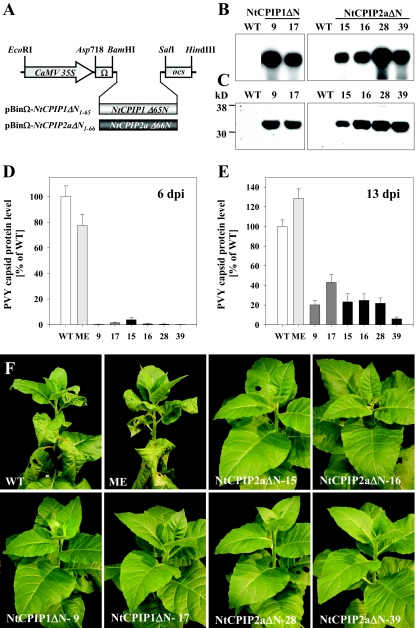
For ectopic expression of dominant-negative mutants of NtCPIP1 and NtCPIP2a, truncated cDNA fragments lacking aa 1 to 65 (NtCPIP1Δ65N) and aa 1 to 66 (NtCPIP2aΔ66N) were amplified by PCR using the gene-specific primers D249 (5′-GGATCCTATATACGGCGATGAGGCGTTGAAATC-3′) and D251 (5′-GTCGACTTAGTCAACAGTCCTGCCCAGCAC-3′) for NtCPIP1Δ65N and D250 (5′-GGATCCTGACGTGTACGGTGATGATGCATTG-3′) and D210 (5′-GTCGACTTAGTCAGCGCTCCTGCACAGTAC-3′) for NtCPIP2aΔ66N, respectively. After subcloning into pCR-Blunt, the fragments were inserted into pBinAR between the CaMV 35S promoter and ocs terminator. In order to improve translation efficiency, the 5′-untranslated overdrive sequence (Ω) of TMV U1 (32) was placed between the promoter and the NtCPIP1Δ65N or NtCPIP2aΔ66N coding sequences, respectively. This also introduced an ATG start codon in an optimized plant consensus sequence within an NcoI cloning site. Stable transformation of tobacco plants with the resulting pBinΩ-NtCPIP1ΔN1-65 and pBinΩ-NtCPIP2aΔN1-66 constructs was performed by Agrobacterium-mediated gene transfer as described previously (60).
RNA and protein analysis of transgenic plants.
Extraction of total RNA from leaf material and Northern blot analysis was performed as described by Chen et al. (17). NtCPIP1 and NtCPIP2a specific transcripts were detected by using random-primed 32P-labeled cDNA fragments, respectively.
Protein extraction and Western blot analysis followed the protocol described by Hofius et al. (40). Leaf material was homogenized in 2× SDS sample buffer containing 50 mM Tris-HCl, 5% (vol/vol) β-mercaptoethanol, 10% (vol/vol) glycerin, and 2% (wt/vol) SDS (pH 6.8). After heat denaturation, equal amounts of protein were separated on 12.5% (vol/vol) SDS-polyacrylamide gels and transferred to nitrocellulose membrane (Porablot). An immunoreaction was carried out with either rabbit polyclonal anti-Myc antibody (1:3,000 dilution; Santa Cruz Biotechnology, Santa Cruz, CA) or polyclonal anti-NtCPIP1 (1:3,000) and anti-NtCPIP2a antiserum (1:5,000), generated against affinity-purified His6-tagged NtCPIP1 and NtCPIP2a proteins (see above) in rabbits using custom service from Eurogentec (Seraing, Belgium).
BiFC assay.
Protein-protein interaction studies using the BiFC technique were carried out as described previously (72). For construction of the binary plasmids, full-length cDNAs of PVY CP and both NtCPIPs were PCR amplified using appropriate oligonucleotides and fused as BamHI/SalI fragments to either the N-terminal portion of YFP (YFPN) in the binary vector pSPYNE-35S (NtCPIP1 and NtCPIP2a) or to the C-terminal part of yellow fluorescent protein (YFPC) in pSPYCE-35S (PVY CP). The resulting constructs were introduced into A. tumefaciens strain C58C1(pGV2260) and agroinfiltrated in pairwise combinations together with the p19 silencing suppressor (optical density at 600 nm of 1.0:1.0:1.0) into leaves of 3-week-old N. benthamiana plants according to the procedure described above. Three days after infiltration, coexpression of proteins was assured by Western blot analysis with anti-HA and anti-Myc antibodies (data not shown). For microscopic analysis, sections from agroinfiltrated leaves were manually cut, incubated in 50 mM phosphate buffer (pH 7.2), and scanned in the epidermal cell layer for reconstituted YFP fluorescence by using the confocal microscope LSM 510 META (Zeiss, Göttingen, Germany). An excitation light of 488 nm produced by the krypton/argon laser and an emission filter of 516 to 537 nm allowed the detection of YFP-specific fluorescence, which was finally superimposed with the Nomarski scan by means of the Zeiss LSM version 3.0.
Virus infections and movement studies.
PVY infection of tobacco plants and immunological detection of PVY CP was performed as described previously (37) using virus-specific enzyme-linked immunosorbent assay (ELISA) reagents provided by BIOREBA (Reinach, Switzerland).
For analysis of viral movement, PVY was tagged with the green fluorescent protein (GFP) by using the intron stabilized PVY-123 full-length cDNA clone (11; kindly provided by E. Johansen, University of Copenhagen, before publication) for inserting smRS-GFP (18) according to the same cloning strategy as published by Dietrich and Maiss (20). The full-length clone was propagated in E. coli NM522 (Pharmacia), and DNA was prepared by using a QIAGEN maxikit (QIAGEN). Biolistic delivery of the GFP-labeled PVY cDNA into tobacco leaves was performed with a hand-held particle gun (Bio-Rad Helios gene gun system). Detection and visualization of initial infection sites was achieved by cobombardment of the GFP-tagged PVY cDNA together with a DsRed expression vector (pe35AscIoptRed) (21). Fluorescently tagged virus was imaged by confocal laser scanning microscopy (Leica TCS SP2) 4 days postinfection (dpi). GFP and DsRed were excited with the argon laser (488 nm) and the helium-neon laser (543 nm), respectively. Cross talk was eliminated by previous lambda-scanning or manual regulation of laser intensity.
Sequence data from the present study have been deposited with the EMBL/GenBank data libraries under accession numbers AY319647 (PVY CP), AY319648 (NtCPIP1), AY319649 (NtCPIP2a), AY319650 (NtCPIP2b), respectively.
RESULTS
Isolation of PVY CP interacting tobacco proteins.
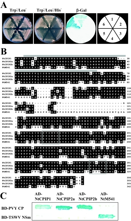
To identify plant proteins that interact with PVY CP, the GAL4-based yeast two-hybrid system (30) was used. The entire PVY CP coding region was amplified by reverse transcription-PCR from PVYN (N-strain)-infected leaf material (37) and used in fusion with the GAL4 BD as bait to screen an AD-tagged cDNA library constructed from tobacco source leaves. Out of a total number of approximately 8 × 10⁷ transformants, four positive clones were identified showing activity of both reporter genes HIS3 and lacZ. Library plasmids were rescued, and the specificity of the interaction was verified by retransformation into the reporter strain in combination with the bait BD-PVY CP or control plasmids (Fig. 1A). Sequence analysis revealed that the isolated cDNA clones encoded for an identical protein designated NtCPIP1. Among these clones, the longest AD-fused open reading frame (ORF) coded for 270 aa. The full-length NtCPIP1 cDNA clone encoding a protein of 306 aa was obtained by screening a cDNA tobacco leaf library (38) using the NtCPIP1 cDNA fragment as a probe. Remarkably, two additional cDNA clones different from NtCPIP1 but highly similar to each other were isolated and subsequently designated NtCPIP2a and NtCPIP2b. Alignment of the protein sequences showed that NtCPIP1 shared substantial similarity of 81.3 and 82.6% identical amino acid residues with NtCPIP2a and NtCPIP2b, respectively, whereas NtCPIP2a and NtCPIP2b were 97.4% identical to each other (Fig. 1B). Yeast two-hybrid analyses with AD-tagged constructs of NtCPIP2a and NtCPIP2b in combination with BD-PVY CP (Fig. 1C) or appropriate control plasmids (data not shown) verified a similar binding of NtCPIP2a and NtCPIP2b to PVY CP as shown for NtCPIP1. Due to the presence of a conserved J-domain (44) at the N terminus (aa 4 to 68 for NtCPIP1 and aa 4 to 70 for NtCPIP2a/2b, respectively), the CP-interacting NtCPIPs could be assigned to the large and diverse family of DnaJ-like proteins, and highest similarity was detected to several DnaJ-like genes from Arabidopsis thaliana (GenBank accession numbers AAD39315, AAS32885, AAF07844, AAD25656, and T48181). Interestingly, NtCPIPs also shared substantial similarity with a subset of J-domain proteins encoded by the N. tabacum gene NtDnaJ_M541 (59.2% identity on amino acid level) and its Lycopersicon esculentum Le19/8 (59.5%) and Arabidopsis thaliana AtA39 (59.8%) orthologues that were previously identified to interact with the TSWV movement protein NSm (66, 70). However, PVY CP was unable to bind NtDnaJ_M541 and consistently TSWV NSm did not interact with the different NtCPIPs in yeast (Fig. 1C).
FIG. 1.
Isolation of NtCPIPs from N. tabacum that interact with PVY CP. (A) Specific interaction between PVY CP and NtCPIP1 in the yeast two-hybrid system. Yeast cells transformed with bait and prey vectors were plated on Trp−/Leu− medium to test for double transformation and on Trp−/Leu−/His− medium for protein interaction. As a second reporter of the interaction, lacZ activity was tested using a β-galactosidase filter assay (β-Gal). Reporter gene activation was observed only for colonies cotransformed with BD-PVY CP and AD-NtCPIP1 (1) or with BD-SNF1 and BD-SNF4 representing a positive control (6). No interaction was detectable for any of the other transformations. Combinations of transformed plasmid: 1, BD-PVY CP/AD-NtCPIP1; 2, pGBT9 vector/AD-NtCPIP1; 3, BD-p53/AD-NtCPIP1; 4, BD-SNF1/AD-NtCPIP1; 5, BD-PVY CP/AD-SNF4; 6, BD-SNF1/AD-SNF4. (B) Alignment of deduced amino acid sequences of NtCPIPs with a DnaJ-like protein from N. tabacum (NtM541) using the CLUSTAL W program (DNASTAR, Madison, WI). NtCPIP2a and NtCPIP2b, isolated by cDNA library screening by using NtCPIP1 as a probe, were both shown to specifically interact with PVY CP in the yeast two-hybrid system (data not shown; see Fig. 1C), whereas NtM541 was previously identified to bind to the TSWV NSm movement protein (66). Regions of identity are shaded in black and gaps introduced for alignment are indicated by dashes. The predicted J domain is marked by a gray horizontal bar, and two conserved motifs (K-X-X-X-K-E/K) indicative for a lysine-enriched domain are boxed. (C) Interaction ability of PVY CP and TSWV NSm with NtCPIPs or NtM541.Yeast cells expressing combinations of the indicated viral bait and DnaJ-like prey proteins were grown on Trp−/Leu−/His− medium and analyzed qualitatively for β-galactosidase activity.
Mutations in the core region of PVY CP abolish yeast interaction with NtCPIPs.
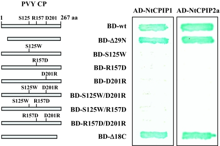
To analyze whether the interaction between PVY CP and NtCPIPs can be mapped either to the core region or to the N- and C-terminal regions of the CP, a series of PVY CP mutants as GAL4-binding domain fusions were generated. To this end, N-terminal (29 aa, PVY CPΔ29N) and C-terminal (18 aa, PVY CPΔ18C) deletions were made or single and double amino acid substitutions were generated by site-directed mutagenesis. The amino acids targeted in these substitution mutants were three highly conserved residues in the core region of potyviral CPs (25), which have been demonstrated to be essential for successful cell-to-cell movement of TEV (23, 24), as well as for PD-mediated trafficking of pressure-injected recombinant Bean common mosaic necrosis virus and Lettuce mosaic virus CPs (59). Qualitative yeast two-hybrid assays were performed with the single amino acid substitutions PVY CP S125W, PVY CP R157W, and PVY CP D201R, as well as the double amino acid substitutions PVY CP S125W/R157D, PVY CP S125W/D201R, and PVY CP R157D/D201R (Fig. 2). Binding of PVY CP to NtCPIP1, as well as to NtCPIP2a, was not influenced by N- and C-terminal deletions; however, lacZ reporter gene expression was completely abolished by the three single amino acid substitutions and all combinations of double amino acid substitution mutants, suggesting an essential role of the CP core region for interaction with NtCPIPs.
FIG. 2.
Identification of the PVY CP interaction domain in the yeast two-hybrid system. PVY CP deletion and various single and double amino acid substitution mutants were individually cotransformed with NtCPIP1 or NtCPIP2a into yeast cells and qualitatively assayed for β-galactosidase activity. Mutations introduced into the PVY CP gene are indicated. Amino acids (S125, R157, and D201) targeted in the substitution mutants refer to highly conserved residues in the core domain of potyviral CPs (22).
NtCPIPs interact with PVY CP in vitro and in planta.
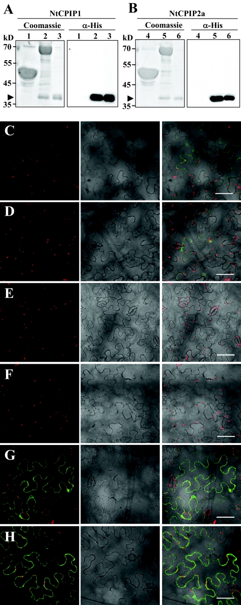
To confirm the interaction ability of NtCPIPs with PVY CPs, an independent in vitro protein binding assay was used. To this end, PVY CP was expressed as MBP fusion in E. coli cells and coupled to an amylose affinity matrix. After incubation with either His-tagged NtCPIP1 or NtCPIP2a proteins, matrix-bound protein complexes were eluted and subsequently analyzed for the presence of NtCPIPs by Western blotting with anti-His antibodies. In agreement with the results obtained by the two-hybrid analysis, NtCPIP1 (Fig. 3A) and NtCPIP2a (Fig. 3B) showed binding to PVY CP, whereas MBP alone did not interact.
FIG. 3.
PVY CP interacts with NtCPIPs in vitro and in planta. (A and B) In vitro binding assay between PVY CP and NtCPIP1 (A) or NtCPIP2a (B). MBP alone (lanes 1 and 4) or in fusion with PVY CP (lanes 2 and 5) was expressed in E. coli, coupled to an amylose matrix, and incubated with 25 μg of affinity-purified His6-tagged NtCPIP1 or NtCPIP2a protein. Aliquots of the eluates (75% of total amount) were separated by SDS-PAGE and tested for the presence of NtCPIPs (arrowheads) by Coomassie blue staining or Western blot analysis with anti-His antibodies. Then, 2 μg of His6-NtCPIP1 (lane 3) or His6-NtCPIP2a (lane 6) was loaded onto the respective gels as input controls. (C to H) BiFC analysis of PVYCP/NtCPIP interaction in plant cells. The coding regions of NtCPIPs and PVY CP were fused with the N-terminal (YFPN, in pSPYNE-35S) or C-terminal (YFPC, in SPYCE-35S) region of YFP, respectively. Plasmids were Agrobacterium infiltrated in N. benthamiana leaves, and the reconstructed YFP signal was detected in the epidermal cell layer by confocal microscopy. Coexpression of empty vectors pSPYNE/pSPYCE (C), PVY CP:YFPC/pSPYNE (D), pSPYCE/NtCPIP1:YFPN (E), pSPYCE/NtCPIP2a:YFPN (F) PVY CP:YFPC/NtCPIP1:YFPN (G), or PVY CP:YFPC/NtCPIP2a:YFPN (H) reveals specific YFP complementation only by PVY CP/NtCPIP interactions. YFP-derived fluorescence signals (in green) of single confocal sections (left) and the transmission mode (middle) were superimposed in the merged image (right). Bars, 50 μm.
To verify the association of PVY CP with NtCPIPs in planta, a BiFC assay was performed in Agrobacterium-infiltrated N. benthamiana plants (72). NtCPIP1 and NtCPIP2a were fused to the N-terminal YFP fragment (YFPN), respectively, whereas PVY CP was merged with the C-terminal YFP fragment (YFPC). Pairwise expression of unfused YFPN and YFPC or their combination with the respective PVY CP or NtCPIP fusions induced very weak or no YFP fluorescence signals in agroinfiltrated epidermal cells (Fig. 3C to F). In contrast, strong YFP fluorescence was observed when combinations of PVY CP and NtCPIP1 or NtCPIP2a were expressed, respectively, indicating the capability of PVY CP/NtCPIP complex formation in plant cells (Fig. 3G and H).
Ectopic expression of dominant-negative mutants of NtCPIPs confers resistance to PVY.
To assess the in planta role of NtCPIP binding to PVY CP during virus infection, we sought to generate transgenic plants with impaired NtCPIP function. An obvious approach was the downregulation of NtCPIPs via posttranscriptional gene silencing by stable expression of hairpin RNA interference (RNAi) constructs (65). However, such a strategy might be limited by an insufficient degree of suppression, the functional complementation of RNAi-silenced NtCPIPs by unkown and nontargeted DnaJ isoforms, and/or the reversion of silencing by the potent potyviral silencing suppressor HC-Pro after virus infection (9, 61). Indeed, infection of transgenic plants specifically silenced for either NtCPIP1 or NtCPIP2a revealed an enhanced local but only transient resistance to PVY (data not shown).
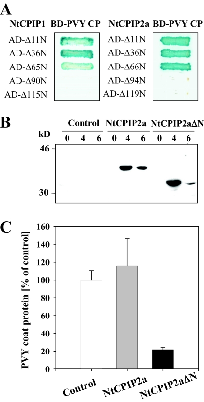
To circumvent such potential constraints of a silencing approach, we intended to ectopically express dominant interfering variants of NtCPIP proteins in transgenic plants. These mutants should retain their ability to bind to PVY CP but should be unable to serve their cellular function. Generally, DnaJ proteins are assumed to function as cochaperones and regulators of heat shock protein 70 (HSP70) proteins by stimulating their ATPase activity via interaction of the J domain (44). Therefore, we analyzed in the yeast two-hybrid system whether deletion of the main portion of the J domain from NtCPIP1 and NtCPIP2a would also affect the interaction with PVY CP. N-terminal deletions of aa 1 to 65 and aa 1 to 66 of NtCPIP1 and NtCPIP2a, respectively, did not abolish lacZ reporter gene activity, suggesting that the J domain is dispensable for binding to PVY CP. However, extending the N-terminal truncations to more than 90 aa resulted in a complete loss of binding (Fig. 4A).
FIG. 4.
Identification of dominant-negative NtCPIP mutants and functional analysis in planta. (A) Interaction of N-terminal deletion mutants of NtCPIP1 and NtCPIP2a with PVY CP in the yeast two-hybrid system. Yeast cells cotransformed with the indicated bait and prey plasmids were grown on Trp−/Leu−/His− medium and qualitatively assayed for β-galactosidase activity. (B) Transient expression of 3x-Myc epitope-tagged full-length and J-domain-deficient NtCPIP2a proteins in N. benthamiana leaves via agroinfiltration. Western blot analysis was performed with identical amounts of total protein extracts from leaves 0, 4, and 6 days after coinfiltration of NtCPIP2a-myc and NtCPIP2aΔN-myc with the p19 silencing suppressor, respectively. Infiltration of p19 alone served as a negative control. (C) Effect of transient expression of full-length and J-domain-deficient NtCPIP2a proteins on susceptibility to PVY infection in N. benthamiana. Leaves were infected with PVY 24 h after agroinfiltration and assayed for accumulation of viral coat protein 4 dpi by ELISA. Values represent means (n = 12) ± the standard error (SE) and are given as the percentage of the p19 control. The results indicate that expression of the dominant-negative mutant but not the full-length variant strongly interferes with the spread of infection.
To test the effect of N-terminally deleted NtCPIP mutant versus the full-length protein on PVY spread in planta, we combined agroinfiltration-mediated protein expression of NtCPIP2a variants with a virus infection assay in N. benthamiana leaves. To this end, full-length NtCPIP2a and J-domain-deficient NtCPIP2aΔN cDNA fragments were placed into a binary vector between the constitutive CaMV 35S promoter and a 3x-Myc epitope tag. The resulting constructs were coinfiltrated with the silencing suppressor p19 into N. benthamiana leaves, leading to considerable protein expression until 4 to 6 days postinfiltration (Fig. 4B). Leaves were challenged with PVY 24 h after agroinfiltration and assayed for virus accumulation at 4 dpi by using an ELISA. As demonstrated in Fig. 4C, only expression of NtCPIP2aΔN and not of full-length NtCPIP2a strongly affected the establishment of virus infection in the local leaf, supporting the concept that J-domain-deleted but CP-interacting NtCPIP variants function as dominant-interfering mutants in planta.
Based on these results, binary constructs for stable expression of dominant-negative variants both for NtCPIP1 and for NtCPIP2a were generated. In order to reach a preferably high expression level in transgenic plants, N-terminal deleted fragments of NtCPIPs were fused to the translational enhancer (Ω) from TMV U1 (32) downstream of the CaMV 35S promoter, resulting in the plasmids pBinΩ-NtCPIP1ΔN1-65 and pBinΩ-NtCPIP2aΔN1-66 (Fig. 5A). After Agrobacterium-mediated transformation, 26 primary transformants for each construct were transferred to the greenhouse and screened for expression of the transgene by Northern analysis. Several plants accumulating considerable amounts of the respective transcripts could be identified (data not shown). Two plants bearing the construct pBinΩ-NtCPIP1ΔN1-65 (designated NtCPIP1ΔN-9 and -17) and four plants transgenic for the pBinΩ-NtCPIP2aΔN1-66 construct (NtCPIP2aΔN-15, -16, -28, and -39) were chosen for further analysis (Fig. 5B). To verify the accumulation of J-domain truncated NtCPIP proteins, leaf samples of the selected lines were subjected to Western analysis with either NtCPIP1- or NtCPIP2a-specific antibodies. As demonstrated in Fig. 5C, transgenic plants accumulated high amounts of the dominant-negative NtCPIP variants, which, however, did not result in detectable morphological changes or growth defects compared to wild-type (WT) or transgenic controls (data not shown).
FIG. 5.
Effect of stable expression of dominant-negative NtCPIP mutants on susceptibility to PVY infection. (A) Schematic representation of binary overexpression constructs used for transformation of N. tabacum. J-domain-deficient NtCPIP1Δ65N and NtCPIP2aΔ66N fragments shown to retain their interaction ability with PVY CP (Fig. 4A) were fused to the 5′-untranslated TMV U1 overdrive sequence (Ω) and placed between the CaMV 35S promoter and ocs terminator in the Bin19-derived vector. (B) Northern analysis of NtCPIP1Δ65N and NtCPIP2aΔ66N specific transcripts. Each lane contains 30 μg of total RNA isolated from WT plants and transgenic lines NtCPIP1Δ-9 and -17 and NtCPIP2aΔ-15, -16, -28, and -39. Northern blots were hybridized with NtCPIP1Δ65N or NtCPIP2aΔ66N cDNAs, respectively. (C) Immunoblot analysis of NtCPIP1Δ65N and NtCPIP2aΔ66N protein accumulation. Identical amounts of total protein extracted from leaf material of WT and transgenic lines were separated by SDS-PAGE and analyzed by Western blotting with rabbit-derived polyclonal anti-NtCPIP1 (dilution 1:3,000) or anti-NtCPIP2a (1:5,000) antibodies and goat-derived secondary antibody conjugated to horseradish peroxidase (dilution 1:100,000). (D) PVY titer in systemic leaves (five or six leaves above the inoculated leaf) of WT (n = 22) and transgenic ME-4 controls (ME, n = 24), as well as of the transgenic lines NtCPIP1ΔN-9 (n = 25) and -17 (n = 25) and NtCPIP2aΔN-15 (n = 23), -16 (n = 24), -28 (n = 24), and -39 (n = 24) at 6 dpi. Values represent means ± the SE and are given as the percentage of the WT level. Plants had developed six to eight leaves prior to PVY inoculation. (E) PVY coat protein levels in systemic leaves (seven or eight leaves above the inoculated leaf) of WT and transgenic lines at 13 dpi. Values represent means ± the SE and are given as the percentage of the WT level. (F) Development of virus-induced symptoms in PVY-infected transgenic lines compared to controls (WT, ME) at 13 dpi, indicating a dramatically increased virus resistance due to the expression of dominant-negative mutants of NtCPIPs.
To investigate whether accumulation of the truncated NtCPIP1 and NtCPIP2a mutant proteins would interfere with PVY multiplication and systemic spread, the kanamycin-resistant T1 progeny of the selected transgenic plants were challenged with PVY and compared to the wild type and a transgenic control (ME). A total of 22 to 25 plants of the candidate lines, as well as a similar number of WT (n = 22) and ME (n = 24) plants were mechanically inoculated at the 7-leaf-stage, and the development of virus infection was monitored by visual symptom analysis and immunological determination of coat protein levels. All inoculated individuals of the transgenic (ME) and nontransgenic control lines (WT) started to display typical disease symptoms in systemic leaves at 5 to 6 dpi, whereas the different transgenic lines did not show any visible virus-induced symptoms at this time point (data not shown). Consistently, the virus titer in systemic leaves was strongly reduced to almost complete resistance in the transgenic lines (Fig. 5D). The durable increase in virus resistance was confirmed at 13 dpi, where all transgenic lines showed dramatically reduced or absent viral symptoms in comparison to the control lines (Fig. 5F). Analysis of the virus titer in systemic leaves verified the strongly reduced systemic invasion by PVY, which was most pronounced in line NtCPIP2aΔN-39 (Fig. 5E). However, viral particles were detectable in systemic leaves of all transgenic lines, indicating that the resistance conferred by expression of dominant-negative NtCPIP mutants did not provide immunity to PVY infection.
To exclude the possibility that the increased virus resistance was caused by interference of the TMV-derived Ω sequence, transgenic plants expressing an unrelated viral protein (HPV L1 CP) under control of the CaMV 35 promoter and the identical Ω translation enhancer (6) were challenged with PVY and compared to WT and NtCPIPΔN transgenic lines. Determination of virus titer in systemic leaves at 6 dpi confirmed a strong reduction of virus spread in NtCPIPΔN plants, which was not seen in L1 transgenic and WT plants, indicating no major impact of the Ω sequence on resistance parameters (data not shown).
Expression of dominant-negative NtCPIP mutants impairs the local spread of PVY-gfp.
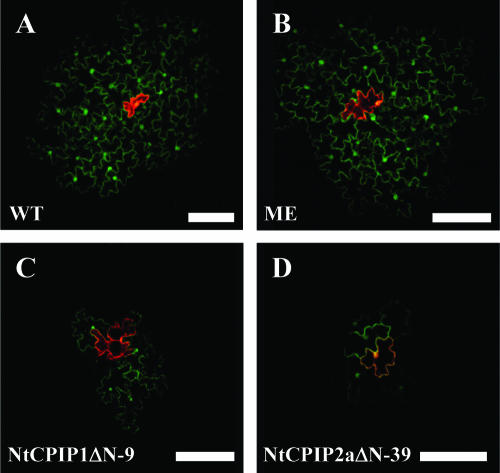
Our finding that mutations in the core region of CP abolished the binding to NtCPIPs in the yeast system suggested the potential contribution of PVY CP-NtCPIP interaction to virion assembly and cell-to-cell movement rather than to long-distance transport. Therefore, we tested whether the NtCPIPΔN transgenic lines were affected in viral cellular spread by using a GFP-labeled PVY cDNA clone that was delivered into epidermal cells of fully expanded source leaves via particle bombardment. As shown in Fig. 6A and B, WT and transgenic control (ME) plants showed uniform GFP fluorescence in initial infection loci 4 days after bombardment that reached an area up to three cells beyond the primarily bombarded cell (visualized by red fluorescence derived from expression of the cobombarded CaMV 35S-DsRed plasmid). In contrast, transgenic lines NtCPIP1ΔN-9 and NtCPIP2aΔN-39 showed considerably smaller zones of GFP fluorescence, which reached only approximately one to two cells in diameter beyond the primary target cell (Fig. 6C and D). These results suggested that impaired NtCPIP function primarily affected viral cell-to-cell transport, thereby causing an overall increase in resistance to PVY infection.
FIG. 6.
Effect of dominant-negative NtCPIP mutants on cellular spread of PVY-gfp. PVY-gfp was cobombarded with a DsRed expression vector into leaves of WT (A) and transgenic (B) controls, as well as leaves of the transgenic lines NtCPIP1ΔN-9 (C) and NtCPIP2aΔN-30 (D). At 4 dpi, bombarded leaves were scanned for GFP- and DsRed-derived fluorescence by confocal microscopy. Representative lesions demonstrated that infections in transgenic lines NtCPIP1ΔN-9 and NtCPIP2aΔN-39 reached a considerably smaller area beyond the primarily bombarded cells (indicated in red) than in the control lines. Bars, 100 μm.
DISCUSSION
In many aspects, cell-to-cell spread of potyviruses still remains an enigma because the intercellular transfer of viral genomes is not mediated by specialized MPs but relies on various proteins with multifunctional properties (57, 67). The CP has been suggested to be implicated in cell-to-cell movement due to its requirement for virion formation and the capacity to traffic viral RNA through modified PD into adjacent cells (24, 59). However, the molecular pathway by which potyviral CPs contribute to intra- and intercellular transport of infectious material is largely unknown. To unravel these processes, it would be necessary to isolate plant proteins that directly interact with the CP in the host cell during infection. Here, we have performed a yeast two-hybrid assay by using PVY CP as bait resulting in the isolation of a subset of DnaJ-like proteins from tobacco, termed NtCPIPs, which were demonstrated to specifically bind to PVY CP in yeast, in vitro and in planta. The functional significance of the interaction could be verified by virus infection of agroinfiltrated N. benthamiana leaves or transgenic plants that ectopically overexpressed dominant-negative mutants of NtCPIPs. These plants showed significantly enhanced virus resistance to PVY, most likely due to strongly reduced cell-to-cell transport. Thus, it can be concluded that recruitment of host chaperones is required for efficient spread of PVY, presumably by interaction of the viral CP with a specific subset of plant DnaJ-like proteins.
Members of the DnaJ (HSP40) multigene family are generally defined by the presence of a N-terminally located J domain and assist as cochaperones of HSP70s in various cellular processes such as protein folding, trafficking, and secretion and also in stress response signaling (10, 44, 50, 56, 71). DnaJ proteins are structurally diverse and grouped according to the combination of three additional domains initially identified in the E. coli DnaJ ortholog: a Gly/Phe-rich domain, a Cys-rich zinc finger domain and a less well conserved C-terminal domain possibly involved in substrate binding (56). However, the identified NtCPIPs fall into a distinct subclass that contain only the J domain. Strikingly, the group of DnaJ-like proteins from tobacco (NtDnaJ_M541), tomato (Le19/8) and Arabidopsis (AtA39), which were recently isolated as TSWV MP (NSm)-binding proteins, also lack, except for the common J domain, domain structures typically found in DnaJ (66, 70). Instead, motifs for a Lys-rich domain (4x K-X-X-X-K-E/K) were identified that are also partially present in the NtCPIP proteins (aa 172 to 177 and aa 269 to 274 for NtCPIP1 and aa 171 to 176 and aa 168 to 273 for NtCPIP2a and -2b, Fig. 1B). However, despite significant sequence and structural similarities, NtCPIPs and NtDnaJ_M541 were not interchangeable for interaction with PVY CP and TSWV MP (NSm), respectively (Fig. 1C), suggesting some specificity of viral proteins in their interaction capability with members of this DnaJ subclass. Independent binding assays verified the ability of PVY CP/NtCPIP complex formation in vitro and in planta (Fig. 3), which significantly strengthened the likelihood that these DnaJ-like protein family members indeed represent novel and relevant binding partners of the potyviral CP.
Unequivocal evidence for a critical role of NtCPIPs in potyviral pathogenesis was provided by resistance analysis of transgenic tobacco plants impaired in NtCPIP function due to overexpression of dominant interfering variants of NtCPIP1 and NtCPIP2a. These deletion mutants lacked the main portion of the J domain which is required for interaction with cellular HSP70 proteins (44) but is dispensable for association with the PVY CP, as revealed by yeast two-hybrid analysis (Fig. 4A). Initially, proof of concept for the inhibitory effect of J-domain deletion mutants versus the full-length protein on PVY infection could be obtained by a combined agroinfiltration and infection assay in N. benthamiana leaves (Fig. 4B). Local PVY accumulation was severely inhibited by transient expression of J-domain-deficient but not of full-length NtCPIP2a (Fig. 4C), suggesting the requirement of NtCPIP function during the initial phases of virus infection. Indeed, mutant analysis in the yeast two-hybrid system identified the CP core region as an essential interaction domain (Fig. 2), thereby linking the PVY CP/NtCPIP association to assembly and plasmodesmal trafficking rather than to long-distance transport processes. Circumstantial evidence for this notion was additionally provided by movement studies using biolistically delivered PVY-gfp, which showed strongly delayed spreading from the primarily infected cell in NtCPIP1ΔN-9 and NtCPIP2aΔN-39 lines compared to the WT and transgenic controls (Fig. 6). Hence, the dominant-negative NtCPIP proteins might have primarily interfered with cell-to-cell transport processes, which finally resulted in strongly enhanced and durable resistance of various independent NtCPIPΔN expressing lines to PVY infection (Fig. 5D to F). Our observation that the increased resistance did not provide immunity to PVY might indicate that the obtained expression level of dominant-negative mutants did not completely suppress the functioning of endogenous NtCPIPs. Nonetheless, the movement and resistance data clearly demonstrate the in vivo relevance of the interaction between the CP and host proteins from the DnaJ family and thus suggest the involvement of HSP70-related mechanisms in PVY infection.
The previously observed binding of the TSWV NSm to the NtCPIP related J-domain proteins from different plant species indicate that different plant viruses might have evolved a similar strategy to exploit HSP70-related chaperone activity in various virulence functions (66). However, in contrast to the present study, experimental data are still lacking demonstrating the in planta role of the NSm-DnaJ protein interaction during TSWV infection. Thus, direct evidence for the importance of HSP70 class proteins in plant virus infection has thus far only been provided by the family of closteroviruses, which encode the only known virus-specific HSP70 homologs (HSP70h) (1). The HSP70h of beet yellows virus has been shown to associate with PD (51) and to function as one of the closteroviral MPs (55). In addition, HSP70h was demonstrated to be essential for virion assembly and stability (2). Interestingly, the basic morphology of the filamentous virions of closteroviruses seems to be similar to that of potyviruses and some other plant virus genera. Accordingly, capsid proteins of these viruses are structurally and evolutionarily related to each other (25). However, closteroviruses are exceptionally long, a feature which was suggested to be the reason for the evolution of two specialized CPs and the integration of HSP70h into the viral genome, thereby providing additional energy for assembly and translocation (2). Due to the dual role of HSP70h in closteroviral virion formation and transport, as well as its concerted action with CPs, it is tempting to speculate that potyviruses may have adopted a similar movement strategy by incorporation of host cellular HSP70s into or in association with the potyviral transport complex. Interestingly, a subclass of plant HSP70 proteins from Cucurbita maxima was previously identified showing plasmodesmal targeting and translocation capacity (3). Members of this subclass might be potential HSP70 candidates recruited by the binding of host NtCPIPs to viral CP and assumed to be required for the chaperone-aided transport of virus particles toward and through PD. A major challenge for the future will be to identify those HSP70 proteins which are able to interact with NtCPIPs.
In summary, our work identifies NtCPIPs as novel potyviral susceptibility factors and also provides a strong in vivo confirmation for the essential role of plant chaperones in virus movement. Taking this into account, the recruitment of molecular chaperones is emerging as a widespread mechanism by which certain plant viruses conquer the host.
Acknowledgments
This study was supported by a grant from the Deutsche Forschungsgemeinschaft (SO 300/6-1).
We especially thank Anita Winger and Lara Lintl for excellent technical assistance, Bernhard Claus for skillful help with the confocal microscope, and Andrea Knospe for plant transformation. We are grateful to T.-R. Soellick and J.-W. Kellmann for providing pAD-NtDnaJ_M541 and pBD-NSm two-hybrid plasmids and to Klaus Harter (Tuebingen, Germany) for the BiFC vectors. We also thank E. Johansen (Copenhagen, Denmark) for providing the full-length PVY cDNA clone prior to publication.
Footnotes
Published ahead of print on 22 August 2007.
REFERENCES
- 1.Agranovsky, A. A., V. P. Boyko, A. V. Karasev, E. V. Koonin, and V. V. Dolja. 1991. Putative 65 kDa protein of beet yellows closterovirus is a homologue of HSP70 heat shock proteins. J. Mol. Biol. 217:603-610. [DOI] [PubMed] [Google Scholar]
- 2.Alzhanova, D. V., A. J. Napuli, R. Creamer, and V. V. Dolja. 2001. Cell-to-cell movement and assembly of a plant closterovirus: roles for the capsid proteins and Hsp70 homolog. EMBO J. 20:6997-7007. [DOI] [PMC free article] [PubMed] [Google Scholar]
- 3.Aoki, K., F. Kragler, B. Xoconostle-Cazares, and W. J. Lucas. 2002. A subclass of plant heat shock cognate 70 chaperones carries a motif that facilitates trafficking through plasmodesmata. Proc. Natl. Acad. Sci. USA 99:16342-16347. [DOI] [PMC free article] [PubMed] [Google Scholar]
- 4.Aranda, M. A., M. Escaler, D. Wang, and A. J. Maule. 1996. Induction of HSP70 and polyubiquitin expression associated with plant virus replication. Proc. Natl. Acad. Sci. USA 93:15289-15293. [DOI] [PMC free article] [PubMed] [Google Scholar]
- 5.Bartel, P. L., and S. Fields. 1995. Analyzing protein-protein interactions using two-hybrid system. Methods Enzymol. 254:241-263. [DOI] [PubMed] [Google Scholar]
- 6.Biemelt, S., U. Sonnewald, P. Galmbacher, L. Willmitzer, and M. Muller. 2003. Production of human papillomavirus type 16 virus-like particles in transgenic plants. J. Virol. 77:9211-9220. [DOI] [PMC free article] [PubMed] [Google Scholar]
- 7.Boevenik, P., and K. Oparka. 2005. Virus-host interactions during movement process. Plant Physiol. 138:1815-1821. [DOI] [PMC free article] [PubMed] [Google Scholar]
- 8.Boyko, V., Q. Hu, M. Seemanpillai, J. Ashby, and M. Heinlein. 2007. Validation of microtubule-associated Tobacco mosaic virus RNA movement and involvement of microtubule-aligned particle trafficking. Plant J. 51:589-603. [DOI] [PubMed] [Google Scholar]
- 9.Brigneti, G., O. Voinnet, W. X. Li, L. H. Ji, S. W. Ding, and D. C. Baulcombe. 1998. Viral pathogenicity determinants are suppressors of transgene silencing in Nicotiana benthamiana. EMBO J. 17:6739-6746. [DOI] [PMC free article] [PubMed] [Google Scholar] [Retracted]
- 10.Bukau, B., and A. L. Horwich. 1998. The Hsp70 and Hsp60 chaperone machines. Cell 92:351-366. [DOI] [PubMed] [Google Scholar]
- 11.Bukovinszki, A., R. Gotz, E. Johansen, E. Maiss, and E. Balazs. 2007. The role of the coat protein region in symptom formation on Physalis floridana varies between PVY strains. Virus Res. 127:122-125. [DOI] [PubMed] [Google Scholar]
- 12.Callaway, A., D. Giesman-Cookmeyer, E. T. Gillock, T. L. Sit, and S. A. Lommel. 2001. The multifunctional capsid proteins of plant RNA viruses. Annu. Rev. Phytopathol. 39:419-460. [DOI] [PubMed] [Google Scholar]
- 13.Carrington, J. C., P. Jensen, and M. C. Schaad. 1998. Genetic evidence for an essential role for potyvirus Cl protein in cell-to-cell movement. Plant J. 14:393-400. [DOI] [PubMed] [Google Scholar]
- 14.Carrington, J. C., K. D. Kasschau, S. K. Mahajan, and M. C. Schaad. 1996. Cell-to-cell and long-distance transport of viruses in plants. Plant Cell 8:1669-1681. [DOI] [PMC free article] [PubMed] [Google Scholar]
- 15.Chen, M. H., and V. Citovsky. 2003. Systemic movement of a tobamovirus requires host cell pectin methylesterase. Plant J. 35:386-392. [DOI] [PubMed] [Google Scholar]
- 16.Chen, M. H., G. W. Tian, Y. Gafni, and V. Citovsky. 2005. Effects of calreticulin on viral cell-to-cell movement. Plant Physiol. 138:1866-1876. [DOI] [PMC free article] [PubMed] [Google Scholar]
- 17.Chen, S., D. Hofius, U. Sonnewald, and F. Bornke. 2003. Temporal and spatial control of gene silencing in transgenic plants by inducible expression of double-stranded RNA. Plant J. 36:731-740. [DOI] [PubMed] [Google Scholar]
- 18.Davis, S. J., and R. D. Vierstra. 1998. Soluble, highly fluorescent variants of green fluorescent protein (GFP) for use in higher plants. Plant Mol. Biol. 36:521-528. [DOI] [PubMed] [Google Scholar]
- 19.Desvoyes, B., S. Faure-Rabasse, M. H. Chen, J. W. Park, and H. B. Scholthof. 2002. A novel plant homeodomain protein interacts in a functionally relevant manner with a virus movement protein. Plant Physiol. 129:1521-1532. [DOI] [PMC free article] [PubMed] [Google Scholar]
- 20.Dietrich, C., and E. Maiss. 2003. Fluorescent labeling reveals spatial separation of potyvirus populations in mixed infected Nicotiana benthamiana plants. J. Gen. Virol. 84:2871-2876. [DOI] [PubMed] [Google Scholar]
- 21.Dietrich, C., and E. Maiss. 2002. Red fluorescent protein DsRed from Discosoma sp. as a reporter protein in higher plants. BioTechniques 32:286-293. [DOI] [PubMed] [Google Scholar]
- 22.Dolja, V. V., V. P. Boyko, A. A. Agranovsky, and E. V. Koonin. 1991. Phylogeny of capsid proteins of rod-shaped and filamentous RNA plant viruses: two families with distinct patterns of sequence and probably structure conservation. Virology 184:79-86. [DOI] [PubMed] [Google Scholar]
- 23.Dolja, V. V., R. Haldeman-Cahill, A. E. Montgomery, K. A. Vandenbosch, and J. C. Carrington. 1995. Capsid protein determinants involved in cell-to-cell and long distance movement of tobacco etch potyvirus. Virology 206:1007-1016. [DOI] [PubMed] [Google Scholar]
- 24.Dolja, V. V., R. Haldeman, N. L. Robertson, W. G. Dougherty, and J. C. Carrington. 1994. Distinct functions of capsid protein in assembly and movement of tobacco etch potyvirus in plants. EMBO J. 13:1482-1491. [DOI] [PMC free article] [PubMed] [Google Scholar]
- 25.Dolja, V. V., and E. V. Koonin. 1991. Phylogeny of capsid proteins of small icosahedral RNA plant viruses. J. Gen. Virol. 72:1481-1486. [DOI] [PubMed] [Google Scholar]
- 26.Dorokhov, Y. L., K. Mäkinen, O. Y. Frolova, A. Merits, J. Saarinen, N. Kalkkinen, J. G. Atabekov, and M. Saarma. 1999. A novel function for a ubiquitous plant enzyme pectin methylesterase: the host-cell receptor for the tobacco mosaic virus movement protein. FEBS Lett. 461:223-228. [DOI] [PubMed] [Google Scholar]
- 27.Dunoyer, P., C. Thomas, S. Harrison, F. Revers, and A. Maule. 2004. A cysteine-rich plant protein potentiates Potyvirus movement through an interaction with the virus genome-linked protein VPg. J. Virol. 78:2301-2309. [DOI] [PMC free article] [PubMed] [Google Scholar]
- 28.Ebneth, M. 1996. Expressionsanalyse des Promotors einer cytosolischen Fruktose-1,6-bisphosphatase aus Kartoffel in transgenen Tabak- und Kartoffelpflanzen. Ph.D. thesis. Free University of Berlin, Berlin, Germany.
- 29.Escaler, M., M. A. Aranda, C. L. Thomas, and A. J. Maule. 2000. Pea embryonic tissues show common responses to the replication of a wide range of viruses. Virology 267:318-325. [DOI] [PubMed] [Google Scholar]
- 30.Fields, S., and O. Song. 1989. A novel genetic system to detect protein-protein interactions. Nature 340:245-246. [DOI] [PubMed] [Google Scholar]
- 31.Fridborg, I., J. Grainger, A. Page, M. Coleman, K. Findlay, and S. Angell. 2003. TIP, a novel host factor linking callose degradation with the cell-to-cell movement of Potato virus X. Mol. Plant-Microbe Interact. 16:132-140. [DOI] [PubMed] [Google Scholar]
- 32.Gallie, D. R., D. E. Sleat, J. W. Watts, P. C. Turner, and T. M. Wilson. 1987. The 5′-leader sequence of tobacco mosaic virus RNA enhances the expression of foreign gene transcripts in vitro and in vivo. Nucleic Acids Res. 15:3257-3273. [DOI] [PMC free article] [PubMed] [Google Scholar]
- 33.Gao, Z., E. Johansen, S. Eyers, C. L. Thomas, T. H. Noel Ellis, and A. J. Maule. 2004. The potyvirus recessive resistance gene, sbm1, identifies a novel role for translation initiation factor eIF4E in cell-to-cell trafficking. Plant J. 40:376-385. [DOI] [PubMed] [Google Scholar]
- 34.Harper, J. W., G. R. Adami, N. Wei, K. Keyomarsi, and S. J. Elledge. 1993. The p21 Cdk-interacting protein Cip1 is a potent inhibitor of G1 cyclin-dependent kinases. Cell 75:805-816. [DOI] [PubMed] [Google Scholar]
- 35.Heinlein, M. 2002. Plasmodesmata: dynamic regulation and role in macromolecular cell-to-cell signaling. Curr. Opin. Plant Biol. 5:543-552. [DOI] [PubMed] [Google Scholar]
- 36.Heinlein, M., M. R. Wood, T. Thiel, and R. N. Beachy. 1998. Targeting and modification of prokaryotic cell-cell junctions by tobacco mosaic virus cell-to-cell movement protein. Plant J. 14:345-351. [DOI] [PubMed] [Google Scholar]
- 37.Herbers, K., P. Meuwly, J. P. Metraux, and U. Sonnewald. 1996. Salicylic acid-independent induction of pathogenesis-related protein transcripts by sugars is dependent on leaf developmental stage. FEBS Lett. 397:239-244. [DOI] [PubMed] [Google Scholar]
- 38.Herbers, K., G. Monke, R. Badur, and U. Sonnewald. 1995. A simplified procedure for the subtractive cDNA cloning of photoassimilate-responding genes: isolation of cDNAs encoding a new class of pathogenesis-related proteins. Plant Mol. Biol. 29:1027-1038. [DOI] [PMC free article] [PubMed] [Google Scholar]
- 39.Höfgen, R., and L. Willmitzer. 1990. Biochemical and genetic analysis fo different patatin isoforms expressed in various organs of potato (Solanum tuberosum). Plant Sci. 66:221-230. [Google Scholar]
- 40.Hofius, D., K. Herbers, M. Melzer, A. Omid, E. Tacke, S. Wolf, and U. Sonnewald. 2001. Evidence for expression level-dependent modulation of carbohydrate status and viral resistance by the potato leafroll virus movement protein in transgenic tobacco plants. Plant J. 28:529-543. [DOI] [PubMed] [Google Scholar]
- 41.Huang, Z., V. M. Andrianov, Y. Han, and S. H. Howell. 2001. Identification of Arabidopsis proteins that interact with the cauliflower mosaic virus (CaMV) movement protein. Plant Mol. Biol. 47:663-675. [DOI] [PubMed] [Google Scholar]
- 42.Iwabuchi, K., B. Li, P. Bartel, and S. Fields. 1993. Use of the two-hybrid system to identify the domain of p53 involved in oligomerization. Oncogene 8:1693-1696. [PubMed] [Google Scholar]
- 43.Kaido, M., Y. Inoue, Y. Takeda, K. Sugiyama, A. Takeda, M. Mori, A. Tamai, T. Meshi, T. Okuno, and K. Mise. 2007. Downregulation of the NbNACa1 gene encoding a movement-protein-interacting protein reduces cell-to-cell movement of Brome mosaic virus in Nicotiana benthamiana. Mol. Plant-Microbe Interact. 20:671-681. [DOI] [PubMed] [Google Scholar]
- 44.Kelley, W. L. 1998. The J-domain family and the recruitment of chaperone power. Trends Biochem. Sci. 23:222-227. [DOI] [PubMed] [Google Scholar]
- 45.Laporte, C., G. Vetter, A. M. Loudes, D. G. Robinson, S. Hillmer, C. Stussi-Garaud, and C. Ritzenthaler. 2003. Involvement of the secretory pathway and the cytoskeleton in intracellular targeting and tubule assembly of Grapevine fanleaf virus movement protein in tobacco BY-2 cells. Plant Cell 15:2058-2075. [DOI] [PMC free article] [PubMed] [Google Scholar]
- 46.Lazarowitz, S. G., and R. N. Beachy. 1999. Probing plant cell structure and function with viral movement proteins. Curr. Opin. Plant Biol. 2:332-338. [DOI] [PubMed] [Google Scholar]
- 47.Lee, J. Y., B. C. Yoo, M. R. Rojas, N. Gomez-Ospina, L. A. Staehelin, and W. J. Lucas. 2003. Selective trafficking of non-cell-autonomous proteins mediated by NtNCAPP1. Science 299:392-396. [DOI] [PubMed] [Google Scholar]
- 48.Lucas, W. J. 2006. Plant viral movement proteins: agents for cell-to-cell trafficking of viral genomes. Virology 344:169-184. [DOI] [PubMed] [Google Scholar]
- 49.Maule, A., V. Leh, and C. Lederer. 2002. The dialogue between viruses and hosts in compatible interactions. Curr. Opin. Plant Biol. 5:279-284. [DOI] [PubMed] [Google Scholar]
- 50.Mayer, M. P., and B. Bukau. 2005. Hsp70 chaperones: cellular functions and molecular mechanism. Cell. Mol. Life Sci. 62:670-684. [DOI] [PMC free article] [PubMed] [Google Scholar]
- 51.Medina, V., V. V. Peremyslov, Y. Hagiwara, and V. V. Dolja. 1999. Subcellular localization of the HSP70-homolog encoded by beet yellows closterovirus. Virology 260:173-181. [DOI] [PubMed] [Google Scholar]
- 52.Nelson, R. S., and A. J. E. Bel. 1998. The mystery of virus trafficking into, through, and out of vascular tissue. Prog. Bot. 59:476-533. [Google Scholar]
- 53.Nelson, R. S., and V. Citovsky. 2005. Plant viruses. Invaders of cells and pirates of cellular pathways. Plant Physiol. 138:1809-1814. [DOI] [PMC free article] [PubMed] [Google Scholar]
- 54.Oparka, K. J. 2004. Getting the message across: how do plant cells exchange macromolecular complexes? Trends Plant Sci. 9:33-41. [DOI] [PubMed] [Google Scholar]
- 55.Peremyslov, V. V., Y. Hagiwara, and V. V. Dolja. 1999. HSP70 homolog functions in cell-to-cell movement of a plant virus. Proc. Natl. Acad. Sci. USA 96:14771-14776. [DOI] [PMC free article] [PubMed] [Google Scholar]
- 56.Qiu, X. B., Y. M. Shao, S. Miao, and L. Wang. 2006. The diversity of the DnaJ/Hsp40 family, the crucial partners for Hsp70 chaperones. Cell. Mol. Life Sci. 63:2560-2570. [DOI] [PMC free article] [PubMed] [Google Scholar]
- 57.Revers, F., O. Le Gall, T. Candresse, and A. J. Maule. 1999. New advances in understanding the molecular biology of plant/potyvirus interactions. Mol. Plant-Microbe Interact. 12:367-376. [Google Scholar]
- 58.Robaglia, C., M. Durand-Tardif, M. Tronchet, G. Boudazin, S. Astier-Manifacier, and F. Casse-Delbart. 1989. Nucleotide sequence of potato virus Y (N strain) genomic RNA. J. Gen. Virol. 70:935-947. [DOI] [PubMed] [Google Scholar]
- 59.Rojas, M. R., F. M. Zerbini, R. F. Allison, R. L. Gilbertson, and W. J. Lucas. 1997. Capsid protein and helper component-proteinase function as potyvirus cell-to-cell movement proteins. Virology 237:283-295. [DOI] [PubMed] [Google Scholar]
- 60.Rosahl, S., J. Schell, and L. Willmitzer. 1987. Expression of a tuber-specific storage protein in transgenic tobacco plants: demonstration of an esterase activity. EMBO J. 6:1155-1159. [DOI] [PMC free article] [PubMed] [Google Scholar]
- 61.Savenkov, E. I., and J. P. Valkonen. 2001. Potyviral helper-component proteinase expressed in transgenic plants enhances titers of Potato leaf roll virus but does not alleviate its phloem limitation. Virology 283:285-293. [DOI] [PubMed] [Google Scholar]
- 62.Schaad, M. C., R. Haldeman-Cahill, S. Cronin, and J. C. Carrington. 1996. Analysis of the VPg-proteinase (NIa) encoded by tobacco etch potyvirus: effects of mutations on subcellular transport, proteolytic processing, and genome amplification. J. Virol. 70:7039-7048. [DOI] [PMC free article] [PubMed] [Google Scholar]
- 63.Schiestl, R. H., and R. D. Gietz. 1989. High efficiency transformation of intact yeast cells using single stranded nucleic acids as a carrier. Curr. Genet. 16:339-346. [DOI] [PubMed] [Google Scholar]
- 64.Scholthof, H. B. 2005. Plant virus transport: motions of functional equivalence. Trends Plant Sci. 10:376-382. [DOI] [PubMed] [Google Scholar]
- 65.Smith, N. A., S. P. Singh, M. B. Wang, P. A. Stoutjesdijk, A. G. Green, and P. M. Waterhouse. 2000. Total silencing by intron-spliced hairpin RNAs. Nature 407:319-320. [DOI] [PubMed] [Google Scholar]
- 66.Soellick, T. R., J. F. Uhrig, G. L. Bucher, J. W. Kellmann, and P. H. Schreier. 2000. The movement protein NSm of tomato spotted wilt tospovirus (TSWV): RNA binding, interaction with the TSWV N protein, and identification of interacting plant proteins. Proc. Natl. Acad. Sci. USA 97:2373-2378. [DOI] [PMC free article] [PubMed] [Google Scholar]
- 67.Urcuqui-Inchima, S., A. L. Haenni, and F. Bernardi. 2001. Potyvirus proteins: a wealth of functions. Virus Res. 74:157-175. [DOI] [PubMed] [Google Scholar]
- 68.Vogel, F., D. Hofius, and U. Sonnewald. 2007. Intracellular trafficking of potato leaf roll virus movement protein in transgenic Arabidopsis. Traffic doi: 10.1111/j.1600-0854.2007.00608.x. [DOI] [PubMed]
- 69.Voinnet, O., S. Rivas, P. Mestre, and D. Baulcombe. 2003. An enhanced transient expression system in plants based on suppression of gene silencing by the p19 protein of tomato bushy stunt virus. Plant J. 33:949-956. [DOI] [PubMed] [Google Scholar]
- 70.von Bargen, S., K. Salchert, M. Paape, B. Piechulla, and J. W. Kellmann. 2001. Interactions between the tomato spotted wilt virus movement protein and plant proteins showing homologies to myosin, kinesin and DnaJ-like chaperones. Plant Physiol. Biochem. 39:1083-1093. [Google Scholar]
- 71.Walsh, P., D. Bursac, Y. C. Law, D. Cyr, and T. Lithgow. 2004. The J-protein family: modulating protein assembly, disassembly and translocation. EMBO Rep. 5:567-571. [DOI] [PMC free article] [PubMed] [Google Scholar]
- 72.Walter, M., C. Chaban, K. Schutze, O. Batistic, K. Weckermann, C. Nake, D. Blazevic, C. Grefen, K. Schumacher, C. Oecking, K. Harter, and J. Kudla. 2004. Visualization of protein interactions in living plant cells using bimolecular fluorescence complementation. Plant J. 40:428-438. [DOI] [PubMed] [Google Scholar]
- 73.Whitham, S. A., S. Quan, H. S. Chang, B. Cooper, B. Estes, T. Zhu, X. Wang, and Y. M. Hou. 2003. Diverse RNA viruses elicit the expression of common sets of genes in susceptible Arabidopsis thaliana plants. Plant J. 33:271-283. [DOI] [PubMed] [Google Scholar]
- 74.Wright, K. M., N. T. Wood, A. G. Roberts, S. Chapman, P. Boevink, K. M. Mackenzie, and K. J. Oparka. 2007. Targeting of TMV movement protein to plasmodesmata requires the actin/ER network: evidence from FRAP. Traffic 8:21-31. [DOI] [PubMed] [Google Scholar]


