Abstract
Ubiquitin ligases of the Nedd4 family regulate membrane protein trafficking by modifying both cargo proteins and the transport machinery with ubiquitin. Here, we investigate the role of the yeast Nedd4 homologue, Rsp5, in protein sorting into vesicles that bud into the multivesicular endosome (MVE) en route to the vacuole. A mutant lacking the Rsp5 C2 domain is unable to ubiquitinate or sort biosynthetic cargo into MVE vesicles, whereas endocytic cargo is ubiquitinated and sorted efficiently. The C2 domain binds specifically to phosphoinositides in vitro and is sufficient for localization to membranes in intact cells. Mutation of a lysine-rich patch on the surface of the C2 domain abolishes membrane interaction and disrupts sorting of biosynthetic cargo. Translational fusion of ubiquitin to a biosynthetic cargo protein alleviates the requirement for the C2 domain in its MVE sorting. These results demonstrate that the C2 domain specifies Rsp5-dependent ubiquitination of endosomal cargo and suggest that Rsp5 function is regulated by membrane phosphoinositides.
Keywords: multivesicular endosome; phospholipid; E3; protein sorting; endocytosis
Introduction
The polypeptide ubiquitin regulates the trafficking of transmembrane proteins in the endocytic pathway and at the point of exit from the trans-Golgi network (for review see Katzmann et al., 2002; Hicke and Dunn, 2003). Ubiquitin influences sorting decisions at these locations by at least two mechanisms. First, when conjugated to the cytosolic domain of a cargo protein, ubiquitin serves as a cis signal to promote sorting into nascent vesicles. Second, ubiquitination regulates the activity of trans-acting components of the transport machinery. The cis and trans functions of ubiquitin control the trafficking of a wide array of signaling receptors and other proteins in all eukaryotic cells, thereby regulating processes as essential as growth control and development.
A cascade of three factors catalyzes protein ubiquitination: a ubiquitin-activating enzyme (E1), a ubiquitin-conjugating enzyme (E2), and a ubiquitin ligase (E3; Hershko et al., 2000; Pickart, 2001). E3s are responsible for substrate selection and are characterized by a HECT catalytic domain or a RING finger (or structurally related) domain. HECT domain E3s of the Nedd4 family regulate the trafficking of diverse proteins in both the endocytic and biosynthetic pathways (Rotin et al., 2000; Helliwell et al., 2001; Soetens et al., 2001; Myat et al., 2002). Nedd4 family proteins exhibit a characteristic structural organization, with two to four WW protein–protein interaction domains and a carboxyl-terminal HECT catalytic domain. Most members also carry an amino-terminal C2 domain, a conserved lipid and protein interaction module that is often regulated by calcium (Nalefski and Falke, 1996; Hurley and Misra, 2000). The sole member of the Nedd4 family in Saccharomyces cerevisiae is Rsp5. Rsp5 is an essential peripheral membrane protein that regulates the internalization step of endocytosis and sorting from the secretory pathway to the lysosome-like vacuole (Galan et al., 1996; Helliwell et al., 2001; Soetens et al., 2001). In addition to its roles in membrane protein transport, Rsp5 modifies a variety of proteins involved in other cellular processes (Beaudenon et al., 1999; Hoppe et al., 2000).
We have investigated the role of Rsp5 in protein sorting into the lumen of the late endosome, also called the multivesicular endosome (MVE) or body. The MVE forms when portions of the late endosome membrane invaginate and pinch off into the lumen, thus forming intralumenal vesicles (Katzmann et al., 2002; Raiborg et al., 2003). When the MVE fuses with the lysosome/vacuole, the intralumenal vesicles are conveyed to the hydrolytic lumen where their lipid and protein constituents are degraded. Sorting of biosynthetic and endocytic transmembrane proteins into MVE vesicles is controlled by the addition of a single ubiquitin moiety to a cytoplasmic domain of these proteins (Katzmann et al., 2001; Reggiori and Pelham, 2001; Urbanowski and Piper, 2001). Trans-acting factors required for MVE vesicle budding include ESCRT (endosomal sorting complex required for transport) complexes I–III and phosphoinositides (PIs; Odorizzi et al., 1998; Bishop and Woodman, 2001; Futter et al., 2001; Katzmann et al., 2001; Babst et al., 2002a,b). Membrane proteins at the late endosome that are excluded from MVE vesicles remain at the limiting membrane or recycle to the plasma membrane (Babst et al., 2000). Thus, this process is critical for the down-regulation of internalized plasma membrane proteins such as activated signaling receptors. As an additional mechanism to down-regulate cell surface activity, some newly synthesized plasma membrane proteins are diverted from the biosynthetic pathway to a late endosome by ubiquitin signals appended by Nedd4 family ligases (Helliwell et al., 2001; Soetens et al., 2001; Keleman et al., 2002; Myat et al., 2002).
In the present work, we demonstrate a role for Rsp5 in the selection of MVE cargo. We provide evidence linking the requirement for the Rsp5 C2 domain with the ubiquitination state of cargo at the late endosome. Furthermore, we describe an interaction between the C2 domain and PIs, and we identify a surface of the C2 domain required for both membrane interaction and MVE sorting. Together, the results suggest a model in which the C2 domain of Rsp5 tethers it to endosomal membranes, poising the ligase for subsequent modification of newly arrived cargo from the biosynthetic pathway.
Results
Role of Rsp5 in MVE sorting
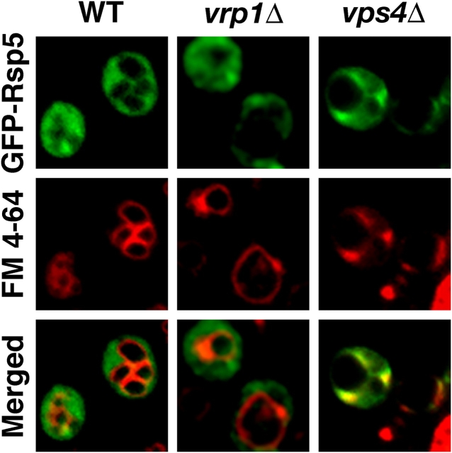
Previous works suggested that Rsp5 functions at a postinternalization step of endocytosis (Dunn and Hicke, 2001; Wang et al., 2001). One characteristic of proteins that function in MVE sorting is that they accumulate on the membrane of an exaggerated perivacuolar endosome observed in cells that lack the AAA ATPase Vps4 (Raymond et al., 1992; Babst et al., 1998). To determine whether or not Rsp5 exhibits this characteristic, we compared the localization of a functional GFP-tagged Rsp5 (GFP-Rsp5) with FM 4-64, a fluorescent dye that labels perivacuolar endosomes and the limiting membrane of the vacuole (Vida and Emr, 1995). In wild-type cells, GFP-Rsp5 was localized diffusely in the cytoplasm and did not overlap with the FM 4-64 marker (Fig. 1) . In cells blocked at the internalization step of endocytosis (vrp1Δ), GFP-Rsp5 was localized diffusely in the cytoplasm and accumulated in punctate spots adjacent to the plasma membrane. In contrast, in vps4Δ cells, GFP-Rsp5 accumulated at perivacuolar endosomes that colabeled with FM 4-64, suggesting that Rsp5 may function in MVE sorting.
Figure 1.
Rsp5 localizes to an exaggerated endosomal compartment in vps4Δ cells. Localization of GFP-tagged Rsp5 in wild-type (LHY4013), vrp1Δ (LHY4014), and vps4Δ (LHY4012) cells was visualized by fluorescence microscopy. The red fluorescent dye FM 4-64 labels perivacuolar endosomes and the limiting membrane of the vacuole. Yellow regions in the merged images indicate areas of GFP-Rsp5 and FM 4-64 colocalization.
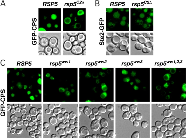
To test whether or not Rsp5 is required for protein sorting into MVE vesicles, we used two markers for this pathway, GFP-tagged carboxypeptidase S (GFP-CPS) and GFP-tagged α-factor receptor (Ste2-GFP). GFP-CPS serves as a biosynthetic marker for the MVE pathway. After exiting the trans-Golgi network, CPS is monoubiquitinated and sorted into MVE vesicles, resulting in its delivery to the vacuole lumen where it functions as a resident hydrolase (Odorizzi et al., 1998; Katzmann et al., 2001). Thus, in wild-type cells expressing GFP-CPS, the vacuole lumen is labeled by GFP fluorescence. When GFP-CPS sorting into the MVE pathway is disrupted (e.g., by mutation of the ubiquitination site), the protein is found at the late endosome and the limiting membrane of the vacuole, detectable as a fluorescent perivacuolar spot and ring, respectively (Katzmann et al., 2001). Ste2-GFP is a chimeric variant of Ste2, a signaling receptor that undergoes ubiquitination at the plasma membrane before its internalization into the endocytic pathway (Hicke and Riezman, 1996). Ste2-GFP is packaged into MVE vesicles on arrival at the late endosome, resulting in its delivery to the vacuole lumen (Odorizzi et al., 1998). Thus, Ste2-GFP serves as an endocytic marker for the MVE pathway.
Our previous work indicated a possible role for the Rsp5 C2 domain in endosomal sorting (Dunn and Hicke, 2001). Therefore, we analyzed MVE sorting in a mutant lacking the C2 domain of Rsp5 (rsp5 C2Δ). These cells grow normally at all temperatures and exhibit normal internalization of Ste2 from the plasma membrane (Dunn and Hicke, 2001). Wild-type RSP5 cells sorted GFP-CPS to the vacuole lumen normally, whereas rsp5 C2Δ cells missorted GFP-CPS to the vacuole limiting membrane (Fig. 2 A). In contrast, rsp5 C2Δ cells effectively sorted Ste2-GFP to the vacuole lumen (Fig. 2 B). Thus, the C2 domain of Rsp5 is important for the MVE sorting of a biosynthetic cargo protein but not of an endocytic cargo protein.
Figure 2.
Rsp5 C2 and WW domains are required for sorting biosynthetic MVE cargo into the vacuole. (A) Sorting of GFP-CPS in RSP5 (LHY2920) and rsp5 C2Δ (LHY2922) cells was visualized by fluorescence microscopy. Differential interference contrast (DIC) microscopy revealed the location of vacuoles. (B) Fluorescence and DIC images of RSP5 (LHY3924) and rsp5 C2Δ (LHY3925) cells expressing Ste2-GFP. (C) Fluorescence and DIC images of RSP5 (LHY4429), rsp5 ww1 (LHY4430), rsp5 ww2 (LHY4431), rsp5 ww3 (LHY4432), and rsp5 ww1,2,3 (LHY4433) cells expressing GFP-CPS.
To determine whether or not the WW domains of Rsp5 contribute to its role in MVE sorting, we analyzed the localization of GFP-CPS in characterized mutants harboring a tryptophan to alanine point mutation in one of the three WW domains or in all three (Dunn and Hicke, 2001). Sorting of GFP-CPS to the vacuole lumen was partially defective in each individual WW domain mutant and was severely defective in the triple rsp5 ww1,2,3 mutant (Fig. 2 C), indicating that the WW domains also regulate MVE sorting of biosynthetic cargo.
The C2 domain is important for localization of Rsp5 to endosomal membranes
Many C2 domains bind to membranes through electrostatic interactions between basic amino acids and negatively charged lipids (Cho, 2001). We noticed clusters of two (KK44–45) and five (KKFKKK74–79) lysines in the sequence of Rsp5 that map to a surface homologous to that used for membrane docking by the protein kinase Cα C2 domain (Kohout et al., 2003; Fig. 3 A), suggesting that the Rsp5 C2 domain might bind to membranes through a direct protein–lipid interaction. The association of Rsp5 with membranes appears to be mediated by multiple regions of the protein (unpublished data), and deletion of the C2 domain does not dramatically affect sedimentation of the protein with membranes during differential centrifugation (Dunn and Hicke, 2001). Therefore, we investigated if the isolated Rsp5 C2 domain is sufficient for membrane binding. To this end, we compared the subcellular fractionation behavior of a chimeric GFP-C2 protein with a mutant GFP-C2 in which we converted five of the seven clustered lysines to glutamine (5K→Q) to neutralize the positive surface (GFP-C25K→Q). The majority of GFP-C2 fractionated with the 13,000 g pellet fraction of a yeast cell lysate and was partially released by treatment of the lysate with nonionic detergents, a treatment that also partially released the integral membrane protein Ste2 (Fig. 3 B) . This observation indicates that the C2 domain can function as a sufficient membrane-targeting module. In contrast, GFP-C25K→Q was completely soluble (Fig. 3 C), suggesting that the basic patch mediates membrane interaction.
Figure 3.
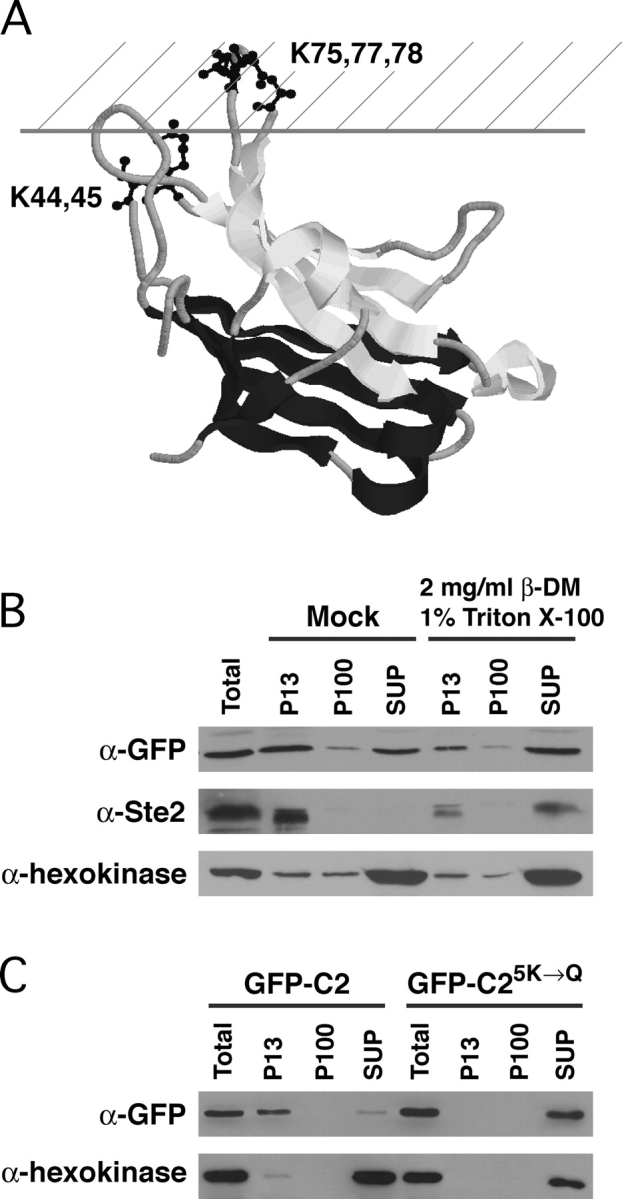
The Rsp5 C2 domain is a membrane-targeting module. (A) Modeled structure of the Rsp5 C2 domain. The locations of five lysines in the Rsp5 C2 domain were aligned with and modeled onto the structure of the protein kinase Cα C2 domain (Protein Data Bank no. 1DSY, http://www.rcsb.org/pdb/) shown in its predicted association with the cytoplasmic surface of a membrane. One loop of the C2 domain is thought to insert into the cytoplasmic leaflet of the lipid bilayer (represented by the hatched area) upon membrane binding (Kohout et al., 2003). (B and C) Subcellular fractionation of wild-type and 5K→Q mutant GFP-C2 chimeric proteins. Lysates from cells expressing GFP-C2 (LHY4488) or GFP-C25K→Q (LHY4489) were separated into 13,000 g pellet (P13), 100,000 g pellet (P100), and supernatant (SUP) fractions by differential centrifugation. (B) Lysate was diluted into buffer alone (Mock) or buffer containing 1% Triton X-100 and 2 mg/ml β-D-maltoside and incubated 30 min on ice before centrifugation. Cell extract equivalents of each fraction and an equivalent unfractionated sample (Total) were resolved by SDS-PAGE and analyzed by immunoblotting with GFP antibodies. Ste2 is an integral membrane protein that served as a control for the solubilization of membranes within the lysate. Hexokinase is a soluble protein that served as a control for the efficiency of cell lysis.
We introduced the 5K→Q mutations into full-length Rsp5 to test if the basic loops in the C2 domain contribute to the localization and function of the native protein. The mutant protein was expressed at a level comparable to wild-type Rsp5 and Rsp5C2Δ (Fig. 4 A). Furthermore, cells carrying the C2Δ or 5K→Q mutations grew at a rate indistinguishable from wild type on rich medium, indicating that the mutants fully support the essential functions of Rsp5 (unpublished data). The rsp5 5K →Q mutant was severely defective for sorting of GFP-CPS to the vacuole (Fig. 4 B), and a GFP-Rsp55K→Q fusion protein failed to localize efficiently to the perivacuolar endosome in vps4Δ cells (Fig. 4 C). Compared with GFP-Rsp5, GFP-Rsp55K→Q was localized to the perivacuolar endosome in fewer cells and with decreased intensity. These data imply that the failure of Rsp5 to interact with specific cellular membranes underlies the MVE sorting defect observed in rsp5 C2Δ and rsp5 5K →Q mutants.
Figure 4.
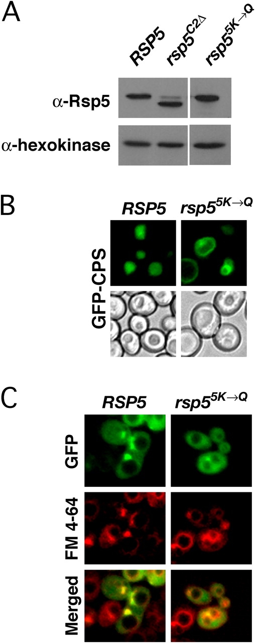
Disruption of basic residues in the C2 domain impairs Rsp5 localization to late endosomes and endosomal sorting of CPS. (A) Protein extract equivalents from rsp5Δ cells expressing Rsp5 (LHY2973), Rsp5C2Δ (LHY2974), and Rsp55K→Q (LHY3923) were analyzed by SDS-PAGE followed by immunoblotting with anti-Rsp5 antiserum. Immunoblotting with hexokinase antiserum was a control for equivalent protein loading in each lane. (B) The location of GFP-CPS in RSP5 (LHY2920) and rsp5 5K →Q (LHY3876) cells was visualized by fluorescence and DIC microscopy. (C) Localization of GFP-Rsp5 (LHY4504) and GFP-Rsp55K→Q (LHY4506) in rsp5Δ vps4Δ cells was visualized by fluorescence microscopy. FM 4-64 marks the location of the exaggerated late endosomal compartment in these cells.
C2 domains from other proteins bind to phospholipids (Hurley and Misra, 2000), and the aforementioned observations suggested the possibility that the Rsp5 C2 domain might also interact directly with membrane phospholipids. To test this idea, we first examined the binding of a recombinant glutathione S-transferase–Rsp5 C2 domain fusion protein (GST-C2) to a commercially available immobilized array of common cellular phospholipids and PIs (“PIP strips”). In this experiment, GST-C2 bound specifically to phosphorylated phosphatidylinositols, whereas the GST-C25K→Q mutant that diminished membrane association in vivo did not bind detectably to any phospholipids (Fig. 5 A). Notably, GST-C2 did not interact with phosphatidylserine (Fig. 5, PS) and phosphatidic acid (Fig. 5, PA), indicating that the C2 domain does not interact with all negatively charged phospholipids. As expected, GST alone and GST fused to a fragment of Rsp5 carrying the WW domains (GST-3×WW) also did not bind to any phospholipids (unpublished data).
Figure 5.
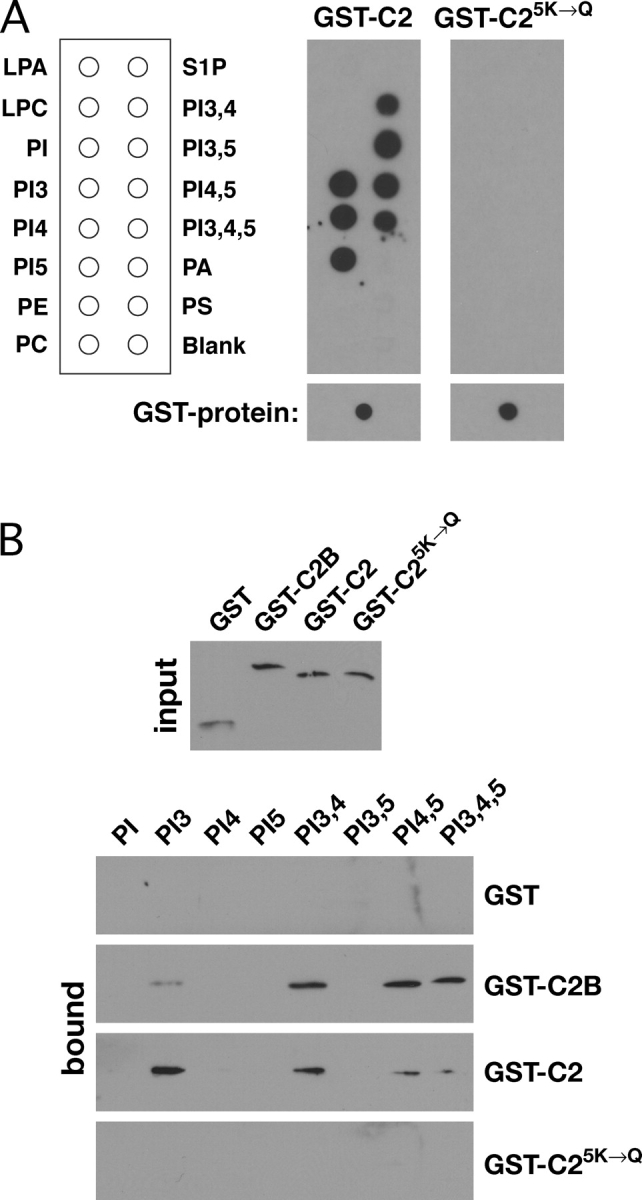
The Rsp5 C2 domain binds to PIs. (A) Equivalent amounts of each GST fusion protein were spotted onto nitrocellulose as a control to demonstrate equivalent levels of each protein in the incubation buffer (bottom). The indicated GST fusion proteins (100 ng/ml) were incubated with PIP strips for 2 h at 4°C. Bound proteins were detected by immunoblotting with anti-GST antibodies. (B) Equivalent amounts (50 ng) of each GST fusion protein (input, top) were incubated with phosphatidylcholine liposomes containing 5% (wt/wt) of the indicated PI. Liposomes were collected by centrifugation, and bound proteins were resolved by SDS-PAGE and detected by immunoblotting with anti-GST antibodies (bound, bottom panel).
To investigate whether or not the C2 domain binds selectively to PIs phosphorylated at specific positions on the inositol ring, we examined the sedimentation of GST-C2 with phosphatidylcholine liposomes reconstituted with individual PI variants (Fig. 5 B). Two fusion proteins served as controls for this experiment: GST fused to the C2B domain of synaptotagmin I (GST-C2B), a C2 domain known to bind PIs in a calcium-independent manner (Fukuda et al., 1994), and GST-C25K→Q. GST-C2 bound to liposomes containing several different PIs, indicating a lack of strong binding specificity in vitro. GST-C2B also bound to multiple PIs, which is consistent with previous observations (Fukuda et al., 1994; Schiavo et al., 1996). GST alone and GST-C25K→Q did not sediment with PI-containing liposomes, confirming that PI interaction is mediated by the C2 domain and requires the charged interaction surface that mediates membrane interactions in vivo.
Role of the C2 domain in cargo ubiquitination
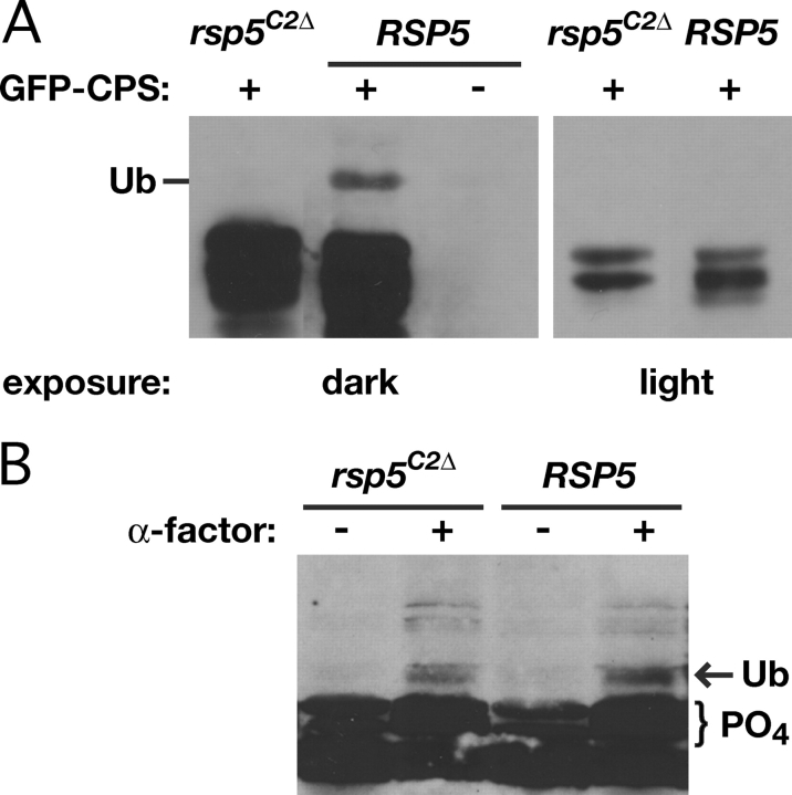
The Rsp5 C2 domain is required for MVE sorting of GFP-CPS but not of Ste2-GFP. Unlike CPS, Ste2 is sorted into transport vesicles by a ubiquitin signal appended at the plasma membrane before arriving at the MVE. This selective sorting requirement for the C2 domain suggested that the domain might promote ubiquitination of newly synthesized biosynthetic cargo. To test this hypothesis, we examined ubiquitination of CPS and Ste2 in RSP5 doa4Δ and rsp5 C2Δ doa4Δ mutant cells. GFP-CPS ubiquitination has been examined previously in cells lacking Doa4 (Katzmann et al., 2001), a protein that removes ubiquitin from cargo before entry into MVE vesicles (Dupré and Haguenauer-Tsapis, 2001), to facilitate the detection of ubiquitinated species. The monoubiquitinated form of GFP-CPS observed in RSP5 doa4Δ cells was severely diminished in rsp5 C2Δ doa4Δ double mutant cells (Fig. 6 A). Ste2 ubiquitination occurred normally in rsp5 C2Δ cells (Fig. 6 B), which is consistent with C2 domain-independent MVE sorting of endocytic cargo. The role of the C2 domain appears to be specific for cargo ubiquitination because the domain is not required for Rsp5-dependent monoubiquitination of Vps27, a trans-acting protein required for MVE cargo sorting (unpublished data).
Figure 6.
The Rsp5 C2 domain is required for CPS ubiquitination. (A) Ubiquitination of GFP-CPS was compared in lysates prepared from rsp5 C2Δ doa4Δ (LHY4088) and RSP5 doa4Δ (LHY4292) cells. A lysate prepared from RSP5 doa4Δ cells without GFP-CPS (LHY4281) was analyzed in parallel as a control. GFP-CPS was detected by immunoblotting with anti-GFP antibodies. Different exposures are shown for rsp5 C2Δ and RSP5 to adjust for minor variations in protein loading. GFP-CPS migrates as a doublet during SDS-PAGE. The monoubiquitinated (Ub) species of GFP-CPS is indicated. This GFP-CPS form was not observed in RSP5 doa4Δ cells carrying the GFP-CPSK8,12R mutant that lacks the lysine ubiquitination site in the CPS cytoplasmic tail (not depicted), confirming that it was the monoubiquitinated species. (B) Ste2 modifications before and after treatment with 10−6 M α-factor for 8 min were compared in rsp5 C2Δ (LHY1101) and RSP5 (LHY1103) cells by immunoblotting with Ste2 antibodies. Ligand-induced hyperphosphorylated (PO4) and monoubiquitinated (Ub) species of the receptor are indicated by the labeled bracket and arrow, respectively.
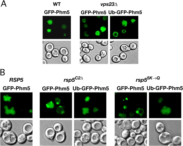
To directly address if the requirement for the C2 domain is explained by its role in biosynthetic cargo ubiquitination, we tested the sorting of an MVE cargo protein that carries an in-frame fusion of ubiquitin to its cytoplasmic domain. Like CPS, Phm5 is a resident vacuolar hydrolase that uses ubiquitin as a signal for entry into the MVE pathway (Reggiori and Pelham, 2001). A translationally fused ubiquitin moiety can direct the sorting of a Phm5 mutant lacking its posttranslational ubiquitination site (Ub-GFP-Phm5). Localization of Ub-GFP-Phm5 to the vacuole lumen required Vps23, a ubiquitin-binding component of the ESCRT-I sorting complex (Katzmann et al., 2001; Fig. 7 A), confirming that sorting of this fusion protein occurs by the canonical MVE sorting pathway. As expected, GFP-Phm5 was sorted to the vacuole lumen in RSP5 cells (Fig. 7 B). In the rsp5 C2Δ and rsp5 5K →Q mutants, GFP-Phm5 localized primarily to the perivacuolar late endosome. In contrast, Ub-GFP-Phm5 was sorted efficiently to the vacuole lumen in these mutants. These observations indicate that prior attachment of ubiquitin to a cargo protein circumvents the requirement for the C2 domain in MVE sorting.
Figure 7.
Translational fusion of ubiquitin to a biosynthetic cargo protein suppresses the requirement for the Rsp5 C2 domain in MVE sorting. Sorting of GFP-Phm5 and Ub-GFP-Phm5 in cells of the indicated genotype was analyzed by fluorescence and DIC microscopy. (A) GFP-Phm5 was expressed in wild-type cells (LHY4227). GFP-Phm5 and Ub-GFP-Phm5 were expressed in vps23Δ cells (LHY4583 and LHY4584). (B) GFP-Phm5 was expressed in RSP5 cells (LHY4492). GFP-Phm5 and Ub-GFP-Phm5 were expressed in rsp5 C2Δ (LHY3834 and LHY3835) and rsp5 5K →Q (LHY4490 and LHY4491) cells. The number of rings versus perivacuolar dots in which GFP-Phm5 accumulated in mutant cells varied from experiment to experiment.
Discussion
Ubiquitin signals the inclusion of transmembrane proteins into vesicles at multiple sites in the cell. A previous paper suggested that a Golgi-localized transmembrane ubiquitin ligase (Tul1) modifies biosynthetic cargo proteins en route to the vacuole before their sorting into the MVE pathway (Reggiori and Pelham, 2002). We do not observe a requirement for Tul1 in the endosomal sorting of two cargo proteins, CPS and Phm5 (Fig. S1, available at http://www.jcb.org/cgi/content/full/jcb.200309026/DC1), raising the possibility that an alternate E3 mediates ubiquitination of these substrates. In the present work, we show that the HECT domain ligase Rsp5, a Nedd4 homologue and central regulator of ubiquitin-dependent transport, is required for the ubiquitination and endosomal sorting of biosynthetic cargo proteins.
Our studies uncover a unique role for the Rsp5 C2 domain in MVE sorting. Subcellular fractionation and fluorescence localization experiments indicate that the C2 domain functions as a membrane-targeting module that facilitates the localization of Rsp5 to perivacuolar endosomes. Furthermore, we observe that the C2 domain is required for ubiquitination and sorting of cargo derived from the biosynthetic pathway, but not of cargo derived from the endocytic pathway, which acquire a ubiquitin sorting signal at the plasma membrane in a manner independent of the C2 domain. An in-frame fusion of ubiquitin to a cargo molecule suppresses the requirement for the C2 domain in its sorting, providing strong genetic evidence that ubiquitination of cargo is the functionally relevant consequence of C2 domain function in MVE sorting. Collectively, the data support a model in which the C2 domain recruits Rsp5 to endosomal membranes, thereby facilitating the ubiquitination of newly arrived cargo from the biosynthetic pathway. To our knowledge, biosynthetic MVE cargo proteins are the first known substrates of Nedd4 family E3s that require the C2 domain for their ubiquitination.
The C2 domain is sufficient for phospholipid interaction in vitro and membrane localization in vivo. Both of these activities are disrupted by mutation of a basic surface of the C2 domain formed by two lysine-rich loops. Consequently, C2 domain interaction with membranes can be explained at least partly by direct interaction with membrane lipids. However, we do not rule out the possibility that C2 domain–protein interactions also contribute to membrane targeting. Interaction with phospholipids in vitro occurs irrespective of the presence of calcium in the binding buffer (unpublished data), which is consistent with observations that the C2 domain does not bind calcium detectably and lacks two calcium-coordinating aspartate residues (Sehgal, 2002). Thus, the Rsp5 C2 domain belongs to the non-Ca2+–binding class of C2 domains that still bind membranes (Lee et al., 1999). Calcium binding by the Nedd4 C2 domain induces localization to the apical plasma membrane in polarized cells (Plant et al., 1997), suggesting that calcium interaction evolved to mediate specialized roles of Rsp5 homologues in different organisms or cell types.
Among an array of cellular phospholipids, the C2 domain interacts specifically with PIs, dynamic lipid second messengers that regulate membrane trafficking by recruiting effector proteins involved in cargo selection and vesicle budding (Odorizzi et al., 2000; Cockcroft and De Matteis, 2001). The C2 domain exhibits promiscuous interactions with multiple PI isoforms in vitro, a characteristic shared between several C2 and plekstrin homology domains (Chung et al., 1998; Kavran et al., 1998; Mehrotra et al., 2000; Catz et al., 2002). Although in most experiments we observed a modest preference for PI(3)P, we hypothesize that high specificity for the physiologically relevant PI variants requires contextual interactions between Rsp5 and other membrane-associated proteins in vivo.
Genetic studies have established a role for specific PIs in entry into the endocytic and MVE pathways (Odorizzi et al., 1998; Itoh et al., 2001). PI(3)P recruits multiple proteins important for MVE cargo sorting to the endosome, including the FYVE domain proteins Vps27/Hrs and the Fab1 PI kinase. Fab1 converts PI(3)P to PI(3,5)P2 and is important for sorting of biosynthetic cargo into MVE vesicles (Odorizzi et al., 1998). Fusion of ubiquitin to Phm5 rescues its missorting in a fab1Δ mutant (Dove et al., 2002; Reggiori and Pelham, 2002), and fab1Δ mutants sort the endocytic marker Ste2-GFP normally (Odorizzi et al., 1998), suggesting that the role of PI(3,5)P2, like that of the Rsp5 C2 domain, is to promote biosynthetic MVE cargo ubiquitination. However, we do not observe a defect in biosynthetic cargo ubiquitination in fab1Δ cells or in cells lacking the PI 3-kinase Vps34 (unpublished data), suggesting that PI(3)P and PI(3,5)P2 are not essential for cargo ubiquitination. Although these observations are difficult to reconcile, one possible explanation is that conversion of PI(3)P to PI(3,5)P2 is necessary to trigger the release of ubiquitinated cargo from an Rsp5 ubiquitin ligase complex, but is unnecessary when ubiquitination of cargo is deregulated, as in the case of cargo–ubiquitin fusions. Additionally or alternatively, cargo ubiquitination may occur in a location that is unproductive in fab1Δ and vps34Δ mutant cells either by Rsp5 or another ubiquitin ligase.
How might Rsp5 recognize endosomal cargo proteins? The WW domains, which are also important for MVE sorting, may contribute indirectly to substrate recognition but probably do not bind cargo proteins directly because CPS and Phm5 lack PPXY motifs, the Rsp5-type WW domain ligand (Chang et al., 2000). Instead, these domains may promote assembly of a multicomponent Rsp5 complex at the endosome, or they may function independently to target trans-acting factors of the MVE sorting machinery (e.g., Vps27) for ubiquitination. Intriguingly, Nedd4 can ubiquitinate a soluble protein in the absence of any detectable physical interaction (Polo et al., 2002), indicating that productive E3–substrate interactions can be remarkably transient or weak. Moreover, for some cargo, features of the transmembrane domain are critical for inclusion into the MVE pathway (Reggiori et al., 2000). An attractive hypothesis emerging from these observations is that Rsp5 targets transmembrane substrates without specific sequence recognition, instead relying heavily on colocalization of enzyme and substrate in small membrane microdomains. We suggest that precise localization mediated by the C2 domain may be a key component of endosomal cargo recognition.
In sum, our results underscore the widespread role of Nedd4 family proteins in the regulation of ubiquitin-dependent sorting and the diversity of substrate-targeting mechanisms used by these E3s. Some of the basic residues important for endosome binding by the Rsp5 C2 domain are conserved in its metazoan homologues, and Nedd4 family E3s have been genetically linked to the MVE sorting machinery in the budding of enveloped viruses from infected cells (Garrus et al., 2001). Therefore, we speculate that the involvement of Rsp5 and its C2 domain in MVE sorting reflects an evolutionarily conserved cellular role for Nedd4 family proteins.
Materials and methods
Strains, plasmids, and reagents
Strains used in this work are listed in Table I . All single deletion strains (except rsp5Δ) were obtained from the EUROSCARF consortium (www.uni-frankfurt.de/fb15/mikro/euroscarf). LHY1101 (rsp5 C2Δ) and LHY1103 (RSP5) have been described previously (Dunn and Hicke, 2001). Other rsp5Δ strains were constructed by sporulation of an rsp5Δ/RSP5 diploid and dissection on medium containing oleic acid (Hoppe et al., 2000). For functional analyses, rsp5Δ cells were transformed with the indicated plasmids, and a representative transformant is shown. The doa4Δ rsp5Δ and vps4Δ rsp5Δ strains were made by sporulation of heterozygous diploids carrying a pRSP5[URA3] plasmid for survival of rsp5Δ haploid progeny. Progeny carrying doa4Δ rsp5Δ or vps4Δ rsp5Δ double disruptions were selected. Mutants were transformed with the specified mutant or wild-type RSP5[TRP1] plasmid, and the pRSP5[URA3] plasmid was eliminated by growth on 5-fluoroorotic acid.
Table I. Yeast strains.
| Strain | Genotypea |
|---|---|
| LHY1101 | pHA-rsp5 C2Δ [TRP1] rsp5Δ::HIS3 trp1 leu2 ura3 lys2 ade2 bar1 or bar1Δ::HIS3 |
| LHY1103 | pHA-RSP5[TRP1] rsp5Δ::HIS3 trp1 leu2 ura3 lys2 ade2 bar1 or bar1Δ::HIS3 |
| LHY2920 | pNotI-RSP5[TRP1] pGFP-CPS[URA3] rsp5Δ::HIS3 his3 leu2 ura3 trp1 bar1 |
| LHY2922 | prsp5 C2Δ [TRP1] pGFP-CPS[URA3] rsp5Δ::HIS3 his3 leu2 ura3 trp1 bar1 |
| LHY2973 | pNotI-RSP5[TRP1] rsp5Δ::HIS3 his3 leu2 ura3 trp1 bar1 |
| LHY2974 | prsp5 C2Δ [TRP1] rsp5Δ::HIS3 his3 leu2 ura3 trp1 bar1 |
| LHY3454 | pGFP-CPS[URA3] ura3Δ leu2Δ his3Δ met15Δ |
| LHY3834 | prsp5 C2Δ [TRP1] pGFP-PHM5[URA3] rsp5Δ::HIS3 his3 leu2 ura3 trp1 bar1 |
| LHY3835 | prsp5 C2Δ [TRP1] pUB-GFP-PHM5[URA3] rsp5Δ::HIS3 his3 leu2 ura3 trp1 bar1 |
| LHY3876 | prsp5 5K→Q [TRP1] pGFP-CPS[URA3] rsp5Δ::HIS3 his3 leu2 ura3 trp1 bar1 |
| LHY3923 | prsp5 5K→Q [TRP1] rsp5Δ::HIS3 his3 leu2 ura3 trp1 bar1 |
| LHY3924 | pNotI-RSP5[TRP1] ura3::STE2-GFP::URA3 rsp5Δ::HIS3 his3 leu2 ura3 trp1 bar1 |
| LHY3925 | prsp5 C2Δ [TRP1] ura3::STE2-GFP::URA3 rsp5Δ::HIS3 his3 leu2 ura3 trp1 bar1 |
| LHY4012 | pGFP-RSP5[URA3] vps4Δ::kanMX4 ura3Δ leu2Δ his3Δ met15Δ |
| LHY4013 | pGFP-RSP5[URA3] ura3Δ leu2Δ his3Δ met15Δ |
| LHY4014 | pGFP-RSP5[URA3] vrp1Δ::kanMX4 ura3Δ leu2Δ his3Δ met15Δ |
| LHY4088 | prsp5 C2Δ [TRP1] pGFP-CPS[URA3] MATα doa4Δ::LEU2 rsp5Δ::HIS3 his3 trp1 ade2 ura3 leu2 lys2 bar1 |
| LHY4227 | pGFP-PHM5[URA3] ura3Δ leu2Δ his3Δ met15Δ |
| LHY4229 | pGFP-PHM5[URA3] tul1Δ::kanMX4 ura3Δ leu2Δ his3Δ met15Δ |
| LHY4281 | pRSP5[TRP1] MATα doa4Δ::LEU2 rsp5Δ::HIS3 his3 trp1 ade2 ura3 leu2 lys2 bar1 |
| LHY4291 | pGFP-CPS[URA3] tul1Δ::kanMX4 ura3Δ leu2Δ his3Δ met15Δ |
| LHY4292 | pRSP5[TRP1] pGFP-CPS[URA3] MATα doa4Δ::LEU2 rsp5Δ::HIS3 his3 trp1 ade2 ura3 leu2 lys2 bar1 |
| LHY4429 | pRSP5[TRP1] pGFP-CPS[URA3] rsp5Δ::HIS3 his3 leu2 ura3 trp1 bar1 |
| LHY4430 | pRSP5 ww1AxxP [TRP1] pGFP-CPS[URA3] rsp5Δ::HIS3 his3 leu2 ura3 trp1 bar1 |
| LHY4431 | pRSP5 ww2AxxP [TRP1] pGFP-CPS[URA3] rsp5Δ::HIS3 his3 leu2 ura3 trp1 bar1 |
| LHY4432 | pRSP5 ww3AxxP [TRP1] pGFP-CPS[URA3] rsp5Δ::HIS3 his3 leu2 ura3 trp1 bar1 |
| LHY4433 | pRSP5 ww1AxxPww2AxxPww3AxxP [TRP1] pGFP-CPS[URA3] rsp5Δ::HIS3 his3 leu2 ura3 trp1 bar1 |
| LHY4488 | pGFP-C2[TRP1] his3 trp1 lys2 ura3 leu2 bar1 |
| LHY4489 | pGFP-C2 5K→Q [TRP1] his3 trp1 lys2 ura3 leu2 bar1 |
| LHY4490 | prsp5 5K→Q [TRP1] pGFP-PHM5[URA3] rsp5Δ::HIS3 his3 leu2 ura3 trp1 bar1 |
| LHY4491 | prsp5 5K→Q [TRP1] pUB-GFP-PHM5[URA3] rsp5Δ::HIS3 his3 leu2 ura3 trp1 bar1 |
| LHY4492 | pNotI-RSP5[TRP1] pGFP-PHM5[URA3] rsp5Δ::HIS3 his3 leu2 ura3 trp1 bar1 |
| LHY4504 | pGFP-RSP5[TRP1] MATα vps4Δ::kanMX4 rsp5Δ::HIS3 his3 leu2 ura3 trp1 |
| LHY4506 | pGFP-RSP5 5K→Q [TRP1] MATα vps4Δ::kanMX4 rsp5Δ::HIS3 his3 leu2 ura3 trp1 |
| LHY4583 | pGFP-PHM5[URA3] vps23Δ::kanMX4 ura3Δ leu2Δ his3Δ met15Δ |
| LHY4584 | pUB-GFP-PHM5[URA3] vps23Δ::kanMX4 ura3Δ leu2Δ his3Δ met15Δ |
All strains are MATa unless otherwise indicated.
Yeast strains were propagated in rich (YPUAD) medium or selective (YNB) medium (Sherman, 1991). To prepare oleic acid medium, 2 mM oleic acid and 0.2% NP-40 were added to YPUAD medium after autoclaving. Anti-Ste2 antibodies were purified from rabbit antiserum as described previously (Hicke and Riezman, 1996). Rsp5 rabbit antiserum was produced with recombinant Rsp5 purified from Escherichia coli and cleaved from a GST affinity tag by PreScission protease (Amersham Biosciences). Anti-GFP antibodies were purchased from Roche Applied Science, and anti-GST antibodies were purchased from Amersham Biosciences. Anti-hexokinase antiserum was a gift from G. Schatz (University of Basel, Basel, Switzerland).
Construction of GST-C2 (encoding Rsp5 amino acids 1–142; LHP1663), rsp5 C2Δ (LHP510), rsp5 ww1 (LHP730), rsp5 ww2 (LHP845), rsp5 ww3 (LHP846), rsp5 ww1,2,3 (LHP952), and STE2-GFP (LHP1921) plasmids has been described previously (Sehgal et al., 2000; Dunn and Hicke, 2001; Shih et al., 2002). pGST-C2B was a gift from H. Godwin (Northwestern University, Evanston, IL). pGFP-CPS was provided by S. Emr (University of California, San Diego, San Diego, CA; Odorizzi et al., 1998), and GFP-PHM5 and UB-GFP-PHM5 plasmids were a gift from H. Pelham (Medical Research Council Laboratory of Molecular Biology, Cambridge, England; Reggiori and Pelham, 2001).
The GFP-RSP5 chimera was constructed by ligating a NotI–GFP fragment, generated by PCR amplification of a plasmid encoding GFP (BD Biosciences) with oligonucleotides containing flanking NotI sites, into pNotI-RSP5 (LHP477). The resulting insert was released by SacI and XhoI digestion and subcloned into SacI- and XhoI-digested pRS424 to generate pGFP-RSP5 (LHP1466). The fusion protein complements the lethality of an rsp5Δ mutation. pGFP-C2 (LHP1527) was made by introducing into pGFP-RSP5 two in-frame stop codons after the Ser140 codon of RSP5 by the Quikchange method (Stratagene). The GST-3×WW plasmid (LHP703) was constructed by subcloning all three WW domains of Rsp5, generated by PCR amplification of RSP5 with oligonucleotides containing nontemplated EcoRI (5′) or XhoI (3′) sites, into EcoRI- and XhoI-digested pGEX-PKT (Sehgal et al., 2000). The 5K→Q mutations (K44Q, K45Q, K75Q, K77Q, and K78Q) were introduced by sequential site-directed mutagenesis of pGST-C2, pNotI-RSP5, pGFP-RSP5, and pGFP-C2 to generate pGST-C2 5K →Q (LHP1709), pRSP5 5K →Q (LHP1710), pGFP-RSP5 5K →Q (LHP1711), and pGFP-C2 5K →Q (LHP1713), respectively. In all cases, fusion proteins and mutants exhibited the expected mobility by SDS-PAGE.
Fluorescence microscopy
Cells were grown at 24 or 30°C to logarithmic phase, harvested by centrifugation, and washed in 1 ml of ice-cold PBS, pH 7.5, containing 10–20 mM NaN3. Cells were mounted on a slide by embedding in 1.67% low-melt agarose and viewed with either a confocal microscope (model DMRXE7; Leica) or a deconvolution microscope (model DMIRE2; Leica). Labeling with FM 4-64 (Molecular Probes) was performed as described previously (Vida and Emr, 1995) with a 20-min chase.
Lipid binding assays
Recombinant proteins were expressed in BL21-Codon Plus E. coli (Stratagene) propagated in LB medium supplemented with 100 μg/ml ampicillin and 20 μg/ml chloramphenicol for plasmid maintenance. Induction and purification of GST fusion proteins was performed essentially as described previously (Dunn and Hicke, 2001). GST-3×WW expression was induced with 0.2 mM IPTG at 17–21°C, and the expression of GST, GST-C2B, GST-C2, and GST-C25K→Q was induced with 0.2–0.25 mM IPTG at 24 or 37°C. The concentration of recombinant proteins was determined by quantitative Coomassie blue staining with protein standard controls.
Membrane-immobilized lipids (PIP-Strips) were purchased from Echelon Research Laboratories. Each membrane was incubated with blocking buffer (3% fatty acid-free BSA, 0.1% [vol/vol] Tween-20, and TBS, pH 7.5) for 1 h at RT. 100 ng/ml of purified GST fusion proteins were added in blocking buffer and incubated for 2 h at 4°C. Bound proteins were detected by immunoblotting with anti-GST antibodies.
Liposomes were prepared by mixing phosphatidylcholine (Avanti Polar Lipids, Inc.) with 5% (wt/wt) of the indicated PIs (Echelon Research Laboratories). Lipids were dried by a flow of argon, kept under vacuum for 10 min, resuspended to 400 μg lipids/ml with binding buffer (50 mM phosphate buffer and 150 mM NaCl, pH 8.0), and sonicated for 10 min to create a homogeneous suspension. Liposomes were added to siliconized microcentrifuge tubes (Fisher Scientific) with binding buffer containing 0.2% fatty acid-free BSA (0.1% final concentration) to a final concentration of 200 μg lipids/ml. 50 μg of purified GST fusion proteins were added and incubated at RT for 20 min. Liposomes were recovered by centrifugation at 8,000 g for 20 min and washed once with binding buffer containing 0.1% BSA. Bound proteins were eluted with Laemmli sample buffer and detected by SDS-PAGE followed by immunoblotting with anti-GST antibodies.
Cell lysates, fractionation, and immunoblotting
To analyze Rsp5 expression levels, whole cell extracts were prepared by alkaline lysis and TCA precipitation as described previously (Horvath and Riezman, 1994). Lysates used for analysis of Ste2 modifications were prepared by glass bead lysis and urea/SDS extraction as described previously (Hicke and Riezman, 1996; Dunn and Hicke, 2001). For detection of CPS ubiquitination, cells were lysed by vortexing with glass beads in native IP buffer (0.2 M sorbitol, 50 mM KOAc, 25 mM KCl, 10 mM Hepes, and 1 mM EDTA, pH 7.0) containing protease inhibitors at 1-min intervals until >90% lysis was achieved. Lysates were extracted with 2 mg/ml n-dodecyl β-D-maltoside for 30 min on ice and clarified by centrifugation. Subcellular fractionation and detergent treatment were performed as described previously (Pryer et al., 1993; Dunn and Hicke, 2001).
Immunoblotting was performed as described previously (Dunn and Hicke, 2001). Secondary antibodies were HRP-conjugated anti–mouse (Pierce Chemical Co.) or HRP-conjugated anti–rabbit (Sigma-Aldrich).
Online supplemental material
Fig. S1 depicts the localization of GFP-CPS and GFP-Phm5 in congenic wild-type and tul1Δ cells as determined by fluorescence microscopy. Online supplemental material is available at http://www.jcb.org/cgi/content/full/jcb.200309026/DC1.
Acknowledgments
We are grateful to Scott Emr, Hilary Godwin, Hugh Pelham, and Jeff Schatz for providing reagents and to Robert Lamb for use of the confocal microscope. We thank Ben Sehgal and Hilary Godwin for helpful discussions.
This work was supported by the National Institutes of Health (grant DK61299).
R. Dunn, D.A. Klos, and A.S. Adler contributed equally to this paper.
The online version of this article includes supplemental material.
R. Dunn's present address is Dept. of Molecular Biology, Massachusetts General Hospital, Boston, MA 02114.
Abbreviations used in this paper: DIC, differential interference contrast; MVE, multivesicular endosome; PI, phosphoinositide.
References
- Babst, M., B. Wendland, E.J. Estepa, and S.D. Emr. 1998. The Vps4p AAA ATPase regulates membrane association of a Vps protein complex required for normal endosome function. EMBO J. 17:2982–2993. [DOI] [PMC free article] [PubMed] [Google Scholar]
- Babst, M., G. Odorizzi, E.J. Estepa, and S.D. Emr. 2000. Mammalian tumor susceptibility gene 101 (TSG101) and the yeast homologue, Vps23p, both function in late endosomal trafficking. Traffic. 1:248–258. [DOI] [PubMed] [Google Scholar]
- Babst, M., D.J. Katzmann, E.J. Estepa-Sabal, T. Meerloo, and S.D. Emr. 2002. a. ESCRT-III: an endosome-associated heterooligomeric protein complex required for MVB sorting. Dev. Cell. 3:271–282. [DOI] [PubMed] [Google Scholar]
- Babst, M., D.J. Katzmann, W.B. Snyder, B. Wendland, and S.D. Emr. 2002. b. Endosome-associated complex, ESCRT-II, recruits transport machinery for protein sorting at the multivesicular body. Dev. Cell. 3:283–289. [DOI] [PubMed] [Google Scholar]
- Beaudenon, S.L., M.R. Huacani, G. Wang, D.P. McDonnell, and J.M. Huibregtse. 1999. Rsp5 ubiquitin-protein ligase mediates DNA damage-induced degradation of the large subunit of RNA polymerase II in Saccharomyces cerevisiae. Mol. Cell. Biol. 19:6972–6979. [DOI] [PMC free article] [PubMed] [Google Scholar]
- Bishop, N., and P. Woodman. 2001. TSG101/mammalian VPS23 and mammalian VPS28 interact directly and are recruited to VPS4-induced endosomes. J. Biol. Chem. 276:11735–11742. [DOI] [PubMed] [Google Scholar]
- Catz, S.D., J.L. Johnson, and B.M. Babior. 2002. The C2A domain of JFC1 binds to 3′-phosphorylated phosphoinositides and directs plasma membrane association in living cells. Proc. Natl. Acad. Sci. USA. 99:11652–11657. [DOI] [PMC free article] [PubMed] [Google Scholar]
- Chang, A., S. Cheang, X. Espanel, and M. Sudol. 2000. Rsp5 WW domains interact directly with the carboxyl-terminal domain of RNA polymerase II. J. Biol. Chem. 275:20562–20571. [DOI] [PubMed] [Google Scholar]
- Cho, W. 2001. Membrane targeting by C1 and C2 domains. J. Biol. Chem. 276:32407–32410. [DOI] [PubMed] [Google Scholar]
- Chung, S.H., W.J. Song, K. Kim, J.J. Bednarski, J. Chen, G.D. Prestwich, and R.W. Holz. 1998. The C2 domains of Rabphilin3A specifically bind phosphatidylinositol 4,5-bisphosphate containing vesicles in a Ca2+-dependent manner. In vitro characteristics and possible significance. J. Biol. Chem. 273:10240–10248. [DOI] [PubMed] [Google Scholar]
- Cockcroft, S., and M.A. De Matteis. 2001. Inositol lipids as spatial regulators of membrane traffic. J. Membr. Biol. 180:187–194. [DOI] [PubMed] [Google Scholar]
- Dove, S.K., R.K. McEwen, A. Mayes, D.C. Hughes, J.D. Beggs, and R.H. Michell. 2002. Vac14 controls PtdIns(3,5)P(2) synthesis and Fab1-dependent protein trafficking to the multivesicular body. Curr. Biol. 12:885–893. [DOI] [PubMed] [Google Scholar]
- Dunn, R., and L. Hicke. 2001. Domains of the Rsp5 ubiquitin protein ligase required for receptor-mediated and fluid-phase endocytosis. Mol. Biol. Cell. 12:421–435. [DOI] [PMC free article] [PubMed] [Google Scholar]
- Dupré, S., and R. Haguenauer-Tsapis. 2001. Deubiquitination step in the endocytic pathway of yeast plasma membrane proteins: crucial role of Doa4p ubiquitin isopeptidase. Mol. Cell. Biol. 21:4482–4494. [DOI] [PMC free article] [PubMed] [Google Scholar]
- Fukuda, M., J. Aruga, M. Niinobe, S. Aimoto, and K. Mikoshiba. 1994. Inositol-1,3,4,5-tetrakisphosphate binding to C2B domain of IP4BP/synaptotagmin II. J. Biol. Chem. 269:29206–29211. [PubMed] [Google Scholar]
- Futter, C.E., L.M. Collinson, J.M. Backer, and C.R. Hopkins. 2001. Human VPS34 is required for internal vesicle formation within multivesicular endosomes. J. Cell Biol. 155:1251–1264. [DOI] [PMC free article] [PubMed] [Google Scholar]
- Galan, J.M., V. Moreau, B. André, C. Volland, and R. Haguenauer-Tsapis. 1996. Ubiquitination mediated by the Npi1p/Rsp5p ubiquitin-protein ligase is required for endocytosis of the yeast uracil permease. J. Biol. Chem. 271:10946–10952. [DOI] [PubMed] [Google Scholar]
- Garrus, J.E., U.K. von Schwedler, O.W. Pornillos, S.G. Morham, K.H. Zavitz, H.E. Wang, D.A. Wettstein, K.M. Stray, M. Cote, R.L. Rich, et al. 2001. Tsg101 and the vacuolar protein sorting pathway are essential for HIV-1 budding. Cell. 107:55–65. [DOI] [PubMed] [Google Scholar]
- Helliwell, S.B., S. Losko, and C.A. Kaiser. 2001. Components of a ubiquitin ligase complex specify polyubiquitination and intracellular trafficking of the general amino acid permease. J. Cell Biol. 153:649–662. [DOI] [PMC free article] [PubMed] [Google Scholar]
- Hershko, A., A. Ciechanover, and A. Varshavsky. 2000. The ubiquitin system. Nat. Med. 6:1073–1081. [DOI] [PubMed] [Google Scholar]
- Hicke, L., and H. Riezman. 1996. Ubiquitination of a yeast plasma membrane receptor signals its ligand-stimulated endocytosis. Cell. 84:277–287. [DOI] [PubMed] [Google Scholar]
- Hicke, L., and R. Dunn. 2003. Regulation of membrane protein transport by ubiquitin and ubiquitin-binding proteins. Ann. Rev. Cell Dev. Biol. 19:141–172. [DOI] [PubMed] [Google Scholar]
- Hoppe, T., K. Matuschewski, M. Rape, S. Schlenker, H.D. Ulrich, and S. Jentsch. 2000. Activation of a membrane-bound transcription factor by regulated ubiquitin/proteasome-dependent processing. Cell. 102:577–586. [DOI] [PubMed] [Google Scholar]
- Horvath, A., and H. Riezman. 1994. Rapid protein extraction from Saccharomyces cerevisiae. Yeast. 10:1305–1310. [DOI] [PubMed] [Google Scholar]
- Hurley, J.H., and S. Misra. 2000. Signaling and subcellular targeting by membrane-binding domains. Annu. Rev. Biophys. Biomol. Struct. 29:49–79. [DOI] [PMC free article] [PubMed] [Google Scholar]
- Itoh, T., S. Koshiba, T. Kigawa, A. Kikuchi, S. Yokoyama, and T. Takenawa. 2001. Role of the ENTH domain in phosphatidylinositol-4,5-bisphosphate binding and endocytosis. Science. 291:1047–1051. [DOI] [PubMed] [Google Scholar]
- Katzmann, D.J., M. Babst, and S.D. Emr. 2001. Ubiquitin-dependent sorting into the multivesicular body pathway requires the function of a conserved endosomal sorting complex, ESCRT-I. Cell. 106:145–155. [DOI] [PubMed] [Google Scholar]
- Katzmann, D.J., G. Odorizzi, and S.D. Emr. 2002. Receptor downregulation and multivesicular-body sorting. Nat. Rev. Mol. Cell Biol. 3:893–905. [DOI] [PubMed] [Google Scholar]
- Kavran, J.M., D.E. Klein, A. Lee, M. Falasca, S.J. Isakoff, E.Y. Skolnik, and M.A. Lemmon. 1998. Specificity and promiscuity in phosphoinositide binding by pleckstrin homology domains. J. Biol. Chem. 273:30497–30508. [DOI] [PubMed] [Google Scholar]
- Keleman, K., S. Rajagopalan, D. Cleppien, D. Teis, K. Paiha, L.A. Huber, G.M. Technau, and B.J. Dickson. 2002. Comm sorts Robo to control axon guidance at the Drosophila midline. Cell. 110:415–427. [DOI] [PubMed] [Google Scholar]
- Kohout, S.C., S. Corbalan-Garcia, J.C. Gomez-Fernandez, and J.J. Falke. 2003. C2 domain of protein kinase Cα: elucidation of the membrane docking surface by site-directed fluorescence and spin labeling. Biochemistry. 42:1254–1265. [DOI] [PMC free article] [PubMed] [Google Scholar]
- Lee, J.O., H. Yang, M.M. Georgescu, A. Di Cristofano, T. Maehama, Y. Shi, J.E. Dixon, P. Pandolfi, and N.P. Pavletich. 1999. Crystal structure of the PTEN tumor suppressor: implications for its phosphoinositide phosphatase activity and membrane association. Cell. 99:323–334. [DOI] [PubMed] [Google Scholar]
- Mehrotra, B., D.G. Myszka, and G.D. Prestwich. 2000. Binding kinetics and ligand specificity for the interactions of the C2B domain of synaptogmin II with inositol polyphosphates and phosphoinositides. Biochemistry. 39:9679–9686. [DOI] [PubMed] [Google Scholar]
- Myat, A., P. Henry, V. McCabe, L. Flintoft, D. Rotin, and G. Tear. 2002. Drosophila Nedd4, a ubiquitin ligase, is recruited by Commissureless to control cell surface levels of the roundabout receptor. Neuron. 35:447–459. [DOI] [PubMed] [Google Scholar]
- Nalefski, E.A., and J.J. Falke. 1996. The C2 domain calcium-binding motif: structural and functional diversity. Protein Sci. 5:2375–2390. [DOI] [PMC free article] [PubMed] [Google Scholar]
- Odorizzi, G., M. Babst, and S.D. Emr. 1998. Fab1p PtdIns(3)P 5-kinase function essential for protein sorting in the multivesicular body. Cell. 95:847–858. [DOI] [PubMed] [Google Scholar]
- Odorizzi, G., M. Babst, and S.D. Emr. 2000. Phosphoinositide signaling and the regulation of membrane trafficking in yeast. Trends Biochem. Sci. 25:229–235. [DOI] [PubMed] [Google Scholar]
- Pickart, C.M. 2001. Mechanisms underlying ubiquitination. Annu. Rev. Biochem. 70:503–533. [DOI] [PubMed] [Google Scholar]
- Plant, P.J., H. Yeger, O. Staub, P. Howard, and D. Rotin. 1997. The C2 domain of the ubiquitin protein ligase Nedd4 mediates Ca2+-dependent plasma membrane localization. J. Biol. Chem. 272:32329–32336. [DOI] [PubMed] [Google Scholar]
- Polo, S., S. Sigismund, M. Faretta, M. Guidi, M.R. Capua, G. Bossi, H. Chen, P. De Camilli, and P.P. Di Fiore. 2002. A single motif responsible for ubiquitin recognition and monoubiquitination in endocytic proteins. Nature. 416:451–455. [DOI] [PubMed] [Google Scholar]
- Pryer, N.K., N.R. Salama, R. Schekman, and C.A. Kaiser. 1993. Cytosolic Sec13p complex is required for vesicle formation from the endoplasmic reticulum in vitro. J. Cell Biol. 120:865–875. [DOI] [PMC free article] [PubMed] [Google Scholar]
- Raiborg, C., T.E. Rusten, and H. Stenmark. 2003. Protein sorting into multivesicular endosomes. Curr. Opin. Cell Biol. 15:446–455. [DOI] [PubMed] [Google Scholar]
- Raymond, C.K., I. Howald-Stevenson, C.A. Vaters, and T.H. Stevens. 1992. Morphological classification of the yeast vacuolar protein sorting mutants: evidence for a prevacuolar compartment in class E vps mutants. Mol. Biol. Cell. 3:1389–1402. [DOI] [PMC free article] [PubMed] [Google Scholar]
- Reggiori, F., and H.R. Pelham. 2001. Sorting of proteins into multivesicular bodies: ubiquitin-dependent and -independent targeting. EMBO J. 20:5176–5186. [DOI] [PMC free article] [PubMed] [Google Scholar]
- Reggiori, F., and H.R. Pelham. 2002. A transmembrane ubiquitin ligase required to sort membrane proteins into multivesicular bodies. Nat. Cell Biol. 4:117–123. [DOI] [PubMed] [Google Scholar]
- Reggiori, F., M.W. Black, and H.R. Pelham. 2000. Polar transmembrane domains target proteins to the interior of the yeast vacuole. Mol. Biol. Cell. 11:3737–3749. [DOI] [PMC free article] [PubMed] [Google Scholar]
- Rotin, D., O. Staub, and R. Haguenauer-Tsapis. 2000. Ubiquitination and endocytosis of plasma membrane proteins: role of Nedd4/Rsp5p family of ubiquitin-protein ligases. J. Membr. Biol. 176:1–17. [DOI] [PubMed] [Google Scholar]
- Schiavo, G., Q.M. Gu, G.D. Prestwich, T.H. Sollner, and J.E. Rothman. 1996. Calcium-dependent switching of the specificity of phosphoinositide binding to synaptotagmin. Proc. Natl. Acad. Sci. USA. 93:13327–13332. [DOI] [PMC free article] [PubMed] [Google Scholar]
- Sehgal, B.U. 2002. Interactions of EF-hand and C2 proteins with calcium and lead. Ph.D. thesis. Northwestern University, Evanston, IL. 129 pp.
- Sehgal, B.U., R. Dunn, L. Hicke, and H.A. Godwin. 2000. High-yield expression and purification of recombinant proteins in bacteria: a versatile vector for glutathione S-transferase fusion proteins containing two protease cleavage sites. Anal. Biochem. 281:232–234. [DOI] [PubMed] [Google Scholar]
- Sherman, F. 1991. Getting started with yeast. Methods Enzymol. 194:3–21. [DOI] [PubMed] [Google Scholar]
- Shih, S.C., K.J. Katzmann, J.D. Schnell, M. Sutanto, S.C. Emr, and L.H. Hicke. 2002. Epsins and Vps27/Hrs contain ubiquitin-binding domains that function in receptor endocytosis. Nat. Cell Biol. 4:389–393. [DOI] [PubMed] [Google Scholar]
- Soetens, O., J.O. De Craene, and B. Andre. 2001. Ubiquitin is required for sorting to the vacuole of the yeast general amino acid permease, Gap1. J. Biol. Chem. 276:43949–43957. [DOI] [PubMed] [Google Scholar]
- Urbanowski, J., and R.C. Piper. 2001. Ubiquitin sorts proteins into the intralumenal degradative compartment of the late endosome/vacuole. Traffic. 2:623–631. [DOI] [PubMed] [Google Scholar]
- Vida, T.A., and S.D. Emr. 1995. A new vital stain for visualizing vacuolar membrane dynamics and endocytosis in yeast. J. Biol. Chem. 128:779–792. [DOI] [PMC free article] [PubMed] [Google Scholar]
- Wang, G., M. McCaffrey, B. Wendland, S. Dupré, R. Haguenauer-Tsapis, and J. Huibregtse. 2001. Localization of the Rsp5p ubiquitin-protein ligase at multiple sites within the endocytic pathway. Mol. Cell. Biol. 21:3564–3575. [DOI] [PMC free article] [PubMed] [Google Scholar]


