Abstract
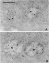
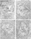
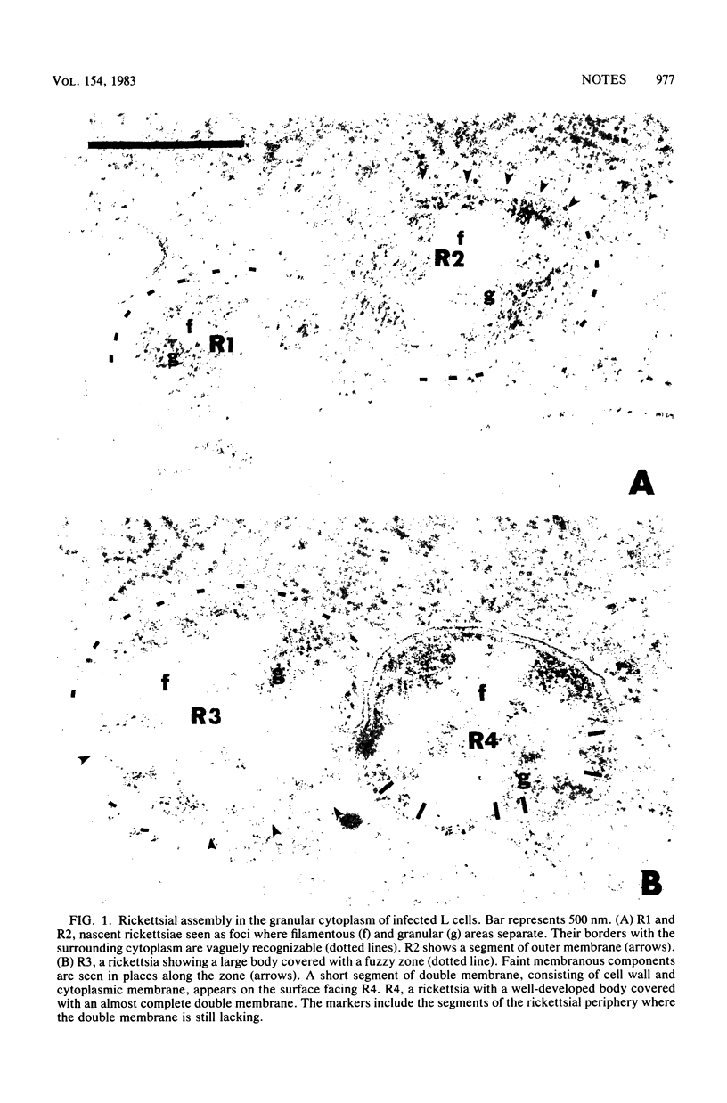
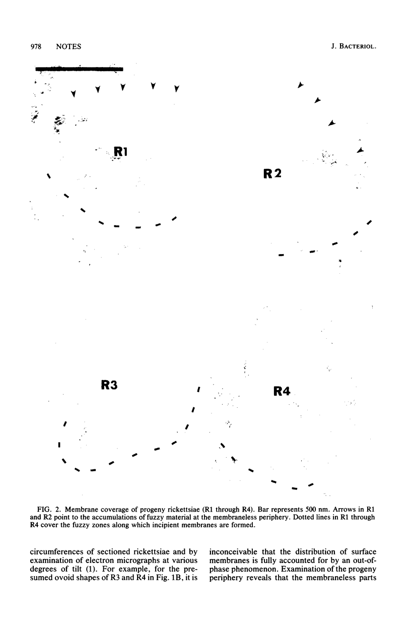
In the assembly of Rickettsia tsutsugamushi progeny in irradiated L cells, nascent forms first appear as undemarcated foci in the host cell granular cytoplasm, in which electron-lucent filamentous (f) and electron-dense granular (g) areas differentiate. Morphological observations indicated that the assembly involves formation of a filamentous network in the f area, manufacture of rickettsial ribosomes in the g area, and formation of mildly electron-dense fuzzy zones, along which a double membrane assembles.
Full text
PDF
Images in this article
Selected References
These references are in PubMed. This may not be the complete list of references from this article.
- Hase T. Growth pattern of Rickettsia tsutsugamushi in irradiated L cells. J Bacteriol. 1983 May;154(2):879–892. doi: 10.1128/jb.154.2.879-892.1983. [DOI] [PMC free article] [PubMed] [Google Scholar]
- Silverman D. J., Wisseman C. L., Jr Comparative ultrastructural study on the cell envelopes of Rickettsia prowazekii, Rickettsia rickettsii, and Rickettsia tsutsugamushi. Infect Immun. 1978 Sep;21(3):1020–1023. doi: 10.1128/iai.21.3.1020-1023.1978. [DOI] [PMC free article] [PubMed] [Google Scholar]
- Weiss E. Growth and physiology of rickettsiae. Bacteriol Rev. 1973 Sep;37(3):259–283. doi: 10.1128/br.37.3.259-283.1973. [DOI] [PMC free article] [PubMed] [Google Scholar]


