Abstract
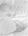
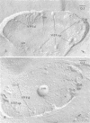
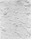
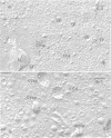
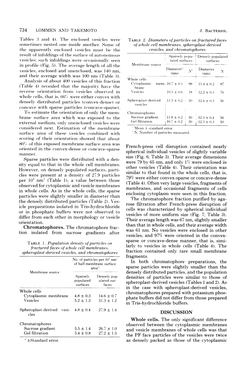
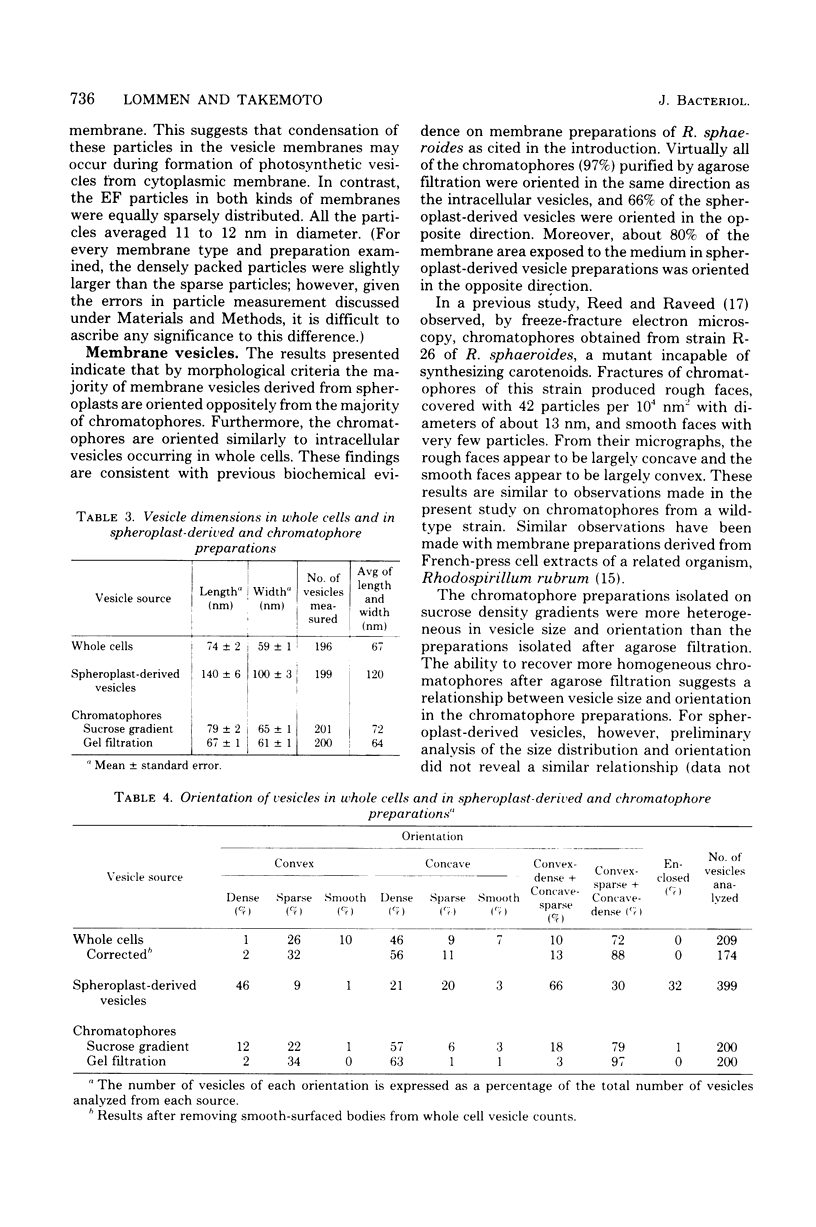
By using freeze-fracture electron microscopy, chromatophores and spheroplast-derived membrane vesicles from photosynthetically grown Rhodopseudomonas sphaeroides were compared with cytoplasmic membrane and intracellular vesicles of whole cells. In whole cells, the extracellular fracture faces of both cytoplasmic membrane and vesicles contained particles of 11-nm diameter at a density of about 5 particles per 10(4) nm2. The protoplasmic fracture faces contained particles of 11 to 12-nm diameter at a density of 14.6 particles per 10(4) nm2 on the cytoplasmic membrane and a density of 31.3 particles per 10(4) nm2 on the vesicle membranes. The spheroplast-derived membrane fraction consisted of large vesicles of irregular shape and varied size, often enclosing other vesicles. Sixty-six percent of the spheroplast-derived vesicles were oriented in the opposite way from the intracellular vesicle membranes of whole cells. Eighty percent of the total vesicle surface area that was exposed to the external medium (unenclosed vesicles) showed this opposite orientation. The chromatophore fractions contained spherical vesicles of uniform size approximately equal to the size of the vesicles in whole cells. The majority (79%) of the chromatophores purified on sucrose gradients were oriented in the same way as vesicles in whole cells, whereas after agarose filtration almost all (97%) were oriented in this way. Thus, on the basis of morphological criteria, most spheroplast-derived vesicles were oriented oppositely from most chromatophores.
Full text
PDF

Images in this article
Selected References
These references are in PubMed. This may not be the complete list of references from this article.
- HOLT S. C., MARR A. G. LOCATION OF CHLOROPHYLL IN RHODOSPIRILLUM RUBRUM. J Bacteriol. 1965 May;89:1402–1412. doi: 10.1128/jb.89.5.1402-1412.1965. [DOI] [PMC free article] [PubMed] [Google Scholar]
- Jackson J. B., Crofts A. R., von Stedingk L. V. Ion transport induced by light and antibiotics IN CHROMATOPHORES FROM Rhodospirillum rubrum. Eur J Biochem. 1968 Oct 17;6(1):41–54. doi: 10.1111/j.1432-1033.1968.tb00417.x. [DOI] [PubMed] [Google Scholar]
- LASCELLES J. Adaptation to form bacteriochlorophyll in Rhodopseudomonas spheroides: changes in activity of enzymes concerned in pyrrole synthesis. Biochem J. 1959 Jul;72:508–518. doi: 10.1042/bj0720508. [DOI] [PMC free article] [PubMed] [Google Scholar]
- Matsuura K., Nishimura M. Sidedness of membrane structures in Rhodopseudomonas sphaeroides. Electrochemical titration of the spectrum changes of carotenoid in spheroplasts, spheroplast membrane vesicles and chromatophores. Biochim Biophys Acta. 1977 Mar 11;459(3):483–491. doi: 10.1016/0005-2728(77)90047-0. [DOI] [PubMed] [Google Scholar]
- Oelze J., Drews G. Membranes of photosynthetic bacteria. Biochim Biophys Acta. 1972 Apr 18;265(2):209–239. doi: 10.1016/0304-4157(72)90003-2. [DOI] [PubMed] [Google Scholar]
- Prince R. C., Baccarini-Melandri A., Hauska G. A., Melandri B. A., Crofts A. R. Asymmetry of an energy transducing membrane the location of cytochrome c2 in Rhodopseudomonas spheroides and Rhodopseudomonas capsulata. Biochim Biophys Acta. 1975 May 15;387(2):212–227. doi: 10.1016/0005-2728(75)90104-8. [DOI] [PubMed] [Google Scholar]
- VATTER A. E., WOLFE R. S. The structure of photosynthetic bacteria. J Bacteriol. 1958 Apr;75(4):480–488. doi: 10.1128/jb.75.4.480-488.1958. [DOI] [PMC free article] [PubMed] [Google Scholar]


