Abstract
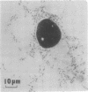
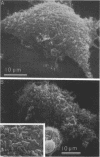
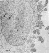
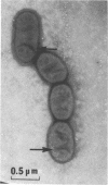
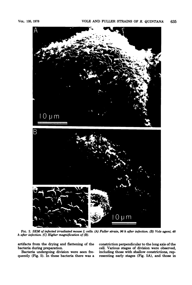
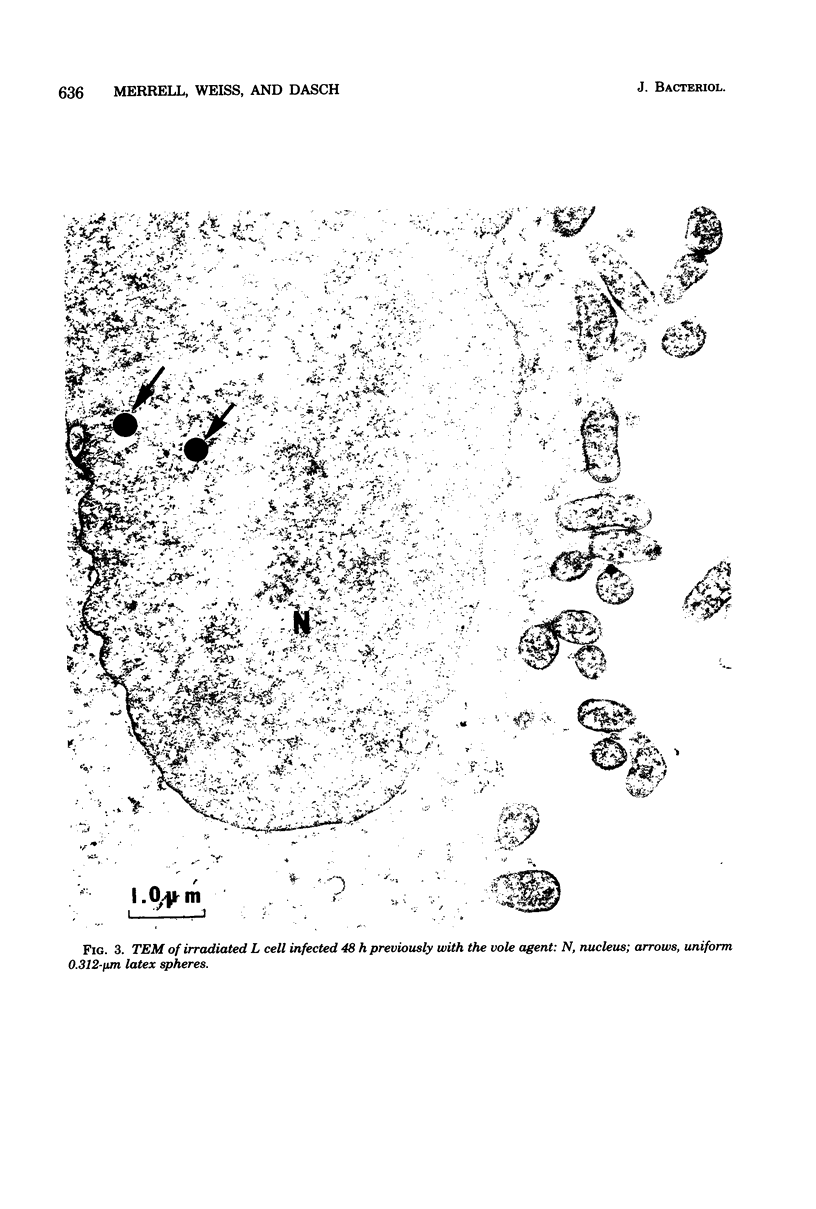
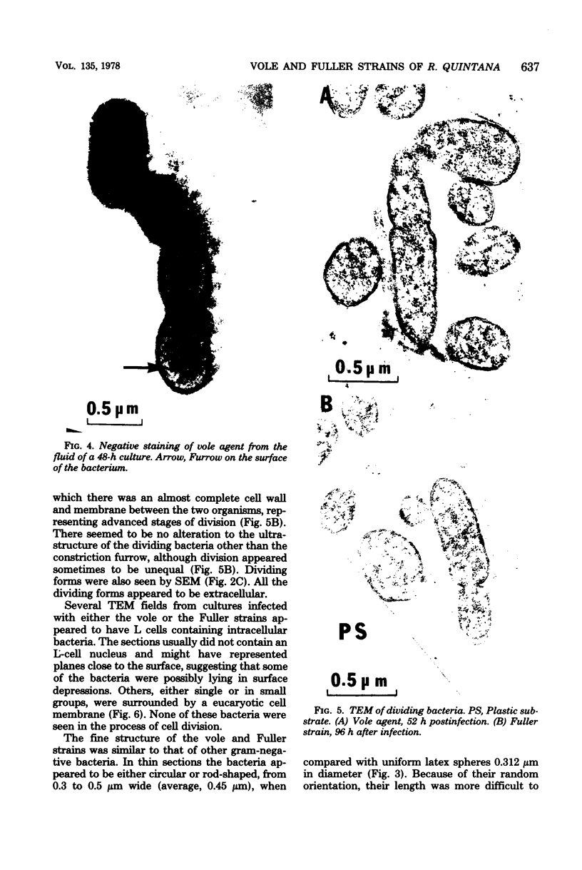
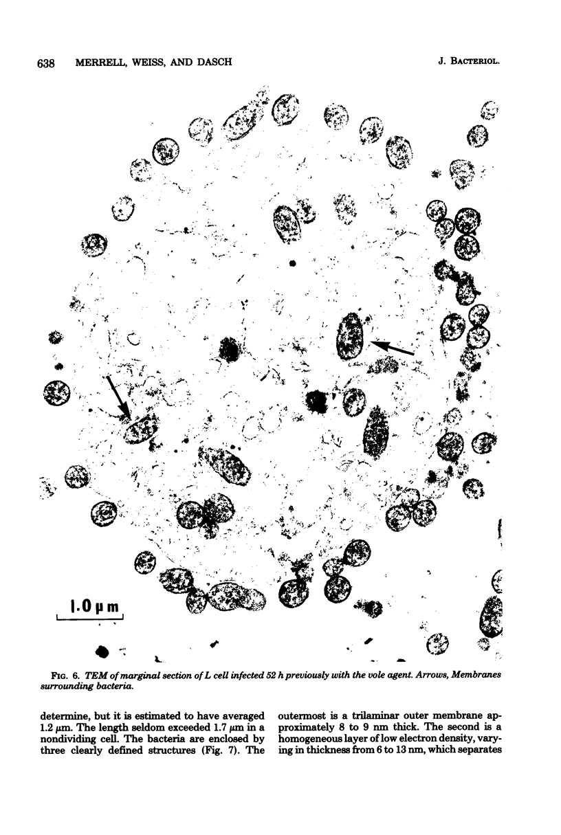
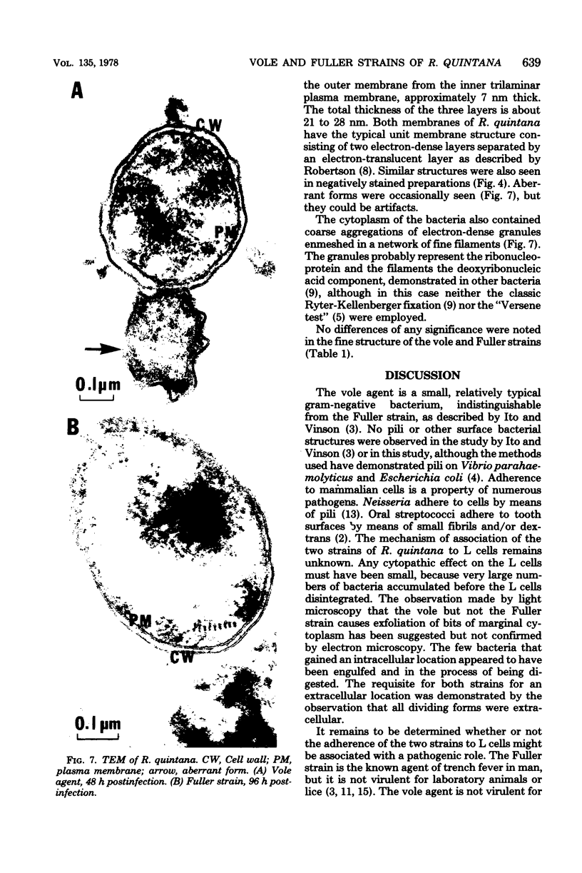
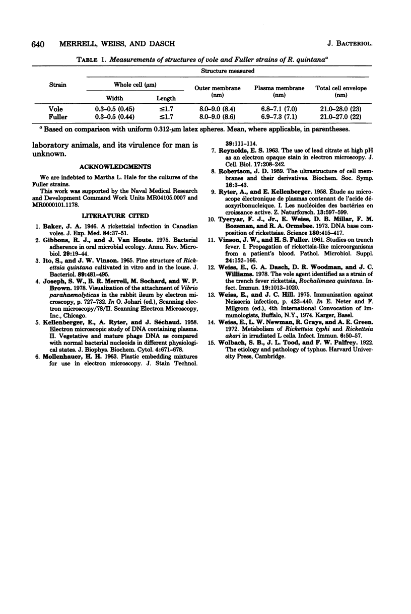
The vole and Fuller strains of Rochalimaea quintana were grown on monolayers of mouse L cells irradiated 7 days previously and examined by light microscopy and scanning and transmission electron microscopy. Most of the bacteria of both strains were shown to adhere to the L cells but remained in an extracellular location. Cell division was frequently seen among the extracellular bacteria. The few intracellular bacteria seemed to be within vacuoles and did not multiply. Attachment to the eucaryotic cell did not seem to involve pili or other bacterial surface structures. The dimensions of the bacteria were approximately 0.45 micron in width by 1.0 to 1.7 micron in length. The cell envelope consisted of the usual trilaminar cell wall and plasma membranes separated by a layer of low electron density, as found in other gram-negative bacteria. No significant differences between the vole and Fuller strains either in morphology or relationship to eucaryotic cells were encountered.
Full text
PDF
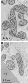
Images in this article
Selected References
These references are in PubMed. This may not be the complete list of references from this article.
- Gibbons R. J., Houte J. V. Bacterial adherence in oral microbial ecology. Annu Rev Microbiol. 1975;29:19–44. doi: 10.1146/annurev.mi.29.100175.000315. [DOI] [PubMed] [Google Scholar]
- ITO S., VINSON J. W. FINE STRUCTURE OF RICKETTSIA QUINTANA CULTIVATED IN VITRO AND IN THE LOUSE. J Bacteriol. 1965 Feb;89:481–495. doi: 10.1128/jb.89.2.481-495.1965. [DOI] [PMC free article] [PubMed] [Google Scholar]
- KELLENBERGER E., RYTER A., SECHAUD J. Electron microscope study of DNA-containing plasms. II. Vegetative and mature phage DNA as compared with normal bacterial nucleoids in different physiological states. J Biophys Biochem Cytol. 1958 Nov 25;4(6):671–678. doi: 10.1083/jcb.4.6.671. [DOI] [PMC free article] [PubMed] [Google Scholar]
- REYNOLDS E. S. The use of lead citrate at high pH as an electron-opaque stain in electron microscopy. J Cell Biol. 1963 Apr;17:208–212. doi: 10.1083/jcb.17.1.208. [DOI] [PMC free article] [PubMed] [Google Scholar]
- ROBERTSON J. D. The ultrastructure of cell membranes and their derivatives. Biochem Soc Symp. 1959;16:3–43. [PubMed] [Google Scholar]
- RYTER A., KELLENBERGER E., BIRCHANDERSEN A., MAALOE O. Etude au microscope électronique de plasmas contenant de l'acide désoxyribonucliéique. I. Les nucléoides des bactéries en croissance active. Z Naturforsch B. 1958 Sep;13B(9):597–605. [PubMed] [Google Scholar]
- Tyeryar F. J., Jr, Weiss E., Millar D. B., Bozeman F. M., Ormsbee R. A. DNA base composition of rickettsiae. Science. 1973 Apr 27;180(4084):415–417. doi: 10.1126/science.180.4084.415. [DOI] [PubMed] [Google Scholar]
- VINSON J. W., FULLER H. S. Studies on trench fever. I. Propagation of Rickettsia-like microorganisms from a patient's blood. Pathol Microbiol (Basel) 1961;24(Suppl):152–166. [PubMed] [Google Scholar]
- Weiss E., Dasch G. A., Woodman D. R., Williams J. C. Vole agent identified as a strain of the trench fever rickettsia, Rochalimaea quintana. Infect Immun. 1978 Mar;19(3):1013–1020. doi: 10.1128/iai.19.3.1013-1020.1978. [DOI] [PMC free article] [PubMed] [Google Scholar]
- Weiss E., Newman L. W., Grays R., Green A. E. Metabolism of Rickettsia typhi and Rickettsia akari in irradiated L cells. Infect Immun. 1972 Jul;6(1):50–57. doi: 10.1128/iai.6.1.50-57.1972. [DOI] [PMC free article] [PubMed] [Google Scholar]


