Abstract
β-Lactamase type I is reported for the first time to occur in the sporulated form in a penicillin-resistant Bacillus species. The enzyme was readily characterized from the B. cereus 5/B line (ATCC 13061) by mass spectrometry and two-dimensional gel electrophoresis.
A common cause of antibiotic resistance in bacteria is an increased abundance of β-lactamases (10). This can be the result of genetic engineering (16), or it can be caused by the selection of resistant variants in the presence of antibiotics. β-Lactamase genes are found in the wild-type genomes of many bacteria, including Bacillus species. These chromosomal β-lactamases do not generally provide effective antibiotic resistance in wild-type bacilli, despite evidence that the genes are not completely silenced (1, 11, 14). Under antibiotic selection pressure, however, a number of strains show increased resistance, suggesting mutation-induced upregulation of β-lactamase expression. The Bacillus cereus 5/B line (ATCC 13061) is stably resistant to penicillin, having been selected by exposure to penicillin 5 decades ago (7, 18). Water-soluble β-lactamase type I has been reported to be expressed in high abundance in vegetative cells of this resistant strain and also to be secreted by the vegetative bacteria (19). The occurrence of β-lactamase in sporulated Bacillus species has been predicted by Saz (17). Terrorist or other antisocial distribution of Bacillus species (e.g., anthrax) selected for drug resistance would likely occur with spores, and B. cereus 5/B (ATCC 13061) is studied here as a model spore type.
The objectives of the present work are to interrogate the presence of β-lactamase in the sporulated form of stably resistant strain ATCC 13061 and to evaluate direct matrix-assisted laser desorption ionization (MALDI)-time of flight analysis for rapid preliminary detection in spores of this indicator of antibiotic resistance. B. cereus T is used here as a control, since it is not resistant to penicillin.
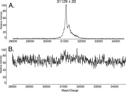
Spores were prepared by following standard procedures (2, 4, 13, 15, 20). Purity was estimated by microscopic examination as >98%. Eight MALDI spectra obtained on a Shimadzu Biosciences Axima-CFR Plus MALDI-time of flight mass spectrometer (Columbia, MD) directly from spores of B. cereus ATCC 13061 (American Type Culture Collection, Manassas, VA) (Fig. 1A) provided an average molecular mass of 31,129 Da ± 20 Da. No ions are detected in this mass range in the spectrum (Fig. 1B) acquired from the control sample, B. cereus T (obtained from H. O. Halvorson), by the use of a roughly equivalent number of spores.
FIG. 1.
Partial MALDI mass spectra obtained from spores of Bacillus cereus ATCC 13061 (A) and Bacillus cereus T (B).
The average molecular mass of unprocessed β-lactamase type I is calculated to be 33,597 Da. Consequently, peptide analysis was carried out to test the hypothesis that the molecular mass detected at around 31,100 Da belongs to a processed isoform of β-lactamase type I. Suspensions of spores of the two B. cereus strains were incubated in 2% sodium dodecyl sulfate (SDS) solutions for 15 min, boiled in a water bath for 2 min, sonicated for 5 min, and centrifuged at 500 × g for 14 min. Eighty micrograms of protein from each supernatant was loaded onto a 15% Tris-HCl gel (Bio-Rad, Hercules, CA), developed in one dimension, and stained with Coomassie blue stain.
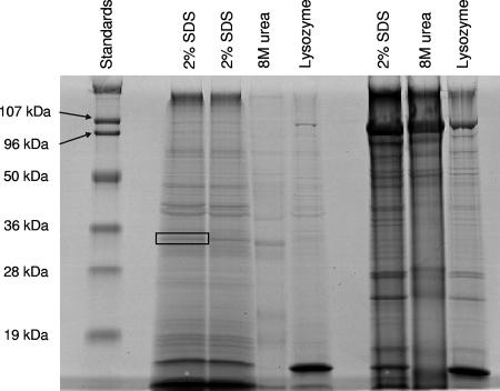
Figure 2 shows the one-dimensional (1-D) gel patterns of proteins recovered from ATCC 13061 (lanes 2 to 5) and B. cereus T (lanes 6 to 9) by the use of 2% SDS solution, 8 M urea, or treatment with aqueous lysozyme. A peak is detected at around 33,000 Da in the SDS and urea extracts from ATCC 13061, while none is detected in comparable extracts from B. cereus T. Artifactual contamination was considered a source for β-lactamase, possibly secreted by vegetative cells and adsorbed on the outside of the spores or originating from vegetative cell debris uncleared from the spores. This was checked by suspending the spores in 40% ethanol, 1% Triton X-100, or 1:1 40% formic acid-30% acetonitrile. After centrifugation, no protein was detected in the supernatants by 1-D gel electrophoresis.
FIG. 2.
Gel electrophoresis of extracts of spores of Bacillus cereus ATCC 13061 (lanes 2 to 5) and extracts of spores of Bacillus cereus T (lanes 6 to 9).
To support the relationship between the protein desorbed by MALDI from the intact spores and the band near 33,000 Da in the 1-D gel, the intact protein was recovered from the gel by following published methods (6, 12) and was characterized by MALDI analysis with a molecular mass at 31,119 ± 20 Da (spectrum not shown). This matches that of the protein desorbed directly from the spores, within experimental uncertainty.
The band of interest was subjected to in-gel digestion with trypsin (5). The peptides recovered were analyzed by collisionally induced dissociation in tandem mass spectrometry experiments using electrospray on a QStar Pulsar tandem mass spectrometer (Applied Biosystems, Foster City, CA). The Mascot search engine (Matrix Science, London, United Kingdom) was used to search collisionally induced dissociation spectra against the Swiss-Prot prokaryote database. Table 1 summarizes the identification of eight peptides from ATCC 13061, all of which match tryptic peptides expected from β-lactamase type I or its precursor protein. The carboxyl-terminal peptide is identified; however, the amino-terminal peptide is not.
TABLE 1.
Peptides from in-gel digestion identified by MS-MS measurements
| Position | Sequence | Scorea | E valuea |
|---|---|---|---|
| 85-92 | R.FAFASTYK.A | 52 | 0.00026 |
| 93-106 | K.ALAAGVLLQQNSTK.K | 26 | 0.087 |
| 117-128 | K.EDLVDYSPVTEK.H | 58 | 5.9 × 10⁻⁵ |
| 146-158 | R.YSDNTAGNILFHK.I | 26 | 0.07 |
| 272-282 | R.SPIIIAILSSK.D | 61 | 2.3 × 10⁻⁵ |
| 272-285 | R.SPIIIAILSSKDEK.E | 38 | 0.0053 |
| 286-295 | K.EATYDNQLIK.E | 43 | 0.0019 |
| 296-306 | K.EAAEVVIDAIK.- | 67 | 8.3 × 10⁻⁶ |
Quantitative evaluations of the reliability of the identification were calculated by the Mascot search engine.
The average molecular mass observed, 31,129 ± 20 Da, falls between the mass of unprocessed β-lactamase type I predicted from its gene, 33,597 Da (Swiss-Prot accession no. P10424) (19), and the mass reported for processed β-lactamase type I (29,063 Da) secreted from recombinant B. subtilis vegetative cells (19). Cleavage between Leu-23 and Val-24 (Fig. 3) would provide a processed protein with an average mass of 31,135 Da, consistent with the mass measured here for β-lactamase type I isolated from sporulated penicillin-resistant B. cereus 5/B.
FIG. 3.
Sequence of β-lactamase type I precursor (Swiss-Prot accession no. P10424). The proposed cleavage is indicated by italics and underlining, while observed peptides are indicated by boldface and underlining.
The absence reported here of detectable amounts of β-lactamase type I in B. cereus T is consistent with recent proteomic-scale studies of proteins in other lactam-susceptible Bacillus spores (3, 8, 9) in which isoforms of β-lactamase were not observed.
The first four identified peptides listed in Table 1 are also found in β-lactamase type I proteins observed in vegetative B. anthracis and B. thuringiensis. This suggests that these peptides might form the basis of a hypothesis-driven approach to the detection of antibiotic resistance in sporulated forms of the entire B. cereus group. The sequence motif used by the PROSITE database (http://www.expasy.org/prosite/) to characterize proteins from the β-lactamase class A active-site protein family (PS00146) begins with Phe in the identified peptide FAFASTYK and continues through the middle of the adjacent identified peptide ALAAGVLLQQNSTK. The remaining four peptides of Table 1 are found only in B. cereus β-lactamase type I, which suggests that they could be used to provide species-specific identification of the bacterium expressing β-lactamase.
Acknowledgments
This work was supported in part by Public Health Service grant CA-126189 from the National Cancer Institute, USDA cooperative agreement 5812757342, a fellowship from the Fulbright Program for Foreign Graduates sponsored by the U.S. Department of State (N.T.), and a fellowship from the Helsingin Sanomat 100th Anniversary Foundation (O.L.).
Footnotes
Published ahead of print on 7 December 2007.
REFERENCES
- 1.Chen, Y., J. Succi, F. C. Tenover, and T. M. Koehler. 2003. β-Lactamase genes of the penicillin-susceptible Bacillus anthracis Sterne strain. J. Bacteriol. 185:823-830. [DOI] [PMC free article] [PubMed] [Google Scholar]
- 2.Hathout, Y., P. A. Demirev, Y.-P. Ho, J. L. Bundy, V. Ryzhov, L. Sapp, J. Stutler, J. Jackman, and C. Fenselau. 1999. Identification of Bacillus spores by matrix-assisted laser desorption ionization-mass spectrometry. Appl. Environ. Microbiol. 65:4313-4319. [DOI] [PMC free article] [PubMed] [Google Scholar]
- 3.Jagtap, P., G. Michailidis, R. Zielke, A. K. Walker, N. Patel, J. R. Strahler, A. Driks, P. C. Andrews, and J. R. Maddock. 2006. Early events of Bacillus anthracis germination identified by time-course quantitative proteomics. Proteomics 6:5199-5211. [DOI] [PubMed] [Google Scholar]
- 4.Jenkinson, H. F., W. D. Sawyer, and J. Mandelstam. 1981. Synthesis and order of assembly of spore coat proteins in Bacillus subtilis. J. Gen. Microbiol. 123:1-16. [Google Scholar]
- 5.Jensen, O. N., M. Wilm, A. Shevchenko, and M. Mann. 1999. Sample preparation methods for mass spectrometric peptide mapping directly from 2-DE gels. Methods Mol. Biol. 112:513-530. [DOI] [PubMed] [Google Scholar]
- 6.Jin, Y., and T. Manabe. 2005. High-efficiency protein extraction from polyacrylamide gels for molecular mass measurement by matrix-assisted laser desorption/ionization-time of flight-mass spectrometry. Electrophoresis 26:1019-1028. [DOI] [PubMed] [Google Scholar]
- 7.Kogut, M., M. R. Pollock, and E. J. Tridgell. 1956. Purification of penicillin-induced penicillinase of Bacillus cereus NRRL 569: a comparison of its properties with those of a similarly purified penicillinase produced spontaneously by a constitutive mutant strain. Biochem. J. 62:391-401. [DOI] [PMC free article] [PubMed] [Google Scholar]
- 8.Kuwana, R., Y. Kasahara, M. Fujibayashi, H. Takamatsu, N. Ogasawara, and K. Watabe. 2002. Proteomics characterization of novel spore proteins of Bacillus subtilis. Microbiology 148:3971-3982. [DOI] [PubMed] [Google Scholar]
- 9.Liu, H., N. H. Bergman, B. Thomason, S. Shallom, A. Hazen, J. Crossno, D. A. Rasko, J. Ravel, T. D. Read, S. Peterson, J. Yates III, and P. C. Hanna. 2004. Formation and composition of the Bacillus anthracis endospore. J. Bacteriol. 186:164-178. [DOI] [PMC free article] [PubMed] [Google Scholar]
- 10.Majiduddin, F. K., I. C. Mateson, and T. G. Palzkill. 2002. Molecular analysis of beta-lactamase structure and function. Int. J. Med. Microbiol. 292:127-137. [DOI] [PubMed] [Google Scholar]
- 11.Materon, I. C., A. M. Queenan, T. M. Koehler, K. Bush, and T. Palzkill. 2003. Biochemical characterization of β-lactamases Bla1 and Bla2 from Bacillus anthracis. Antimicrob. Agents Chemother. 47:2040-2042. [DOI] [PMC free article] [PubMed] [Google Scholar]
- 12.Mirza, U. A., Y. H. Liu, J. T. Tang, F. Porter, L. Bondoc, G. Chen, B. N. Pramanik, and T. L. Nagabhushan. 2000. Extraction and characterization of adenovirus proteins from sodium dodecylsulfate polyacrylamide gel electrophoresis by matrix-assisted laser desorption/ionization mass spectrometry. J. Am. Soc. Mass Spectrom. 11:356-361. [DOI] [PubMed] [Google Scholar]
- 13.Nicholson, W. L., and P. Setlow. 1990. Sporulation, germination, and outgrowth, p. 391-450. In C. R. Harwood and S. M. Cutting (ed.), Molecular biological methods for Bacillus. John Wiley & Sons, Chichester, United Kingdom.
- 14.Ozer, J. H., D. L. Lowery, and A. K. Saz. 1970. Derepression of β-lactamase (penicillinase) in Bacillus cereus by peptidoglycans. J. Bacteriol. 102:52-63. [DOI] [PMC free article] [PubMed] [Google Scholar]
- 15.Phillips, A. P., and J. W. Ezzell. 1989. Identification of Bacillus anthracis by polyclonal antibodies against extracted vegetative cell antigen. J. Appl. Bacteriol. 66:419-432. [DOI] [PubMed] [Google Scholar]
- 16.Russell, S., N. Edwards, and C. Fenselau. 2007. Detection of plasmid insertion in Escherichia coli by MALDI-TOF mass spectrometry. Anal. Chem. 79:5399-5406. [DOI] [PubMed] [Google Scholar]
- 17.Saz, A. K. 1970. An introspective view of penicillinase. J. Cell. Physiol. 76:397-404. [DOI] [PubMed] [Google Scholar]
- 18.Sneath, P. H. 1955. Proof of the spontaneity of a mutation to penicillinase production in B. cereus. J. Gen. Microbiol. 13:561-571. [DOI] [PubMed] [Google Scholar]
- 19.Wang, W., P. S. Mézes, Y. Q. Yang, R. W. Blacher, and J. O. Lampen. 1985. Cloning and sequencing of the β-lactamase I gene of Bacillus cereus 5/B and its expression in Bacillus subtilis. J. Bacteriol. 163:487-492. [DOI] [PMC free article] [PubMed] [Google Scholar]
- 20.Warscheid, B., and C. Fenselau. 2003. Characterization of Bacillus spore species and their mixtures using post-source decay with a curved-field reflectron. Anal. Chem. 75:5618-5627. [DOI] [PubMed] [Google Scholar]


