Abstract
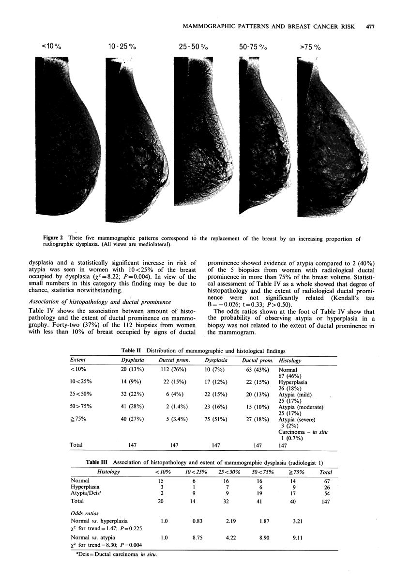
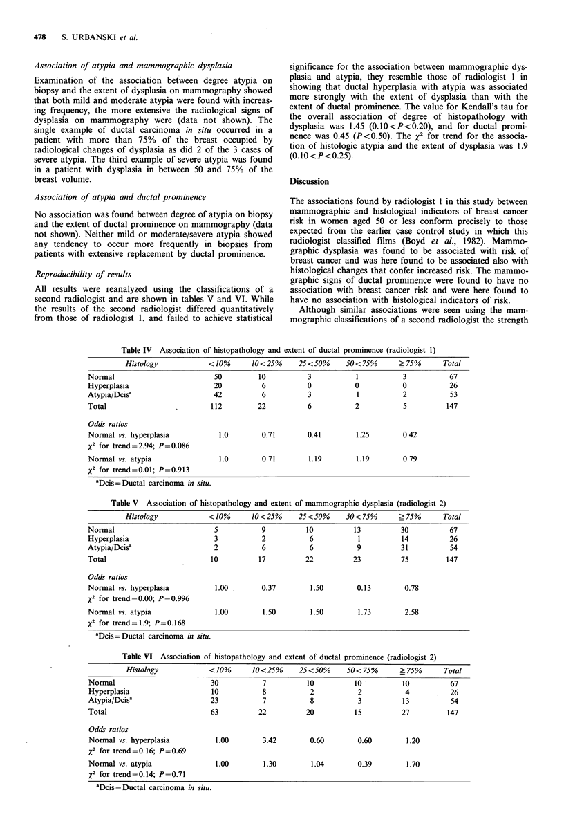
Previous work has shown that extensive mammographic dysplasia in women aged less than 50 was strongly associated with breast cancer but that the radiological appearance of ductal prominence was not associated with risk. In the present paper we examine the association between these mammographic signs in the breast and histological patterns in the terminal ductal lobular unit (TDLU), the region of the breast where breast cancer is believed to originate. Surgical biopsies from a consecutive series of women aged less than 50 were reviewed and classified according to the histopathology of the epithelium in the TDLU. Mammograms from the same subjects were independently classified according to the extent of the radiological signs of dysplasia and ductal prominence. Degree of histopathology and the extent of mammographic dysplasia were associated and atypia of the ductal type was found more frequently in patients with extensive dysplasia. However, the strength and statistical significance of the association varied according to the radiologist who classified the mammograms. No association was found between degree of histopathology and ductal prominence. These results add to the evidence that extensive mammographic dysplasia in women aged less than 50 is a risk factor for breast cancer. They do not indicate that the radiological signs of dysplasia are caused by histological changes in the TDLU.
Full text
PDF
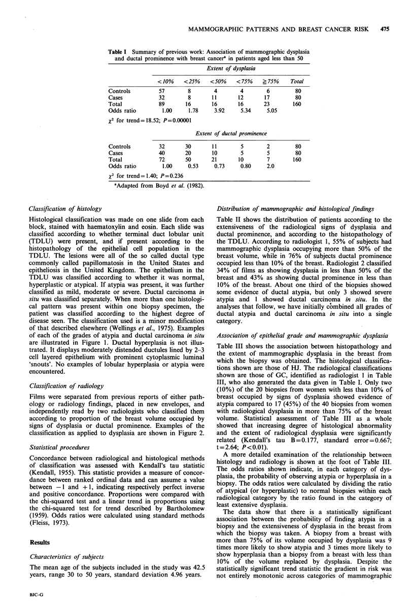
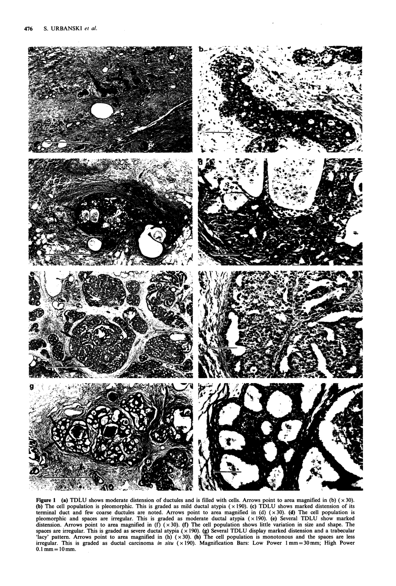
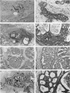
Images in this article
Selected References
These references are in PubMed. This may not be the complete list of references from this article.
- Asch R. H., Greenblatt R. B. The use of an impeded androgen--danazol--in the management of benign breast disorders. Am J Obstet Gynecol. 1977 Jan 15;127(2):130–134. doi: 10.1016/s0002-9378(16)33237-9. [DOI] [PubMed] [Google Scholar]
- Black M. M., Barclay T. H., Cutler S. J., Hankey B. F., Asire A. J. Association of atypical characteristics of benign breast lesions with subsequent risk of breast cancer. Cancer. 1972 Feb;29(2):338–343. doi: 10.1002/1097-0142(197202)29:2<338::aid-cncr2820290212>3.0.co;2-u. [DOI] [PubMed] [Google Scholar]
- Bright R. A., Morrison A. S., Brisson J., Burstein N. A., Sadowsky N. S., Kopans D. B., Meyer J. E. Relationship between mammographic and histologic features of breast tissue in women with benign biopsies. Cancer. 1988 Jan 15;61(2):266–271. doi: 10.1002/1097-0142(19880115)61:2<266::aid-cncr2820610212>3.0.co;2-n. [DOI] [PubMed] [Google Scholar]
- Carlile T., Kopecky K. J., Thompson D. J., Whitehead J. R., Gilbert F. I., Jr, Present A. J., Threatt B. A., Krook P., Hadaway E. Breast cancer prediction and the Wolfe classification of mammograms. JAMA. 1985 Aug 23;254(8):1050–1053. [PubMed] [Google Scholar]
- Dupont W. D., Page D. L. Risk factors for breast cancer in women with proliferative breast disease. N Engl J Med. 1985 Jan 17;312(3):146–151. doi: 10.1056/NEJM198501173120303. [DOI] [PubMed] [Google Scholar]
- Fisher E. R., Palekar A., Kim W. S., Redmond C. The histopathology of mammographic patterns. Am J Clin Pathol. 1978 Apr;69(4):421–426. doi: 10.1093/ajcp/69.4.421. [DOI] [PubMed] [Google Scholar]
- Foote F. W., Stewart F. W. Comparative Studies of Cancerous Versus Noncancerous Breasts. Ann Surg. 1945 Jan;121(1):6–53. doi: 10.1097/00000658-194501000-00002. [DOI] [PMC free article] [PubMed] [Google Scholar]
- Gravelle I. H., Bulstrode J. C., Wang D. Y., Bulbrook R. D., Hayward J. L. The relation between radiographic features and determinants of risk of breast cancer. Br J Radiol. 1980 Feb;53(626):107–113. doi: 10.1259/0007-1285-53-626-107. [DOI] [PubMed] [Google Scholar]
- Hutchinson W. B., Thomas D. B., Hamlin W. B., Roth G. J., Peterson A. V., Williams B. Risk of breast cancer in women with benign breast disease. J Natl Cancer Inst. 1980 Jul;65(1):13–20. [PubMed] [Google Scholar]
- Krook P. M., Carlile T., Bush W., Hall M. H. Mammographic parenchymal patterns as a risk indicator for prevalent and incident cancer. Cancer. 1978 Mar;41(3):1093–1097. doi: 10.1002/1097-0142(197803)41:3<1093::aid-cncr2820410343>3.0.co;2-h. [DOI] [PubMed] [Google Scholar]
- Page D. L., Vander Zwaag R., Rogers L. W., Williams L. T., Walker W. E., Hartmann W. H. Relation between component parts of fibrocystic disease complex and breast cancer. J Natl Cancer Inst. 1978 Oct;61(4):1055–1063. [PubMed] [Google Scholar]
- Wellings S. R., Jensen H. M., Marcum R. G. An atlas of subgross pathology of the human breast with special reference to possible precancerous lesions. J Natl Cancer Inst. 1975 Aug;55(2):231–273. [PubMed] [Google Scholar]
- Wellings S. R., Wolfe J. N. Correlative studies of the histological and radiographic appearance of the breast parenchyma. Radiology. 1978 Nov;129(2):299–306. doi: 10.1148/129.2.299. [DOI] [PubMed] [Google Scholar]
- Wolfe J. N. Risk for breast cancer development determined by mammographic parenchymal pattern. Cancer. 1976 May;37(5):2486–2492. doi: 10.1002/1097-0142(197605)37:5<2486::aid-cncr2820370542>3.0.co;2-8. [DOI] [PubMed] [Google Scholar]
- de Waard F., Rombach J. J. Re: "The relationship between Wolfe's classification of mammograms, accepted breast cancer risk factors, and the incidence of breast cancer". Am J Epidemiol. 1987 Jan;125(1):171–172. doi: 10.1093/oxfordjournals.aje.a114507. [DOI] [PubMed] [Google Scholar]


