

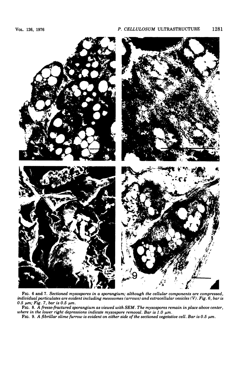
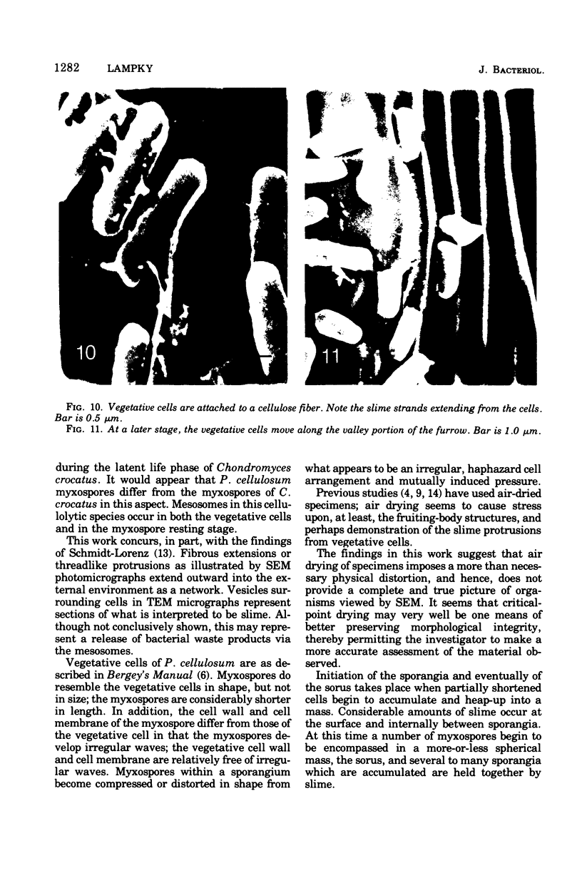
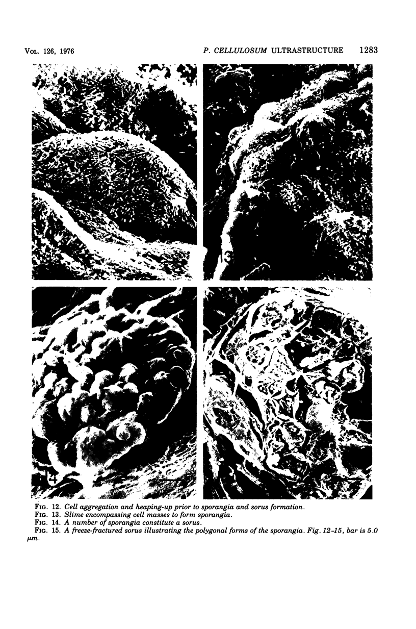
Abstract
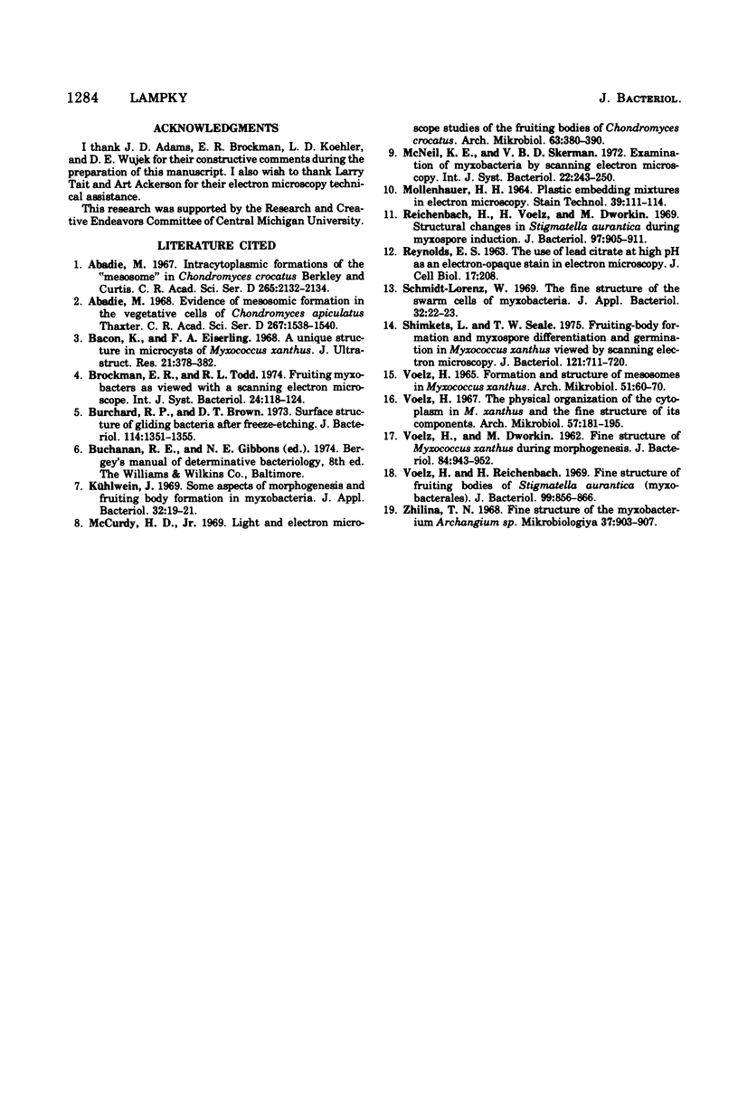
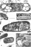
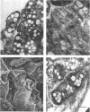
Polyangium cellulosum was examined with the transmission electron microscope and the scanning electron microscope. Freeze-fracturing and critical-point-drying techniques were employed with the latter instrument. Critical-point drying seemed to eliminate the distortion of cells and fruiting bodies. These instruments and techniques allowed for a detailed comparison of cell and fruiting-body ultrastructure. Lipid storage materials and mesosomes were found to be constant cell particulates in both vegetative cells and in the shortened myxospores.
Full text
PDF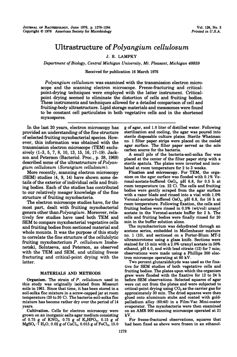


Images in this article
Selected References
These references are in PubMed. This may not be the complete list of references from this article.
- Abadie M. Formations intracytoplasmiques du type "mésosome" chez Chondromyces crocatus Berkeley et Curtis. C R Acad Sci Hebd Seances Acad Sci D. 1967 Dec 18;265(25):2132–2134. [PubMed] [Google Scholar]
- Abadie M. Mise en évidence des formations mésosomiques dans les cellules végétatives du Chondromyces apiculatus Thaxter. C R Acad Sci Hebd Seances Acad Sci D. 1968 Nov 4;267(19):1538–1540. [PubMed] [Google Scholar]
- Bacon K., Eiserling F. A. A unique structure in microcysts of Myxococcus xanthus. J Ultrastruct Res. 1967 Dec;21(5):378–382. doi: 10.1016/s0022-5320(67)80147-3. [DOI] [PubMed] [Google Scholar]
- Burchard R. P., Brown D. T. Surface structure of gliding bacteria after freeze-etching. J Bacteriol. 1973 Jun;114(3):1351–1355. doi: 10.1128/jb.114.3.1351-1355.1973. [DOI] [PMC free article] [PubMed] [Google Scholar]
- Kühlwein H. Some aspects of morphogenesis and fruiting body formation in myxobacteria. J Appl Bacteriol. 1969 Mar;32(1):19–21. doi: 10.1111/j.1365-2672.1969.tb02183.x. [DOI] [PubMed] [Google Scholar]
- MOLLENHAUER H. H. PLASTIC EMBEDDING MIXTURES FOR USE IN ELECTRON MICROSCOPY. Stain Technol. 1964 Mar;39:111–114. [PubMed] [Google Scholar]
- REYNOLDS E. S. The use of lead citrate at high pH as an electron-opaque stain in electron microscopy. J Cell Biol. 1963 Apr;17:208–212. doi: 10.1083/jcb.17.1.208. [DOI] [PMC free article] [PubMed] [Google Scholar]
- Reichenbach H., Voelz H., Dworkin M. Structural changes in Stigmatella aurantiaca during myxospore induction. J Bacteriol. 1969 Feb;97(2):905–911. doi: 10.1128/jb.97.2.905-911.1969. [DOI] [PMC free article] [PubMed] [Google Scholar]
- Schmidt-Lorenz W. The fine structure of the swarm cells of myxobacteria. J Appl Bacteriol. 1969 Mar;32(1):22–23. doi: 10.1111/j.1365-2672.1969.tb02184.x. [DOI] [PubMed] [Google Scholar]
- Shimkets L., Seale T. W. Fruiting-body formation and myxospore differentiation and germination in Mxyococcus xanthus viewed by scanning electron microscopy. J Bacteriol. 1975 Feb;121(2):711–720. doi: 10.1128/jb.121.2.711-720.1975. [DOI] [PMC free article] [PubMed] [Google Scholar]
- VOELZ H., DWORKIN M. Fine structure of Myxococcus xanthus during morphogenesis. J Bacteriol. 1962 Nov;84:943–952. doi: 10.1128/jb.84.5.943-952.1962. [DOI] [PMC free article] [PubMed] [Google Scholar]
- VOELZ H. FORMATION AND STRUCTURE OF MESOSOMES IN MYXOCOCCUS XANTHUS. Arch Mikrobiol. 1965 May 28;51:60–70. doi: 10.1007/BF00406850. [DOI] [PubMed] [Google Scholar]
- Voelz H., Reichenbach H. Fine structure of fruiting bodies of Stigmatella aurantiaca (Myxobacterales). J Bacteriol. 1969 Sep;99(3):856–866. doi: 10.1128/jb.99.3.856-866.1969. [DOI] [PMC free article] [PubMed] [Google Scholar]
- Voelz H. The physical organization of the cytoplasm in Myxococcus zanthus and the fine structure of its components. Arch Mikrobiol. 1967 Jun 6;57(2):181–195. doi: 10.1007/BF00408700. [DOI] [PubMed] [Google Scholar]
- Zhilina T. N. Tonkoe stroenie miksobakterii Archangium sp. Mikrobiologiia. 1968 Sep-Oct;37(5):903–907. [PubMed] [Google Scholar]