

Abstract
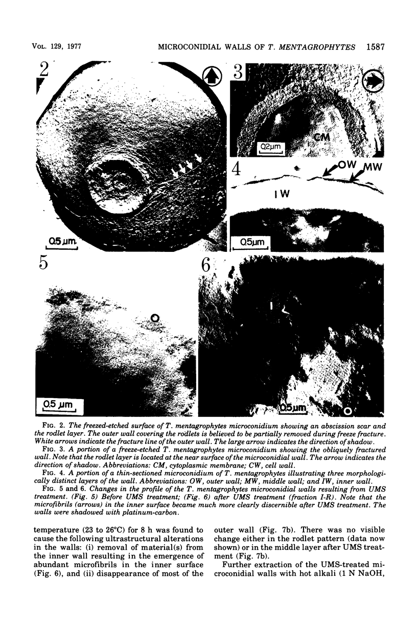
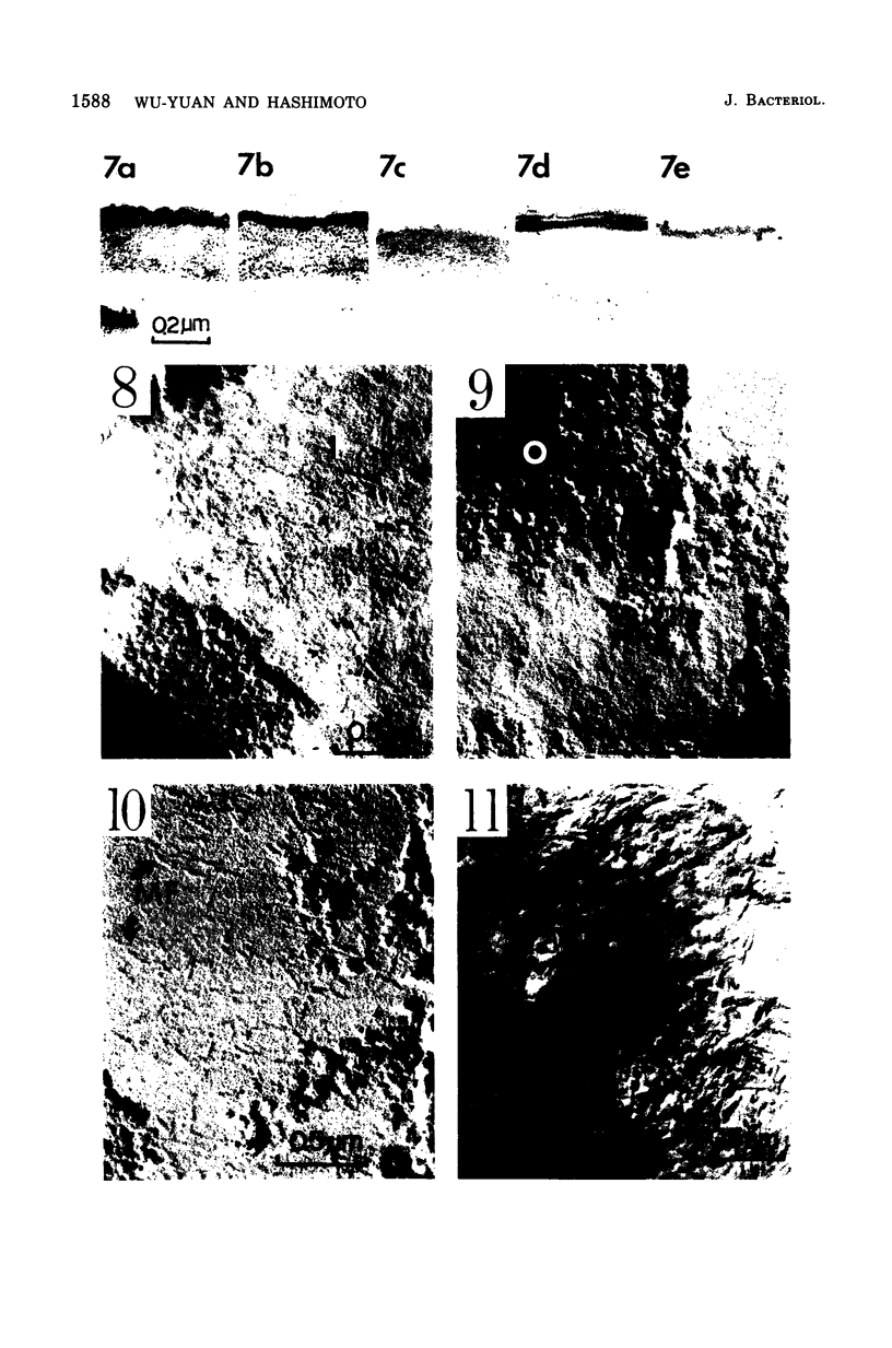
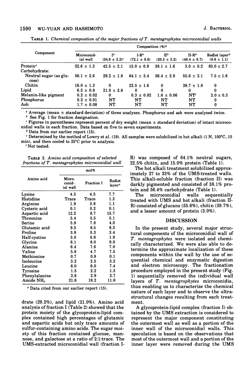
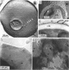
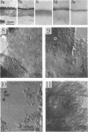
The ultrastructure and chemical composition of the walls of Trichophyton mentagrophytes microconidia were investigated with particular emphasis on the localization of the major structural components within the walls. The walls consisted of carbohydrate (56.1% neutral polysaccharide, and 16.0% chitin), protein (22.6%), lipid (6.5%), ash (1.7%), and trace amounts of melanin (0.2%) and phosphorus (0.2%). in thin sections, three distince layers were recognized. The electron-transparent pellicle (15 to 20 nm thick) covering the outermost surface of the wall consisted of a glycoprotein-lipid complex and was mostly extracted by sodium phosphate buffer (0.1 M, pH 6.5) containing 8 M urea, 1% (vol/vol) mercaptoethanol, and 1% (wt/vol) sodium dodecyl sulfate. The middle electron-dense layer (30 to 50 nm thick) represented the proteinaceous rodlet layer embedded in polysaccharides and could be completely solubilized by hot alkali extraction (1 N NaOH, 100 DEGREES C, 1 h). The thick inner layer (200 to 300 nm thick) was relatively resistant to the above treatments and was found to consist of amorphous glucans and microfibrillar chitin. Approximately half of the inner wall glucans was susceptible to (1 leads to 3)-beta-glucanase.
Full text
PDF

Images in this article

Selected References
These references are in PubMed. This may not be the complete list of references from this article.
- BARTNICKI-GARCIA S., REYES E. CHEMISTRY OF SPORE WALL DIFFERENTIATION IN MUCOR ROUXII. Arch Biochem Biophys. 1964 Oct;108:125–133. doi: 10.1016/0003-9861(64)90363-7. [DOI] [PubMed] [Google Scholar]
- BLUMENTHAL H. J., ROSEMAN S. Quantitative estimation of chitin in fungi. J Bacteriol. 1957 Aug;74(2):222–224. doi: 10.1128/jb.74.2.222-224.1957. [DOI] [PMC free article] [PubMed] [Google Scholar]
- Buckley P. M., Sjaholm V. E., Sommer N. F. Electron microscopy of Botrytis cinerea conidia. J Bacteriol. 1966 May;91(5):2037–2044. doi: 10.1128/jb.91.5.2037-2044.1966. [DOI] [PMC free article] [PubMed] [Google Scholar]
- Cabib E., Bowers B. Chitin and yeast budding. Localization of chitin in yeast bud scars. J Biol Chem. 1971 Jan 10;246(1):152–159. [PubMed] [Google Scholar]
- DAVIS B. J. DISC ELECTROPHORESIS. II. METHOD AND APPLICATION TO HUMAN SERUM PROTEINS. Ann N Y Acad Sci. 1964 Dec 28;121:404–427. doi: 10.1111/j.1749-6632.1964.tb14213.x. [DOI] [PubMed] [Google Scholar]
- FOLCH J., LEES M., SLOANE STANLEY G. H. A simple method for the isolation and purification of total lipides from animal tissues. J Biol Chem. 1957 May;226(1):497–509. [PubMed] [Google Scholar]
- Friedman B. A., Dugan P. R., Pfister R. M., Remsen C. C. Fine structure and composition of the zoogloeal matrix surrounding Zoogloea ramigera. J Bacteriol. 1968 Dec;96(6):2144–2153. doi: 10.1128/jb.96.6.2144-2153.1968. [DOI] [PMC free article] [PubMed] [Google Scholar]
- Gander J. E. Fungal cell wall glycoproteins and peptido-polysaccharides. Annu Rev Microbiol. 1974;28(0):103–119. doi: 10.1146/annurev.mi.28.100174.000535. [DOI] [PubMed] [Google Scholar]
- Ghiorse W. C., Edwards M. R. Ultrastructure of Aspergillus fumigatus conidia development and maturation. Protoplasma. 1973;76(1):49–59. doi: 10.1007/BF01279672. [DOI] [PubMed] [Google Scholar]
- HORIKOSHI K., IIDA S. STUDIES OF THE SPORE COATS OF FUNGI. I. ISOLATION AND COMPOSITION OF THE SPORE COATS OF ASPERGILLUS ORYZAE. Biochim Biophys Acta. 1964 Jul 7;83:197–203. doi: 10.1016/0926-6526(64)90035-7. [DOI] [PubMed] [Google Scholar]
- Hashimoto T., Wu-Yuan C. D., Blumenthal H. J. Isolation and characterization of the rodlet layer of Trichophyton mentagrophytes microconidial wall. J Bacteriol. 1976 Sep;127(3):1543–1549. doi: 10.1128/jb.127.3.1543-1549.1976. [DOI] [PMC free article] [PubMed] [Google Scholar]
- Hashimoto T., Wu C. D., Blumenthal H. J. Characterization of L-leucine-induced germination of Trichophyton mentagrophytes microconidia. J Bacteriol. 1972 Nov;112(2):967–976. doi: 10.1128/jb.112.2.967-976.1972. [DOI] [PMC free article] [PubMed] [Google Scholar]
- Hemmes D. E., Kojima-Buddenhagen E. S., Hohl H. R. Structural and enzymatic analysis of the spore wall layers in Dictyostelium discoideum. J Ultrastruct Res. 1972 Dec;41(5):406–417. doi: 10.1016/s0022-5320(72)90047-0. [DOI] [PubMed] [Google Scholar]
- Jelsma J., Kreger D. R. Ultrastructural observations on (1 leads to 3)-beta-D-glucan from fungal cell-walls. Carbohydr Res. 1975 Aug;43(1):200–203. doi: 10.1016/s0008-6215(00)83988-9. [DOI] [PubMed] [Google Scholar]
- LOWRY O. H., ROSEBROUGH N. J., FARR A. L., RANDALL R. J. Protein measurement with the Folin phenol reagent. J Biol Chem. 1951 Nov;193(1):265–275. [PubMed] [Google Scholar]
- Mahadevan P. R., Mahadkar U. R. Major constituents of the conidial wall of Neurospora crassa. Indian J Exp Biol. 1970 Jul;8(3):207–210. [PubMed] [Google Scholar]
- Morris D. L. Quantitative Determination of Carbohydrates With Dreywood's Anthrone Reagent. Science. 1948 Mar 5;107(2775):254–255. doi: 10.1126/science.107.2775.254. [DOI] [PubMed] [Google Scholar]
- REYNOLDS E. S. The use of lead citrate at high pH as an electron-opaque stain in electron microscopy. J Cell Biol. 1963 Apr;17:208–212. doi: 10.1083/jcb.17.1.208. [DOI] [PMC free article] [PubMed] [Google Scholar]
- WINZLER R. J. Determination of serum glycoproteins. Methods Biochem Anal. 1955;2:279–311. doi: 10.1002/9780470110188.ch10. [DOI] [PubMed] [Google Scholar]