Abstract
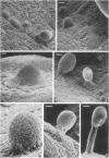
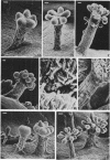
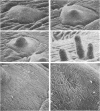
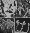
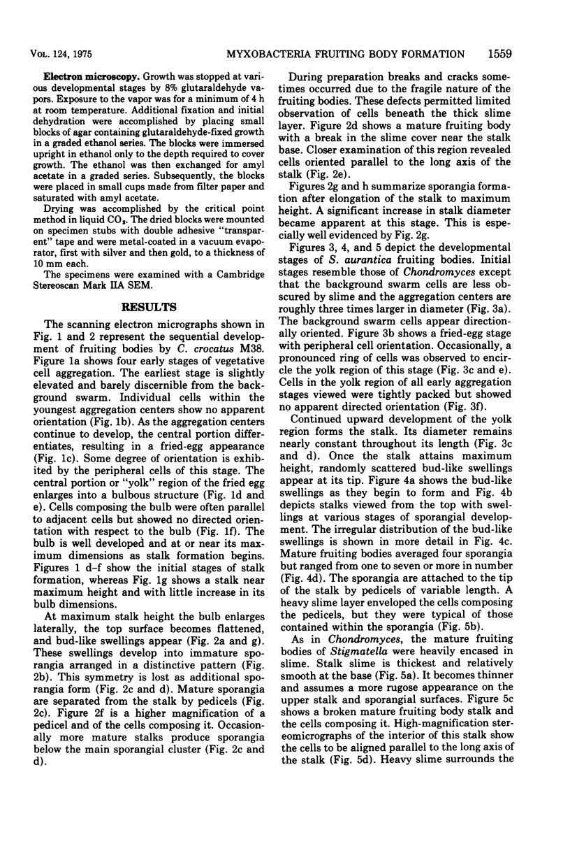
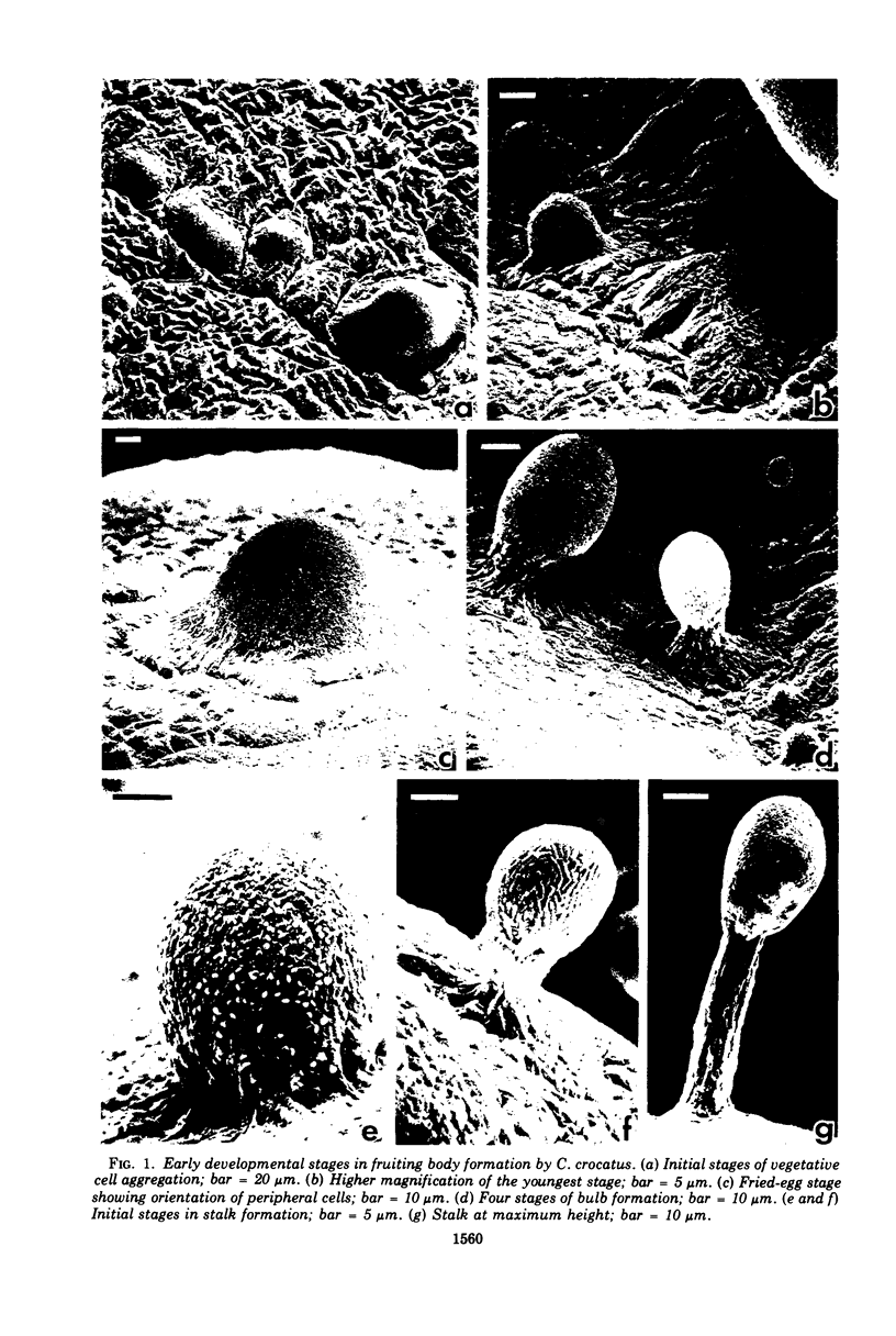
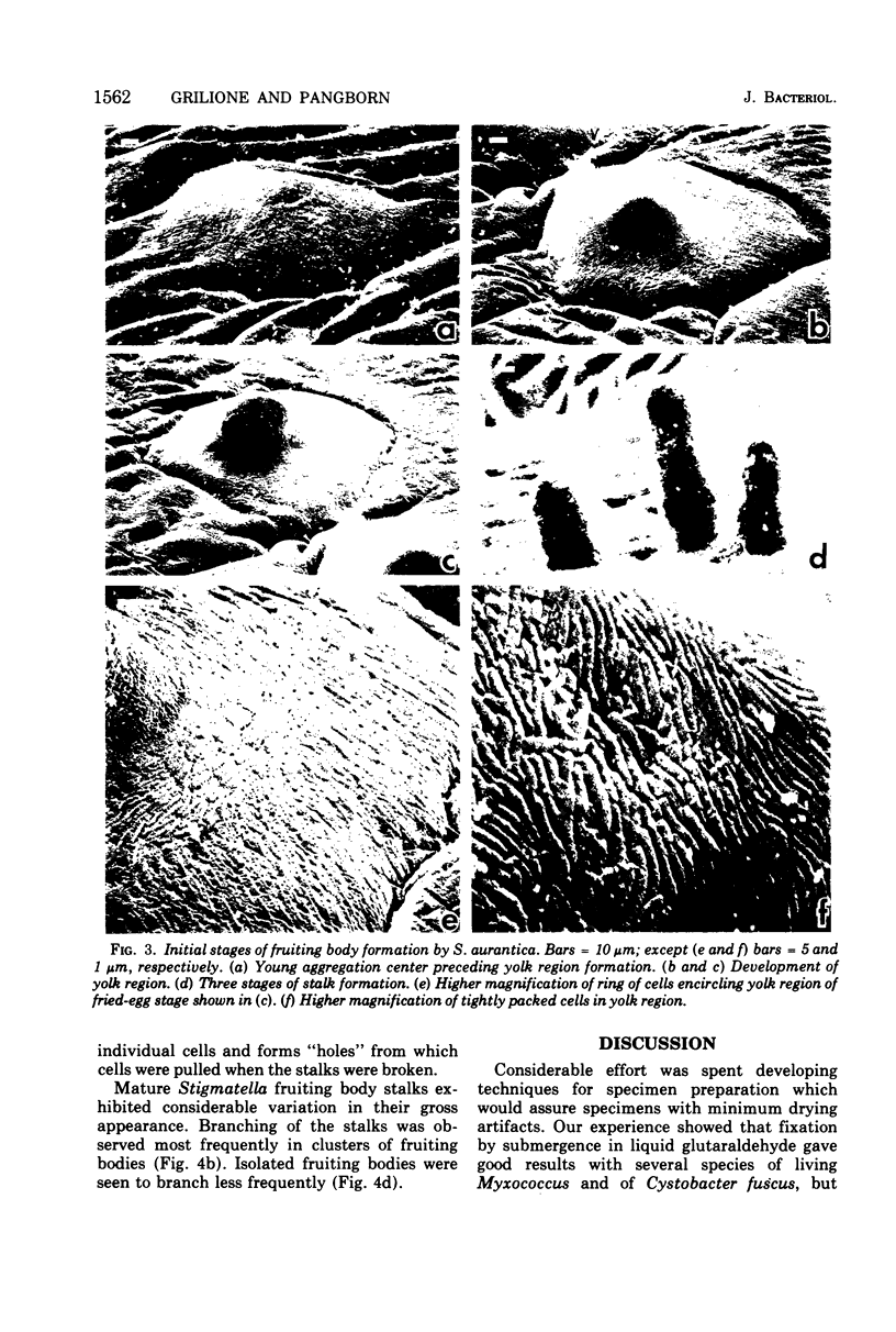
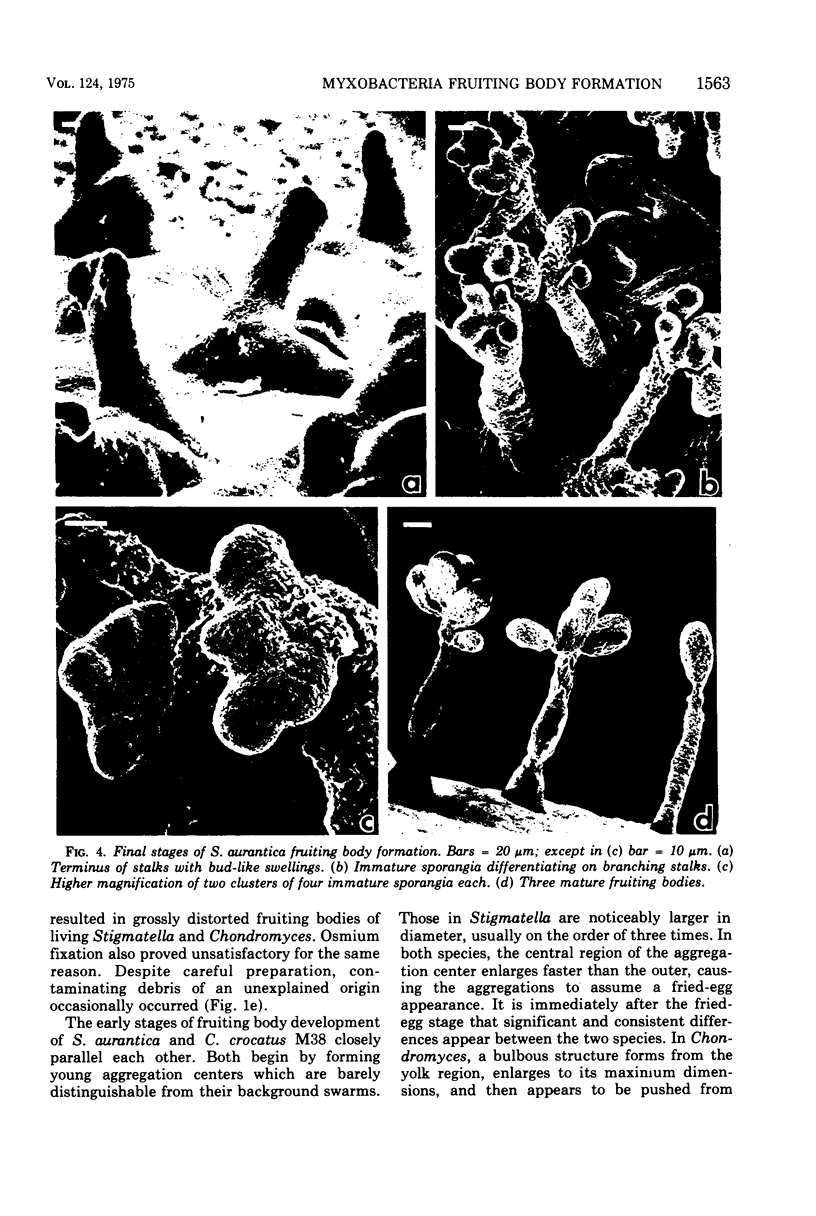
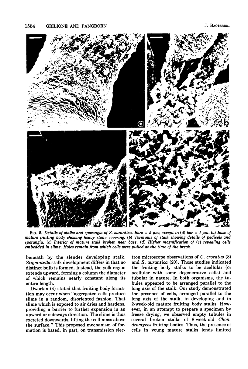
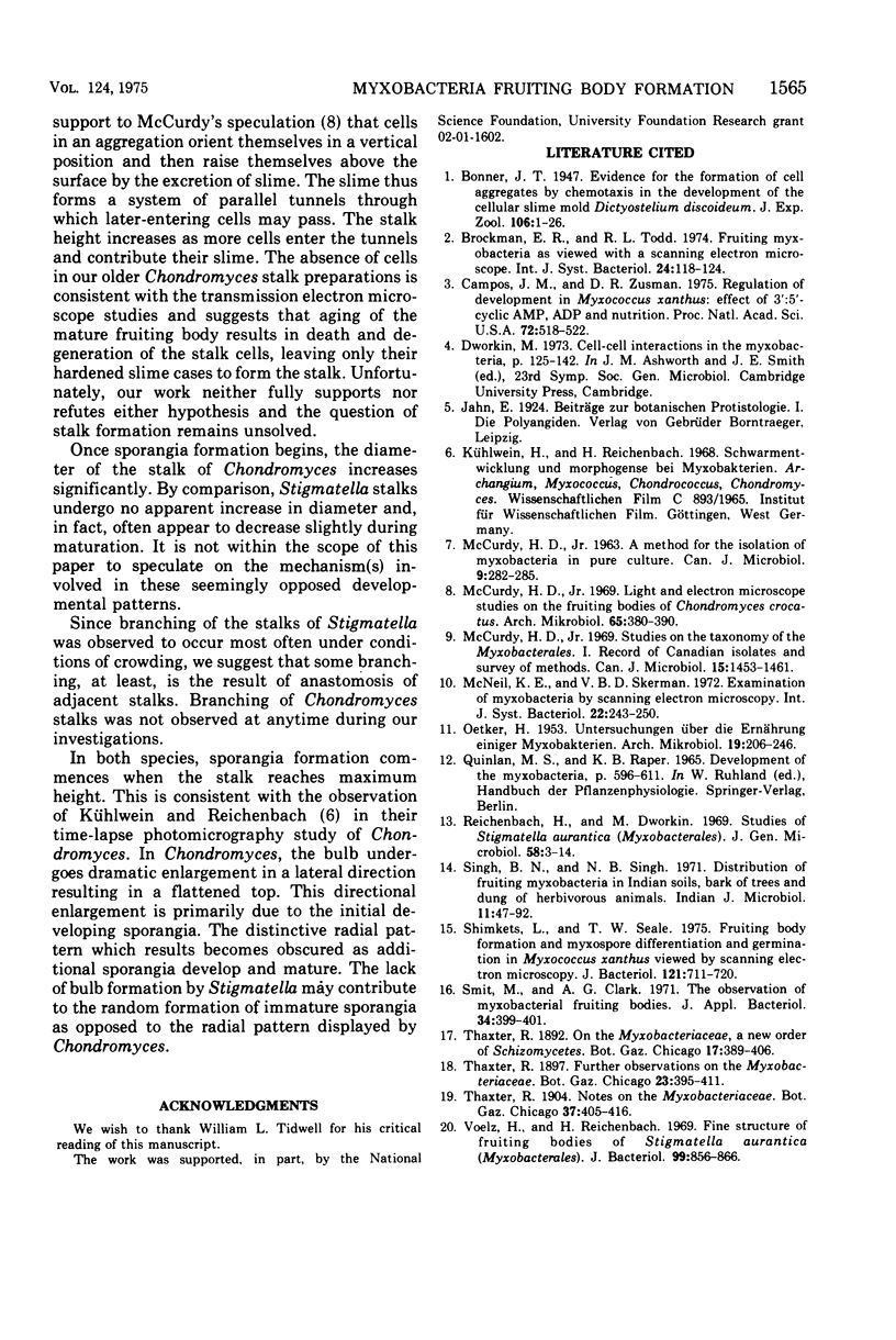
Scanning electron microscopy was used to follow fruiting body formation by pure cultures of Chondromyces crocatus M38 and Stigmatella aurantica. Vegetative cells were grown on SP agar and then transferred to Bonner salts agar for fructification. Fruiting in both species commences with the formation of aggregation centers which resemble a fried egg in appearance. In Chondromyces the elevated center or "yolk" region of the aggregation enlarges into a bulbous structure under which the stalk forms and lengthens. At maximum stalk height the bulb extends laterally as bud-like swellings appear. These are immature sporangia and are arranged in a distintive radial pattern around the top of the stalk. This symmetry is lost as more sporangia are formed. Stigmatella does not form a bulb; rather the yolk region of the aggregation center projects upward to form a column-like stalk which is nearly uniform in diameter throughout its length. At maximum stalk height, the terminus of the stalk develops an irregular pattern of bud-like swellings. These differentiate into sporangia. Stalks of 2-week-old mature fruiting bodies of both species appear to be cellular in composition. Stereomicrographs suggest orientation of these cells parallel to the long axis of the stalk. Stalks of 8-week-old fruiting bodies of Chondromyces were acellular and consisted of empty tubules, suggesting that the cells undergo degeneration with aging of the fruiting body.
Full text
PDF
Images in this article
Selected References
These references are in PubMed. This may not be the complete list of references from this article.
- Campos J. M., Zusman D. R. Regulation of development in Myxococcus xanthus: effect of 3':5'-cyclic AMP, ADP, and nutrition. Proc Natl Acad Sci U S A. 1975 Feb;72(2):518–522. doi: 10.1073/pnas.72.2.518. [DOI] [PMC free article] [PubMed] [Google Scholar]
- McCurdy H. D. Studies on the taxonomy of the Myxobacterales. I. Record of Canadian isolates and survey of methods. Can J Microbiol. 1969 Dec;15(12):1453–1461. doi: 10.1139/m69-259. [DOI] [PubMed] [Google Scholar]
- OETKER H. Untersuchungen über die Ernährung einiger Myxobakterien. Arch Mikrobiol. 1953;19(2):206–246. [PubMed] [Google Scholar]
- Shimkets L., Seale T. W. Fruiting-body formation and myxospore differentiation and germination in Mxyococcus xanthus viewed by scanning electron microscopy. J Bacteriol. 1975 Feb;121(2):711–720. doi: 10.1128/jb.121.2.711-720.1975. [DOI] [PMC free article] [PubMed] [Google Scholar]
- Voelz H., Reichenbach H. Fine structure of fruiting bodies of Stigmatella aurantiaca (Myxobacterales). J Bacteriol. 1969 Sep;99(3):856–866. doi: 10.1128/jb.99.3.856-866.1969. [DOI] [PMC free article] [PubMed] [Google Scholar]


