Abstract
NF-κB is a major transcription factor consisting of 50(p50)- and 65(p65)-kDa proteins that controls the expression of various genes, among which are those encoding cytokines, cell adhesion molecules, and inducible NO synthase (iNOS). After initial activation of NF-κB, which involves release and proteolysis of a bound inhibitor, essential cysteine residues are maintained in the active reduced state through the action of thioredoxin and thioredoxin reductase. In the present study, activation of NF-κB in human T cells and lung adenocarcinoma cells was induced by recombinant human tumor necrosis factor α or bacterial lipopolysaccharide. After lipopolysaccharide activation, nuclear extracts were treated with increasing concentrations of selenite, and the effects on DNA-binding activity of NF-κB were examined. Binding of NF-κB to nuclear responsive elements was decreased progressively by increasing selenite levels and, at 7 μM selenite, DNA-binding activity was completely inhibited. Selenite inhibition was reversed by addition of a dithiol, DTT. Proportional inhibition of iNOS activity as measured by decreased NO products in the medium (NO2− and NO3−) resulted from selenite addition to cell suspensions. This loss of iNOS activity was due to decreased synthesis of NO synthase protein. Selenium at low essential levels (nM) is required for synthesis of redox active selenoenzymes such as glutathione peroxidases and thioredoxin reductase, but in higher toxic levels (>5–10 μM) selenite can react with essential thiol groups on enzymes to form RS–Se–SR adducts with resultant inhibition of enzyme activity. Inhibition of NF-κB activity by selenite is presumed to be the result of adduct formation with the essential thiols of this transcription factor.
Keywords: selenium, human cell lines
The nuclear factor NF-κB is a transcription factor that regulates a number of cellular genes, such as those encoding inflammatory cytokines, adhesion molecules, Rel proteins (1, 2), and inducible NO synthase (iNOS) (3–5). Activation of the provirus form of HIV also involves the transcriptional action of NF-κB (6, 7). NF-κB activation is a complex process that can be triggered initially by many agents, such as inflammatory cytokines, mitogens, bacterial products, protein synthesis inhibitors, reactive oxygen species, UV light, and phorbol esters (1, 2, 8). The actual activation of NF-κB involves a cascade of events that includes phosphorylation–dephosphorylation reactions, release and proteolysis of a bound inhibitor, IκB-α (5), translocation of NF-κB to the nucleus, and maintenance of essential cysteine residues in the protein in the active thiol forms by the thioredoxin–thioredoxin reductase system. Reduced cysteine residue(s) of NF-κB appear to be required for its actual binding to the promotor region of a target gene (11). Dependence of the early activation steps of this cascade on reactive oxygen species is indicated by the inhibitory effects of added N-acetylcysteine and metal chelators (9, 10) and increased intracellular levels of phospholipid hydroperoxide glutathione peroxidase (12).
iNOS, which generates NO by oxidation of l-arginine is induced by exposure of cells to bacterial lipopolysaccharide (LPS) or some cytokines (13) and is controlled at several stages in its synthesis (14). Induction of iNOS in macrophages was shown to be inhibited by agents that block the protease-dependent step of NF-κB activation (5).
Selenium, which is an essential trace element for higher eukaryotes and many bacteria, is also very toxic at elevated levels (15, 16). Low (40–100 nM) levels of added selenite stimulate mammalian cell growth in commonly used culture media, and in serum-free media, selenium supplementation is essential (17). Even under the obligate aerobic growth conditions required for mammalian cells, growth can be inhibited by μM levels of added selenite. In the absence of oxygen, growth of selenium-requiring anerobic bacteria usually is inhibited when selenite levels are increased 10-fold above the required 0.5- to 1-μM levels. Several enzymes that contain essential reactive sulfhydryl groups have been shown to be especially sensitive to selenite treatment (18–22). Inhibition due to formation of a selenotrisulfide type of adduct was established as the mechanism of selenite action in the case of rat brain prostaglandin D synthase (19), and this inhibition was reversed by an excess of DTT. The transcription factor AP-1 also is sensitive to selenite. Spyrou et al. (23) reported that both selenite and selenodiglutathione (GS–Se–SG) inhibited binding of AP-1 to DNA in lymphocyte 3B6 nuclear extracts. Inhibition of AP-1 binding to DNA by selenite was shown by Handel et al. (24) to require Cys272 and Cys154 in the DNA-binding domains of the Jun and Fos components, respectively, of AP1. Recently, Makropoulos et al. (25) suggested that selenium supplementation might be used to modulate the expression of NF-κB target genes and HIV-1. The aim of the present study was to investigate the effects of selenite on iNOS induction by NF-κB.
MATERIALS AND METHODS
Cell Culture.
A human Jurkat T cell line, JPX9 (26), and a human lung adenocarcinoma cell line, NCI-H441 (ATCC HTB-174), were grown in RPMI 1640 medium containing 10% heat-inactivated fetal bovine serum and antibiotic–antimycotic solution under a 90% relative humidity and 5% CO2 at 37°C.
Preparation of Nuclear Extracts from Cells.
JPX9 cells and NCI-H441 cells (2 × 10⁶ cells/ml) were treated with an activator, human tumor necrosis factor α (TNF-α) or bacterial LPS overnight at 37°C. Nuclear extracts were then prepared according to Singh and Aggarwal (27). In brief, 2 × 10⁷ cells were washed with ice-cold PBS (1× PBS) and suspended in 1.0 ml of ice-cold lysis buffer (10 mM Hepes, pH 7.9/10 mM KCl/0.1 mM EDTA/0.1 mM EGTA/1 mM DTT/0.5 mM phenylmethylsulfonyl fluoride/2.0 μg/ml leupeptin/2.0 μg/ml aproprotinin/0.5 μg/ml benzaidine) in a microcentrifuge tube. The cells were held on ice for 15 min and then 31 μl of 10% Nonidet P-40 (Nonidet P-40) was added. The suspension was mixed vigorously for 10 s, and the lysate was centrifuged for 30 s. The nuclear pellet was resuspended in 50 μl of ice-cold nuclear extraction buffer (20 mM Hepes, pH 7.9/400 mM NaCl/0.1 mM EDTA/0.1 mM EGTA/1 mM DTT/1 mM phenylmethylsulfonyl fluoride/2.0 μg/ml leupeptin/2.0 μg/ml aproprotinin/0.5 μg/ml benzaidine) and incubated on ice for 30 min with intermittent mixing. The sample was centrifuged for 5 min, and the supernatant (nuclear extract) was either used immediately or stored at −70°C. The protein concentration was determined by the Coomassie brilliant blue G-250 dye-binding method (28) using the Bio-Rad dye reagent.
Assay of NF-κB DNA Binding Activity.
Electrophoretic mobility shift assays were carried out for determination of NF-κB DNA binding activity using 4 μg of nuclear extract. Extracts were preincubated with various concentrations of sodium selenite at room temperature for 20 min. The treated nuclear extracts were incubated with 10 fmol of 5′ 32P-labeled, 22-mer, double-stranded NF-κB consensus oligonucleotide (5′-AGTTGAGGGGACTTTCCCAGGC-3′; Santa Cruz Biotechnology) for 20 min at room temperature. The reaction mixture contained 3 μg of poly(dI-dC) in a binding buffer [10 mM Tris⋅HCl, pH 7.5/1 mM EDTA/0.5 mM DTT/1% Nonidet P-40/5% glycerol/50 mM NaCl/1 mM MgCl2/3 mM GTP/0.1 mg/ml BSA (29, 30)]. The resulting DNA–protein complex was separated from unbound oligonucleotides on a 6% nondenaturing polyacrylamide gel using 0.5× TBE buffer (45 mM Tris/45 mM boric acid/0.1 mM EDTA, pH 8.3). The gel was dried and the radioactivity visualized using a PhosphoImager (Molecular Dynamics). A double-stranded, mutated oligonucleotide (5′-AGTTGAGGCGACTTTCCCAGGC-3′; Santa Cruz Biotechology) was used to examine the specificity of NF-κB binding to DNA.
iNOS in Cells.
JPX9 cells or NCI-H441 cells grown in RPMI 1640 medium containing 10% fetal bovine serum and 1% antibiotic–antimycotic solution were harvested by centrifugation and resuspended at the density of 1 × 10⁶ cells/ml in the same medium without phenol red. Cells (1 × 10⁷) were plated in 10-cm dishes for NCI-H441 or 75-cm flasks for JPX9, activated by addition of LPS at 75 ng/ml, and incubated overnight. After incubation, various concentrations of sodium selenite were added to the cell suspensions, which were incubated for an additional 3 h. Cells were harvested for preparation of nuclear extracts, and the cell-free medium was analyzed for NO production.
Determination of NO production.
NO production was determined by measuring the sum of both NO2− and NO3−, the final products of NO in vivo, by using the Griess reagent (Cayman Chemicals, Ann Arbor, MI) (31). The absorbance of the deep purple azo compound produced was measured at 540 nm using the plate reader.
RESULTS
Selenite Inhibits NF-κB Binding to DNA.
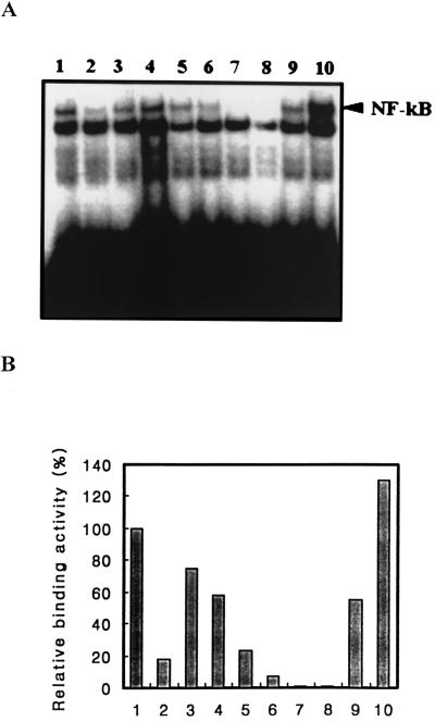
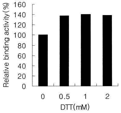
To study the effect of selenite on the DNA-binding activity of transcription factor NF-κB, human Jurkat T cell (JPX9) suspensions (2 × 10⁶ cells/ml) were incubated initially with 0.1 nM human TNF-α to initiate the NF-κB activation cascade. Nuclear extracts were prepared from these treated cells and assayed for NF-κB DNA-binding activity by the electrophoretic mobility shift assay. An initial 20-min incubation of these Jurkat T cell extracts with selenite, before addition of binding buffer and 5′ 32P-labeled, 22-mer, double-stranded NF-κB consensus oligonucleotide, inhibited binding of the labeled oligonucleotide as shown in Fig. 1. Treatment with 7 μM selenite was sufficient to completely inhibit the DNA-binding activity of NF-κB in the extract. When an excess amount of unlabeled NF-κB consensus oligonucleotide was added to the reaction mixture, the retarded radioactive band disappeared (Fig. 1A, lane 2) whereas the mutated consensus oligonucleotide (Fig. 1A, lane 3) failed to compete showing specificity of the binding reaction. Inhibition of NF-κB binding to DNA caused by treatment with 10 μM selenite was reversed by subsequent addition of a reducing agent such as DTT (Fig. 1). Incubation of the selenite-treated extract with 1 mM DTT restored 50% of binding activity, and 2 mM DTT gave complete recovery under the experimental conditions used (Fig. 1, lanes 9 and 10). Addition of the dithiol to the “as prepared” nuclear extract stimulated NF-κB binding activity in the mobility shift assay to the extent of ≈40% (Fig. 2), indicating that essential cysteine residues in the transcription factor were not fully reduced in the preparation studied. In similar experiments carried out with nuclear extracts of human Jurkat T cells, initially activated by treatment with bacterial LPS and with nuclear extracts of human lung adenocarcinoma cells (NCI-H441), and stimulated either with LPS or with human TNF-α, the same sensitivity to selenite treatment was observed (data not shown).
Figure 1.
Effects of sodium selenite on NF-κB DNA binding activity with Jurkat T cell nuclear extracts. (A) Nuclear extracts prepared from human Jurkat T cells preincubated with 0.1 nM human TNF-α at 37°C for 20 min were examined for NF-κB activation by electrophoretic mobility shift assay after treatments with various concentrations of selenite. Lanes: 1, 0 selenite; 2, 0 selenite with wild-type NF-κB consensus oligonucleotide; 3, 0 selenite with mutant NF-κB consensus oligonucleotide; 4, 1 μM selenite; 5, 3 μM selenite; 6, 5 μM selenite; 7, 7 μM selenite; 8, 10 μM selenite; 9, 10 μM selenite with 1 mM DTT; and 10, 10 μM selenite with 2 mM DTT. (B) Quantitative representation of A. The resulting autoradiograms of NF-κB-DNA complex were quantitated by PhosphoImager analysis.
Figure 2.
Effect of DTT on NF-κB DNA binding. Nuclear extracts prepared from human Jurkat T cells preincubated with 0.1 nM human TNF-α at 37°C for 20 min were examined for NF-κB activation by electrophoretic mobility shift assay in the presence of various concentrations of DTT as indicated.
Selenite Inhibition in Vivo.
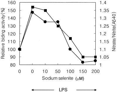
To investigate the effect of selenite treatment in vivo on binding of NF-κB to DNA, Jurkat T cells and human lung adenocarcinoma cells were initially activated by overnight incubation in suitable culture media with bacterial LPS (75 ng/ml) and then incubated an additional 3 h after addition of increasing concentrations of selenite. This treatment did not influence cell viability as determined by the tryptophan blue exclusion method (data not shown). The treated cells were washed with fresh culture medium without selenite, nuclear extracts were prepared, and NF-κB binding activity to DNA was determined by the mobility shift assay. As shown in Fig. 3, there was a dose-dependent inhibition of NF-κB binding to DNA in the nuclear extracts prepared from LPS-activated Jurkat T cells treated with various concentrations of selenite. This DNA-binding activity was abolished by exposure of the intact cells to 100 μM selenite. Similar inhibition was observed in nuclear extracts prepared from human lung adenocarcinoma cells that were treated by the same protocol.
Figure 3.
Selenite-mediated inhibition of NF-κB DNA binding and NO production. The human Jurkat T cells, JPX9, were preincubated overnight with bacterial LPS at 75 ng/ml and then treated with indicated concentrations of sodium selenite for 3 h. For analysis of NF-κB activation (•), nuclear extracts were prepared from untreated and LPS-treated cells, and NF-κB DNA binding was determined by electrophoretic mobility shift assays. The resulting autoradiograms of NF-κB-DNA complex were quantitated by PhosphorImager. For analysis of NO production (▪), the final products of NO in vivo (sum of both NO2− and NO3−) in the culture media of untreated and LPS-treated cells were measured as described in Materials and Methods. ←LPS→ indicates the cultured cells that were retreated with bacterial LPS.
Selenite and iNOS.
The gene for inducible NO synthase has a NF-κB binding sequence motif in the promoter region (3, 4), and the transcriptional activity of NF-κB has been shown by Griscavage et al. (5) to be involved in induction of iNOS by LPS activation and, by Bereta et al. (33), for expression of iNOS in murine endothelial cells. To see if the inhibitory effect of selenite on NF-κB binding to DNA as measured in the mobility shift assay also resulted in decreased NO production by selenite-treated Jurkat T cells, the amounts of NO2− and NO3− present in the culture medium were measured as an estimation of total NO formed. As shown in Fig. 3 (solid squares), the inhibition pattern of NO production is proportional to the inhibition of NF-κB DNA binding activity observed in nuclear extracts of the same selenite-treated cells (Fig. 3, solid circles). The increased DNA binding activity and NO production observed after LPS induction of transcriptional factor activity in the T cells was progressively inhibited by increasing concentrations of selenite. Treatment with 100 μM selenite completely returned both LPS-stimulated activities to the baseline levels.
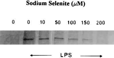
The NO production that decreased as a function of selenite treatment of the cultured T cells was shown to be the result of decreased iNOS protein levels rather than direct inhibition of enzyme activity. Immunoblot assays using polyclonal antibodies elicited to human iNOS protein (Fig. 4) clearly show the induction of enzyme protein resulting from LPS treatment and the progressive decreases in protein levels brought about by subsequent incubation for 3 h with high concentrations of selenite. This decrease in enzyme protein levels during incubation in the presence of selenite was associated with a 25% decrease in the total NO produced in the cultures. As shown in Fig. 4, actual loss of detectable immunoreactive protein occurred in cells exposed to 150–200 μM selenite for 3 h, and NO production in these cultures ceased (Fig. 3). In these cells, selenite treatment not only inhibited further expression of NO synthase but also appears to have caused modification or actual decomposition of enzyme protein already present at the time of selenite addition.
Figure 4.
Selenite-mediated inhibition of iNOS synthesis. Crude extracts were prepared from human Jurkat T cells treated with the indicated concentrations of selenite as described in the legend of Fig. 3 and immunoblotted with a polyclonal antibody against human iNOS, NOS2 (Santa Cruz Biotechnology). ←LPS→ indicates the cells that were treated with bacterial LPS.
DISCUSSION
Numerous studies (11, 23, 24, 32, 34) on the regulation of transcriptional activities of NF-κB and AP-1 have shown that binding of these transcription factors to their target DNA sites depends on the redox state of specific cysteine residues in these proteins. Direct reduction of these cysteine residues by thioredoxin or by thiols such as DTT has been demonstrated in vitro in the case of NF-κB, and, because thioredoxin is known to be transported from the cytoplasm to the nucleus, it should play the same role in vivo (11, 35). In the case of AP-1, reduction of essential cysteine residues in the Fos and Jun subunits is achieved indirectly by a cascade involving the interaction of thioredoxin with a nuclear redox factor, Ref-1, already present in the nucleus (24, 32, 34, 35). Selenite addition to nuclear extracts from human 3B6 lymphocytes was shown to be an efficient inhibitor of the DNA-binding activity of AP-1 (23). It was suggested that this could involve interaction of selenite either with the cysteine residues of Fos and Jun, the Cys-72 residue of thioredoxin, sulfhydryl groups of Ref-1 or all three as possible targets. Partial protection of AP-1 DNA-binding activity from selenite inhibition was observed when a reducing system such as thioredoxin, thioredoxin reductase and NADPH, or DTT was included in the reaction mixture. Because selenite readily forms complexes of the RS–Se–SR and RS–Se− types with reactive sulfhydryl groups in proteins and these adducts are decomposed by various reducing agents, such derivatives could account for the observed inhibition of AP-1 binding to its DNA targets by reactive selenium compounds. In other cases, particularly for rat brain prostaglandin D synthase (19), such a mechanism is a likely explanation of the observed inhibitory effects of selenite and selenodiglutathione (RS–Se–SR) on activity of the isolated enzyme.
In the present study, the inhibitory effect of selenite on the DNA-binding activity of NF-κB was extended to show that the level of a target gene product also was decreased by selenite treatment. The presence of a NF-κB binding sequence in the promoter region of the iNOS gene (3, 4) together with the feasibility of estimating levels of the gene product by immunoblotting and activity of the enzyme in cultured cells by measurement of the reaction product (NO) made this an ideal system of study. Thus, it could be shown that a 3-h treatment of LPS-activated aerobic cultures of human T cells and human lung adenocarcinoma cells with increasing concentrations of selenite, under conditions that had no apparent effect on cell viability, was sufficient (i) to cause a dose-dependent inhibition of the NF-κB DNA binding activity that had been induced by LPS treatment, (ii) to decrease NO production proportionally and (iii), to result in the fall of iNOS protein to a level undetectable in the immunoblot assay using antibodies elicited to the human enzyme. This loss of enzyme protein observed at the highest selenite levels indicates that gene expression ceased and also that protein inactivation, possibly by proteolysis, was induced.
The inhibitory effects of selenite on the expression of a gene under the control of NF-κB shown in the present study offer a possible explanation of the marked toxicity of elevated levels of this form of selenium to animals and many types of bacteria. In addition to the known inhibition of specific isolated enzymes (18–22) by selenite and RS–Se–SR types of thiol adducts, inhibitory effects that can result from inactivation of essential thiol groups of transcription factors such as AP-1 (23) and NF-κB, which control many cellular processes, may be greatly amplified and become global in nature. More detailed study of this type of regulation of cellular activity by selenium compounds may help to explain the well known but poorly understood toxicity of selenium.
ABBREVIATIONS
- iNOS
inducible NO synthase
- TNF
tumor necrosis factor
References
- 1.Baeuerle P A. Biochim Biophys Acta. 1991;1072:63–80. doi: 10.1016/0304-419x(91)90007-8. [DOI] [PubMed] [Google Scholar]
- 2.Grilli M, Jason J-S, Lenardo M J. Int Rev Cytol. 1993;143:1–62. doi: 10.1016/s0074-7696(08)61873-2. [DOI] [PubMed] [Google Scholar]
- 3.Xie Q-W, Kashiwabara Y, Nathan C. J Exp Med. 1993;177:1779–1784. doi: 10.1084/jem.177.6.1779. [DOI] [PMC free article] [PubMed] [Google Scholar]
- 4.Xie Q-W, Whisnant R, Nathan C. J Biol Chem. 1994;269:4705–4708. [PubMed] [Google Scholar]
- 5.Griscavage J M, Wilk S, Ignarro L J. Proc Natl Acad Sci USA. 1996;93:3308–3312. doi: 10.1073/pnas.93.8.3308. [DOI] [PMC free article] [PubMed] [Google Scholar]
- 6.Hazan U, Thomas D, Alcami J, Bachelerie F, Israel N, Yssel H, Virelizier J L, Arenzana-Seisdedos F. Proc Natl Acad Sci USA. 1990;87:7861–7865. doi: 10.1073/pnas.87.20.7861. [DOI] [PMC free article] [PubMed] [Google Scholar]
- 7.Schreck R, Rieber P, Baeuerle P A. EMBO J. 1991;10:2247–2258. doi: 10.1002/j.1460-2075.1991.tb07761.x. [DOI] [PMC free article] [PubMed] [Google Scholar]
- 8.Schulze-Osthoff K, Los M, Baeuerle P A. Biochem Pharmacol. 1995;50:735–741. doi: 10.1016/0006-2952(95)02011-z. [DOI] [PubMed] [Google Scholar]
- 9.Staal F J T, Roederer M, Herzenberg L A, Herzenberg L A. Proc Natl Acad Sci USA. 1990;87:9943–9947. doi: 10.1073/pnas.87.24.9943. [DOI] [PMC free article] [PubMed] [Google Scholar]
- 10.Schreck R, Meier B, Mannel D N, Droge W, Baeuerle P A. J Exp Med. 1992;175:1181–1194. doi: 10.1084/jem.175.5.1181. [DOI] [PMC free article] [PubMed] [Google Scholar]
- 11.Hayashi T, Ueno Y, Okamoto T. J Biol Chem. 1993;268:11380–11388. [PubMed] [Google Scholar]
- 12.Brigelius-Flohe, R., Friedrichs, B., Mauer, S., Schultz, M. & Streicher, R. (1997) Biochem. J., in press. [DOI] [PMC free article] [PubMed]
- 13.Xie Q-W, Nathan C. J Leukocyte Biol. 1994;56:576–582. doi: 10.1002/jlb.56.5.576. [DOI] [PubMed] [Google Scholar]
- 14.Nathan C, Xie Q-W. J Biol Chem. 1994;269:13725–13728. [PubMed] [Google Scholar]
- 15.Stadtman T C. Science. 1973;183:915–921. doi: 10.1126/science.183.4128.915. [DOI] [PubMed] [Google Scholar]
- 16.Stadtman T C. In: Advances in Inorganic Biochemistry. Eichhorn G L, Marzilli L G, editors. Englewood Cliffs, NJ: Prentice–Hall; 1994. pp. 157–175. [Google Scholar]
- 17.McKeehan W L, Hamilton W G, Ham R G. Proc Natl Acad Sci USA. 1976;73:2023–2027. doi: 10.1073/pnas.73.6.2023. [DOI] [PMC free article] [PubMed] [Google Scholar]
- 18.Ganther H E, Corcoran C. Biochemistry. 1969;8:2557–2563. doi: 10.1021/bi00834a044. [DOI] [PubMed] [Google Scholar]
- 19.Islam F, Watanabe Y, Morii H, Hayaishi O. Arch Biochem Biophys. 1991;289:161–166. doi: 10.1016/0003-9861(91)90456-s. [DOI] [PubMed] [Google Scholar]
- 20.Lopez S, Miyashita Y, Simons S S., Jr J Biol Chem. 1990;265:16039–16042. [PubMed] [Google Scholar]
- 21.Frenkel G D, Walcott A, Middleton C. Mol Pharmacol. 1987;31:112–116. [PubMed] [Google Scholar]
- 22.Vernie L N, Collard J G, Eker A P M, de Wildt A, Wilders I T. Biochem J. 1979;180:213–218. doi: 10.1042/bj1800213. [DOI] [PMC free article] [PubMed] [Google Scholar]
- 23.Spyrou G, Bjornstedt M, Kumar S, Holmgren A. FEBS Lett. 1995;368:59–63. doi: 10.1016/0014-5793(95)00599-5. [DOI] [PubMed] [Google Scholar]
- 24.Handel M L, Watts C L, DeFazio A, Day R O, Sutherland R L. Proc Natl Acad Sci USA. 1995;92:4497–4501. doi: 10.1073/pnas.92.10.4497. [DOI] [PMC free article] [PubMed] [Google Scholar]
- 25.Makropoulos V, Bruning T, Schulze-Osthoff K. Arch Toxicol. 1996;70:277–283. doi: 10.1007/s002040050274. [DOI] [PubMed] [Google Scholar]
- 26.Nagata K, Ohtani K, Nakamura M, Sugamura K. J Virol. 1989;63:3220–3226. doi: 10.1128/jvi.63.8.3220-3226.1989. [DOI] [PMC free article] [PubMed] [Google Scholar]
- 27.Singh S, Aggarwal B B. J Biol Chem. 1995;270:24995–25000. doi: 10.1074/jbc.270.42.24995. [DOI] [PubMed] [Google Scholar]
- 28.Bradford M. Anal Biochem. 1976;72:248–254. doi: 10.1016/0003-2697(76)90527-3. [DOI] [PubMed] [Google Scholar]
- 29.Collart M A, Baeuerle P A, Vassalli P. Mol Cell Biol. 1990;10:1498–1506. doi: 10.1128/mcb.10.4.1498. [DOI] [PMC free article] [PubMed] [Google Scholar]
- 30.Hassanain H H, Dai W, Gupta S L. Anal Biochem. 1993;213:162–167. doi: 10.1006/abio.1993.1400. [DOI] [PubMed] [Google Scholar]
- 31.Green L C, Wagner D A, Glogowski J, Skipper P L, Wishnok J S, Tannenaum J S. Anal Biochem. 1982;126:131–138. doi: 10.1016/0003-2697(82)90118-x. [DOI] [PubMed] [Google Scholar]
- 32.Abate C, Patel L, Rauscher F J, III, Curran T. Science. 1990;249:1157–1161. doi: 10.1126/science.2118682. [DOI] [PubMed] [Google Scholar]
- 33.Bereta J, Cohen M C, Bereta M. FEBS Lett. 1995;377:21–25. doi: 10.1016/0014-5793(95)01301-6. [DOI] [PubMed] [Google Scholar]
- 34.Hirota K, Matsui M, Iwata S, Nishiyama A, Mori K, Yodoi J. Proc Natl Acad Sci USA. 1997;94:3633–3638. doi: 10.1073/pnas.94.8.3633. [DOI] [PMC free article] [PubMed] [Google Scholar]
- 35.Nakamura H, Nakamura K, Yodoi J. Ann Rev Immunol. 1996;15:351–369. doi: 10.1146/annurev.immunol.15.1.351. [DOI] [PubMed] [Google Scholar]


