

Abstract
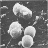
Size frequency distributions of different phototrophic and heterotrophic microorganisms were determined by means of scanning and transmission electron microscopy and electronic particle sizing. Statistically significant differences existed among the three techniques used in this study. Cells processed for electron microscopy showed lower mean cellular volumes than those processed for electronic particle sizing, reflecting a shrinkage by factors ranging from 1.1 to 6.2 (mean, 2.3). Processing of cells for scanning electron microscopy caused higher shrinkage than processing for transmission electron microscopy. Shrinkage was dependent neither on the size nor on the cell wall type of the microorganism. When processed for scanning electron microscopy, phototrophic bacteria were strongly shrunken, whereas heterotrophic microorganisms were less affected. A direct relationship existed among phototrophic bacteria between percentage of shrinkage and specific pigment content. This was probably a consequence of the pigment extraction by organic solvents during the dehydration process, previous to the critical point drying, necessary to examine the specimens under the scanning electron microscope.
Full text
PDF


Images in this article
Selected References
These references are in PubMed. This may not be the complete list of references from this article.
- Aiking H., Sojka G. Response of Rhodopseudomonas capsulata to illumination and growth rate in a light-limited continuous culture. J Bacteriol. 1979 Aug;139(2):530–536. doi: 10.1128/jb.139.2.530-536.1979. [DOI] [PMC free article] [PubMed] [Google Scholar]
- Bowden W. B. Comparison of two direct-count techniques for enumerating aquatic bacteria. Appl Environ Microbiol. 1977 May;33(5):1229–1232. doi: 10.1128/aem.33.5.1229-1232.1977. [DOI] [PMC free article] [PubMed] [Google Scholar]
- Broch-Due M., Ormerod J. G., Fjerdingen B. S. Effect of light intensity of vesicle formation in chlorobium. Arch Microbiol. 1978 Mar;116(3):269–274. doi: 10.1007/BF00417850. [DOI] [PubMed] [Google Scholar]
- Krambeck C., Krambeck H. J., Overbeck J. Microcomputer-assisted biomass determination of plankton bacteria on scanning electron micrographs. Appl Environ Microbiol. 1981 Jul;42(1):142–149. doi: 10.1128/aem.42.1.142-149.1981. [DOI] [PMC free article] [PubMed] [Google Scholar]
- Pedrós-Alió C., Brock T. D. Assessing biomass and production of bacteria in eutrophic lake mendota, wisconsin. Appl Environ Microbiol. 1982 Jul;44(1):203–218. doi: 10.1128/aem.44.1.203-218.1982. [DOI] [PMC free article] [PubMed] [Google Scholar]
- Trentini W. C., Starr M. P. Growth and ultrastructure of Rhodomicrobium vannielii as a function of light intensity. J Bacteriol. 1967 May;93(5):1699–1704. doi: 10.1128/jb.93.5.1699-1704.1967. [DOI] [PMC free article] [PubMed] [Google Scholar]
- Trueba F. J., Woldringh C. L. Changes in cell diameter during the division cycle of Escherichia coli. J Bacteriol. 1980 Jun;142(3):869–878. doi: 10.1128/jb.142.3.869-878.1980. [DOI] [PMC free article] [PubMed] [Google Scholar]
- Watson S. W., Novitsky T. J., Quinby H. L., Valois F. W. Determination of bacterial number and biomass in the marine environment. Appl Environ Microbiol. 1977 Apr;33(4):940–946. doi: 10.1128/aem.33.4.940-946.1977. [DOI] [PMC free article] [PubMed] [Google Scholar]