

Abstract
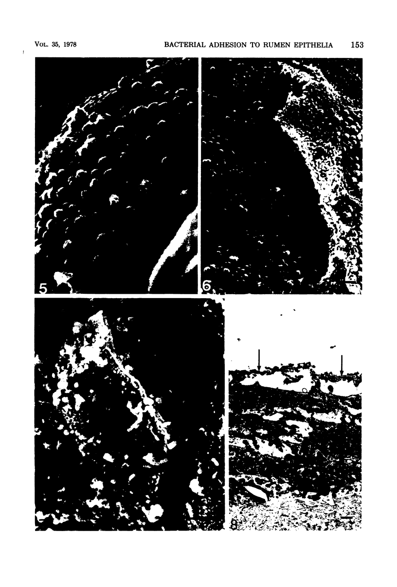
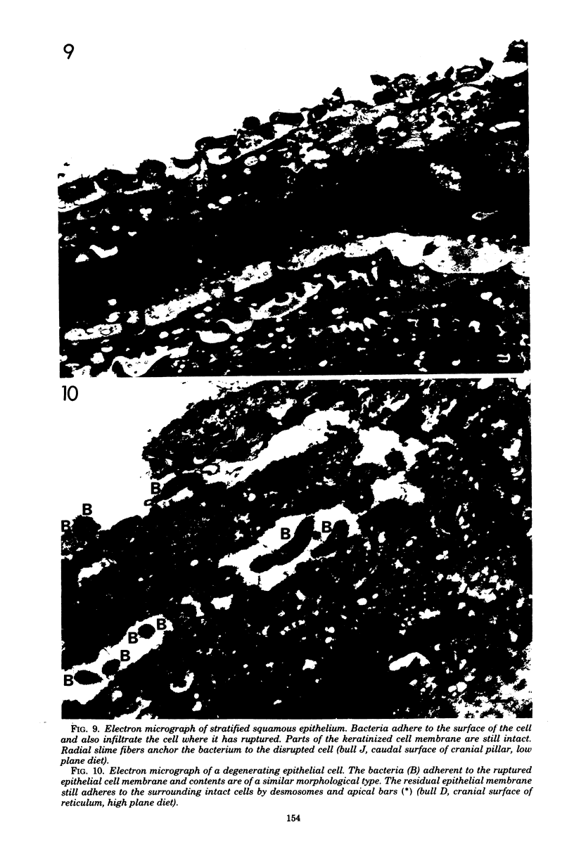
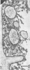
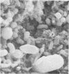
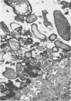
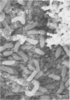
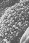
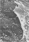
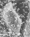
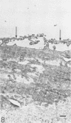
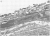
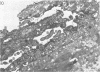
Blocks of tissue were removed from various locations in the bovine digestive tract and fixed and processed for transmission and scanning electron microscopy by techniques that retained adherent bacteria. The distribution of bacteria on the surface of epithelial cells was examined by scanning electron microscopy. This showed intermittent colonization of the epithelia with the formation of occasional microcolonies of morphologically similar bacterial cells. Transmission electron microscopy of ruthenium red-stained material showed the presence of both the glycocalyx of the bovine epithelial cells and fibrous carbohydrate coats surrounding adherent bacteria. The carbohydrate coats appeared to mediate the attachment of bacteria to the epithelium, to food particles, and to each other so that microcolonies were formed. Careful examination of the bacterial colonization of keratinized cells in the process of being sloughed from the surface of the stratified squamous epithelium of the rumen showed that these dead cells were digested by adherent bacteria of a limited number of morphological types. The spatial relationship of this mixed, adherent, microbial population to living and dead epithelial cells and to food particles indicates that digestive processes of some importance may be accomplished by this stationary component of the microbial flora of the digestive tract.
Full text
PDF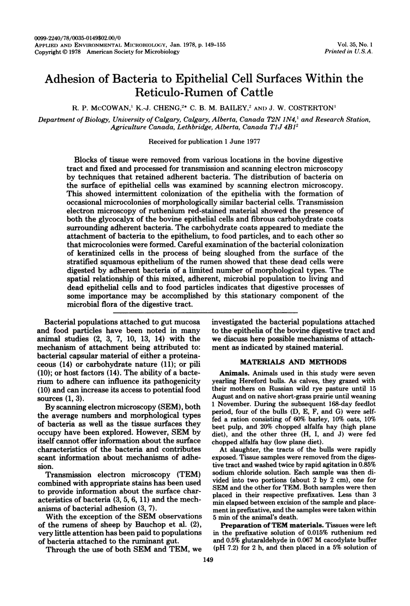
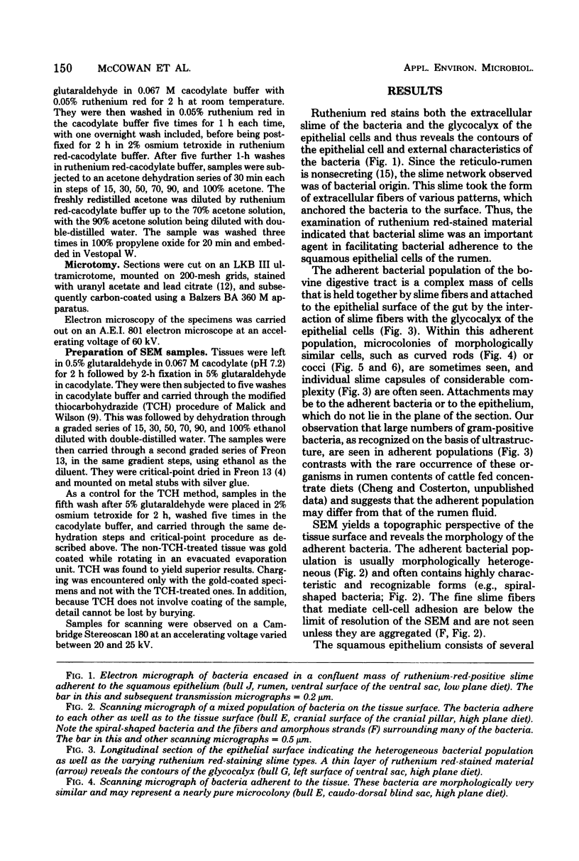
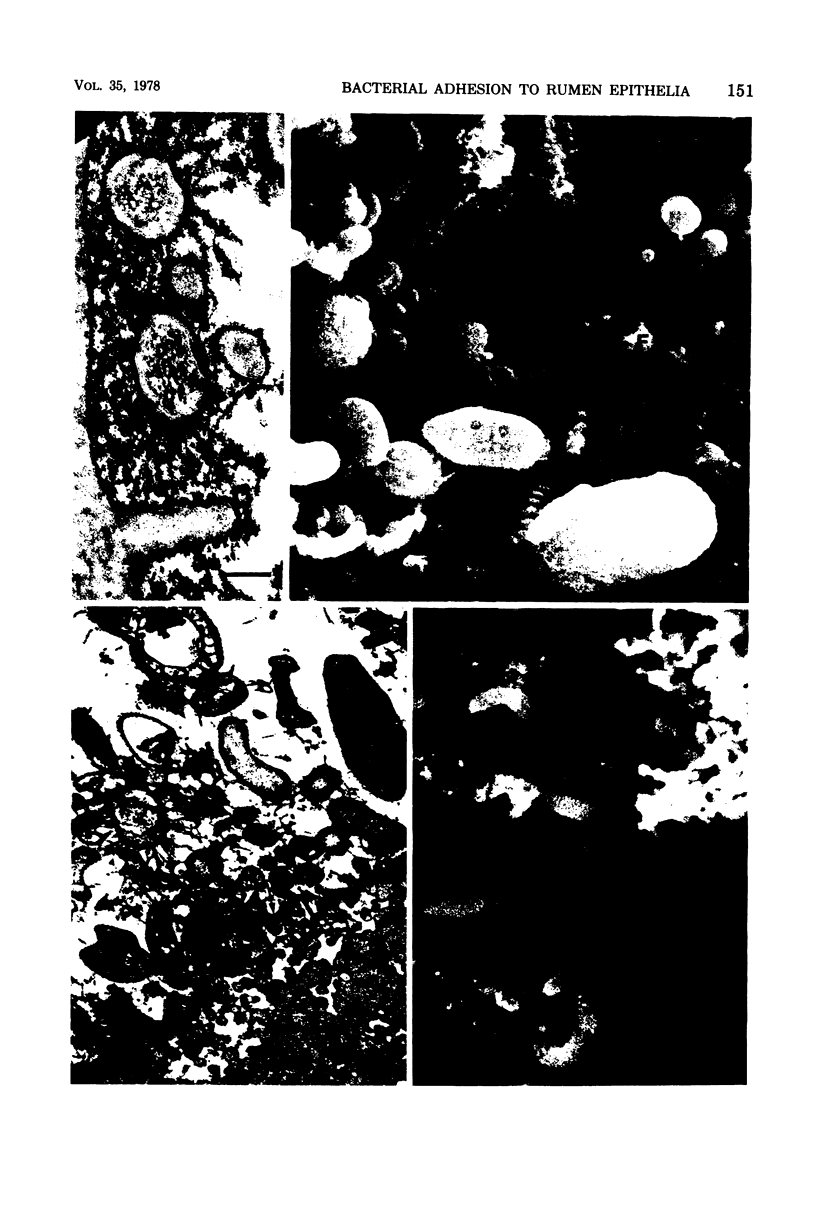
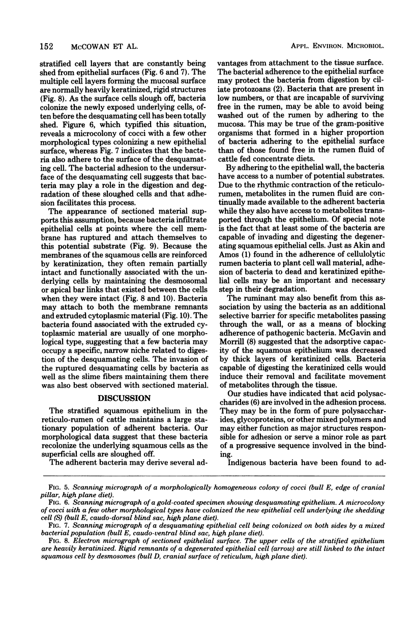
Images in this article
Selected References
These references are in PubMed. This may not be the complete list of references from this article.
- Akin D. E., Amos H. E. Rumen bacterial degradation of forage cell walls investigated by electron microscopy. Appl Microbiol. 1975 May;29(5):692–701. doi: 10.1128/am.29.5.692-701.1975. [DOI] [PMC free article] [PubMed] [Google Scholar]
- Bauchop T., Clarke R. T., Newhook J. C. Scanning electron microscope study of bacteria associated with the rumen epithelium of sheep. Appl Microbiol. 1975 Oct;30(4):668–675. doi: 10.1128/am.30.4.668-675.1975. [DOI] [PMC free article] [PubMed] [Google Scholar]
- Cheng K. J., Akin D. E., Costerton J. W. Rumen bacteria: interaction with particulate dietary components and response to dietary variation. Fed Proc. 1977 Feb;36(2):193–197. [PubMed] [Google Scholar]
- Costerton J. W., Damgaard H. N., Cheng K. J. Cell envelope morphology of rumen bacteria. J Bacteriol. 1974 Jun;118(3):1132–1143. doi: 10.1128/jb.118.3.1132-1143.1974. [DOI] [PMC free article] [PubMed] [Google Scholar]
- Costerton J. W., Ingram J. M., Cheng K. J. Structure and function of the cell envelope of gram-negative bacteria. Bacteriol Rev. 1974 Mar;38(1):87–110. doi: 10.1128/br.38.1.87-110.1974. [DOI] [PMC free article] [PubMed] [Google Scholar]
- Fuller R., Brooker B. E. Lactobacilli which attach to the crop epithelium of the fowl. Am J Clin Nutr. 1974 Nov;27(11):1305–1312. doi: 10.1093/ajcn/27.11.1305. [DOI] [PubMed] [Google Scholar]
- Malick L. E., Wilson R. B. Modified thiocarbohydrazide procedure for scanning electron microscopy: routine use for normal, pathological, or experimental tissues. Stain Technol. 1975 Jul;50(4):265–269. doi: 10.3109/10520297509117069. [DOI] [PubMed] [Google Scholar]
- McGavin M. D., Morrill J. L. Scanning electron microscopy of ruminal papillae in calves fed various amounts and forms of roughage. Am J Vet Res. 1976 May;37(5):497–508. [PubMed] [Google Scholar]
- Nagy B., Moon H. W., Isaacson R. E. Colonization of porcine small intestine by Escherichia coli: ileal colonization and adhesion by pig enteropathogens that lack K88 antigen and by some acapsular mutants. Infect Immun. 1976 Apr;13(4):1214–1220. doi: 10.1128/iai.13.4.1214-1220.1976. [DOI] [PMC free article] [PubMed] [Google Scholar]
- Patterson H., Irvin R., Costerton J. W., Cheng K. J. Ultrastructure and adhesion properties of Ruminococcus albus. J Bacteriol. 1975 Apr;122(1):278–287. doi: 10.1128/jb.122.1.278-287.1975. [DOI] [PMC free article] [PubMed] [Google Scholar]
- Savage D. C., Dubos R., Schaedler R. W. The gastrointestinal epithelium and its autochthonous bacterial flora. J Exp Med. 1968 Jan 1;127(1):67–76. doi: 10.1084/jem.127.1.67. [DOI] [PMC free article] [PubMed] [Google Scholar]
- Suegara N., Morotomi M., Watanabe T., Kawal Y., Mutai M. Behavior of microflora in the rat stomach: adhesion of lactobacilli to the keratinized epithelial cells of the rat stomach in vitro. Infect Immun. 1975 Jul;12(1):173–179. doi: 10.1128/iai.12.1.173-179.1975. [DOI] [PMC free article] [PubMed] [Google Scholar]
- WARNER E. D. The organogenesis and early histogenesis of the bovine stomach. Am J Anat. 1958 Jan;102(1):33–63. doi: 10.1002/aja.1001020103. [DOI] [PubMed] [Google Scholar]