Abstract
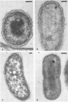
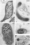
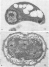
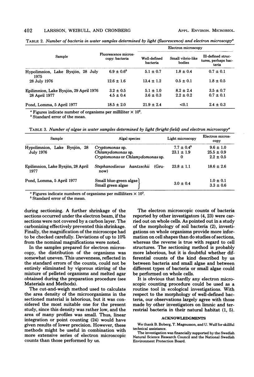
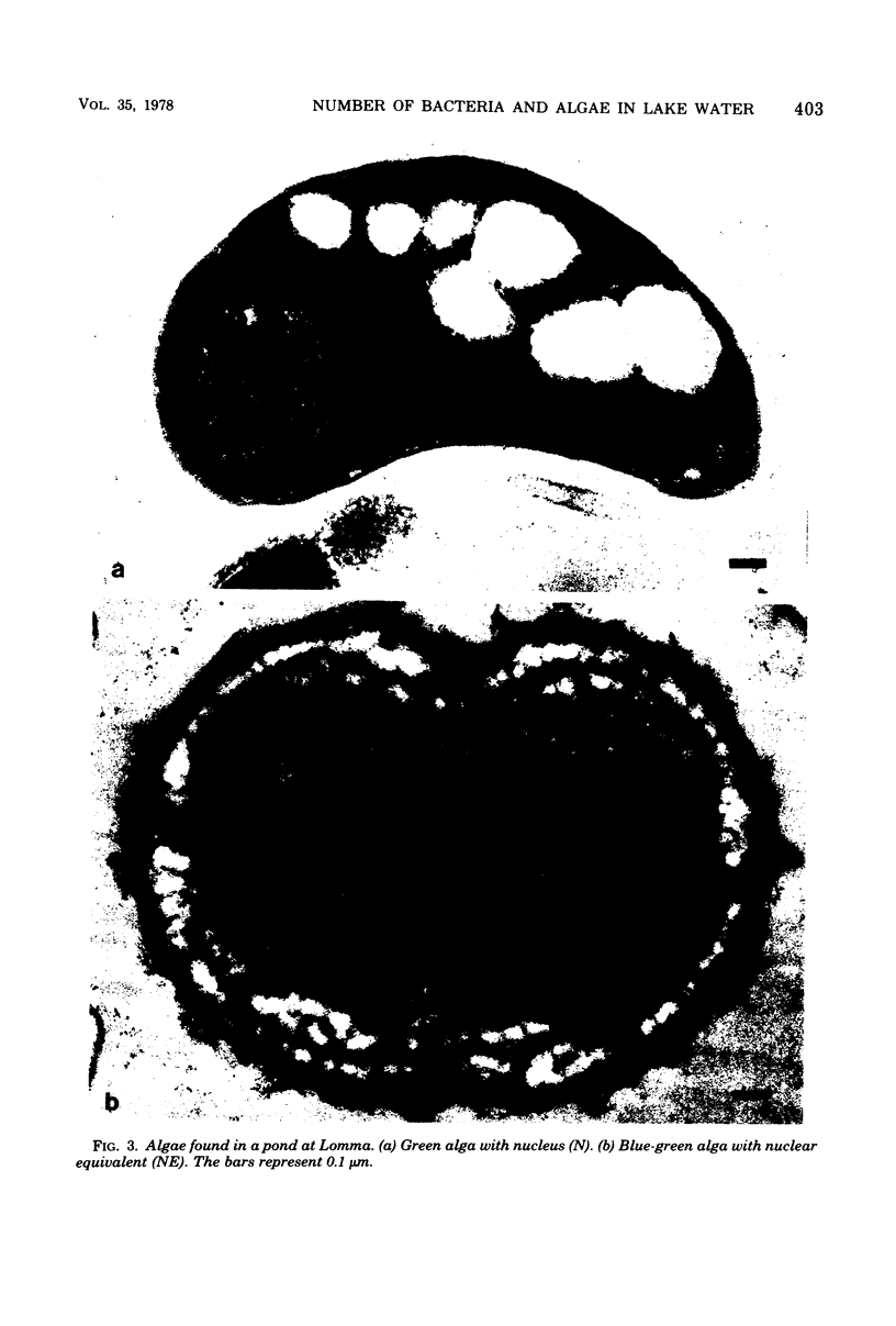
Determinations of the number of microorganisms in lake water samples with the bright-field light microscope were performed using conventional counting chambers. Determinations with the fluorescence microscope were carried out after staining the organisms with acridine orange and filtering them onto Nuclepore filters. For transmission electron microscopy, a water sample was concentrated by centrifugation. The pellet was solidifed in agar, fixed, dehydrated, embedded in Epon, and cut into thin sections. The number and area of organism profiles per unit area of the sections were determined. The number of organisms per unit volume of the pellet was then calculated using stereological formulae. The corresponding number in the lake water was obtained from the ratio of volume of solidified pellet/volume of water sample. Control experiments with pure cultures of bacteria and algae showed good agreement between light and electron microscopic counts. This was also true for most lake water samples, but the electron microscopic preparations from some samples contained small vibrio-like bodies and ill-defined structures that made a precise comparison more difficult. Bacteria and small blue-green and green algae could not always be differentiated with the light microscope, but this was easily done by electron microscopy. Our results show that transmission electron microscopy can be used for checking light microscopic counts of microorganisms in lake water.
Full text
PDF
Images in this article
Selected References
These references are in PubMed. This may not be the complete list of references from this article.
- Bae H. C., Cota-Robles E. H., Casida L. E. Microflora of soil as viewed by transmission electron microscopy. Appl Microbiol. 1972 Mar;23(3):637–648. doi: 10.1128/am.23.3.637-648.1972. [DOI] [PMC free article] [PubMed] [Google Scholar]
- Balkwill D. L., Casida L. E., Jr Microflora of soil as viewed by freeze-etching. J Bacteriol. 1973 Jun;114(3):1319–1327. doi: 10.1128/jb.114.3.1319-1327.1973. [DOI] [PMC free article] [PubMed] [Google Scholar]
- Bowden W. B. Comparison of two direct-count techniques for enumerating aquatic bacteria. Appl Environ Microbiol. 1977 May;33(5):1229–1232. doi: 10.1128/aem.33.5.1229-1232.1977. [DOI] [PMC free article] [PubMed] [Google Scholar]
- Caldwell D. E., Tiedje J. M. A morphological study of anaerobic bacteria from the hypolimnia of two Michigan lakes. Can J Microbiol. 1975 Mar;21(3):362–376. doi: 10.1139/m75-051. [DOI] [PubMed] [Google Scholar]
- Francisco D. E., Mah R. A., Rabin A. C. Acridine orange-epifluorescence technique for counting bacteria in natural waters. Trans Am Microsc Soc. 1973 Jul;92(3):416–421. [PubMed] [Google Scholar]
- Hirsch P. Neue Methoden zur Beobachtung und Isolierung ungewöhnlicher oder wenig bekannter Wasserbakterien. Z Allg Mikrobiol. 1972;12(3):203–218. [PubMed] [Google Scholar]
- Hobbie J. E., Daley R. J., Jasper S. Use of nuclepore filters for counting bacteria by fluorescence microscopy. Appl Environ Microbiol. 1977 May;33(5):1225–1228. doi: 10.1128/aem.33.5.1225-1228.1977. [DOI] [PMC free article] [PubMed] [Google Scholar]
- Jones J. G., Simon B. M. An investigation of errors in direct counts of aquatic bacteria by epifluorescence microscopy, with reference to a new method for dyeing membrane filters. J Appl Bacteriol. 1975 Dec;39(3):317–329. doi: 10.1111/j.1365-2672.1975.tb00578.x. [DOI] [PubMed] [Google Scholar]
- Nikitin D. I., Kuznetsov S. I. Primenenie élektronnoi mikroskopii dlia izucheniia vodnoi mikroflory. Mikrobiologiia. 1967 Sep-Oct;36(5):938–941. [PubMed] [Google Scholar]
- Pfennig N., cohen-Bazire G. Some properties of the green bacterium Pelodictyon clathratiforme. Arch Mikrobiol. 1967;59(1):226–236. doi: 10.1007/BF00406336. [DOI] [PubMed] [Google Scholar]
- RYTER A., KELLENBERGER E., BIRCHANDERSEN A., MAALOE O. Etude au microscope électronique de plasmas contenant de l'acide désoxyribonucliéique. I. Les nucléoides des bactéries en croissance active. Z Naturforsch B. 1958 Sep;13B(9):597–605. [PubMed] [Google Scholar]
- TAUBENECK U. Die Penicillininaktivierung durch Proteus-Arten. Zentralbl Bakteriol Orig. 1956 Dec;167(4):345–346. [PubMed] [Google Scholar]
- Watson S. W., Novitsky T. J., Quinby H. L., Valois F. W. Determination of bacterial number and biomass in the marine environment. Appl Environ Microbiol. 1977 Apr;33(4):940–946. doi: 10.1128/aem.33.4.940-946.1977. [DOI] [PMC free article] [PubMed] [Google Scholar]


