Abstract
Nucleotide excision repair (NER) and DNA mismatch repair are required for some common processes although the biochemical basis for this requirement is unknown. Saccharomyces cerevisiae RAD14 was identified in a two-hybrid screen using MSH2 as “bait,” and pairwise interactions between MSH2 and RAD1, RAD2, RAD3, RAD10, RAD14, and RAD25 subsequently were demonstrated by two-hybrid analysis. MSH2 coimmunoprecipitated specifically with epitope-tagged versions of RAD2, RAD10, RAD14, and RAD25. MSH2 and RAD10 were found to interact in msh3 msh6 and mlh1 pms1 double mutants, suggesting a direct interaction with MSH2. Mutations in MSH2 increased the UV sensitivity of NER-deficient yeast strains, and msh2 mutations were epistatic to the mutator phenotype observed in NER-deficient strains. These data suggest that MSH2 and possibly other components of DNA mismatch repair exist in a complex with NER proteins, providing a biochemical and genetical basis for these proteins to function in common processes.
Eukaryotes contain a DNA mismatch repair (MMR) system involving proteins related to the bacterial MutS and MutL proteins (for a review see ref. 1). The eukaryotic MMR system is more complex than the bacterial system. Instead of involving a single MutS-related protein, eukaryotic MMR involves two different heterodimeric complexes of MutS-related proteins, MSH2-MSH3 and MSH2-MSH6, that each have different mispair recognition specificity (1–7). Similarly, instead of a single MutL-related protein, eukaryotic MMR also involves a heterodimeric complex of two MutL-related proteins, MLH1-PMS1 (PMS2 in humans) (8, 9). Initial characterization of these pathways concentrated on their function in correcting mispaired bases resulting from DNA replication errors and the formation of heteroduplex recombination intermediates. Subsequent studies have suggested that MMR proteins may play more diverse roles in DNA metabolism.
MMR plays roles in genetic recombination beyond the repair of mispaired bases in recombination intermediates. MMR appears to regulate the extent of formation of heteroduplex tracts during recombination (10–12), possibly by regulating the resolution of Holliday junctions (11). MMR also suppresses recombination between divergent sequences (13–16), a process that may be similar to the proposed regulation of heteroduplex tract formation. The MSH2 and MSH3 proteins also act in recombination between duplicated DNA sequences (17, 18) and have been implicated in the removal of nonhomologous DNA strands greater than 30 bases long at the ends of recombining segments (19). This reaction involves the nucleotide excision repair (NER) complex RAD1-RAD10 (20, 21). It is unclear how MMR proteins function in these reactions; however, the ability of MSH2 and the MSH2-MSH6 complex to bind to Holliday junctions and branched DNA structures (ref. 22 and G. T. Marsischky, S. Lee, J. Griffith, and R.D.K., unpublished results) suggest they could bind to branched DNA structures and either target resolution enzymes and endonucleases to these structures or alter their structure making them more susceptible to cleavage.
MMR proteins have been implicated in DNA repair processes requiring NER proteins. Transcription-coupled repair in Escherichia coli and human cells is defective in MMR-defective mutants (23–25) and MMR-defective mutations decrease transcription-coupled repair of thymine glycol adducts but not UV damage in Saccharomyces cerevisiae (26, 27). Mutations in MSH2, MSH3, RAD1, and RAD10 cause a defect in gene conversion of a 26-base insertion mutation, suggesting they could be involved in the repair of large insertion/deletion mispairs (28), and in vitro studies in Drosophila have demonstrated that MEI9, a homologue of S. cerevisiae RAD1, is required for MMR (29). Similarly, it has been observed that NER can repair base-base mispairs in vitro (30), although involvement of MMR proteins in this in vitro reaction was not demonstrated. MSH2 and MSH2-MSH6 complexes bind to DNA damage adducts normally repaired by NER (31–34) although it is unclear whether this binding reflects a role of MMR proteins in the repair of such adducts. In two studies of human MMR in vitro, MMR proteins did not appear to play a role in repair of these types of adducts, whereas in another study MMR proteins did play a role in such repair (35–37). In E. coli, there is evidence that MMR proteins recognize UV and alkylation damage in vivo, although it is not clear whether MMR normally repairs such lesions in E. coli (38–41).
There is a considerable, but sometimes conflicting, body of data suggesting an interaction between MMR and NER. These pathways could have overlapping specificity for DNA damage or alternately some components from each pathway could function together in a hybrid repair pathway. At present, there is little biochemical data concerning how these pathways could interact. In the present study we demonstrate a physical interaction between MMR and NER proteins and provide genetic data supporting an overlapping repair specificity of these pathways.
MATERIALS AND METHODS
General Genetic Methods.
Yeast extract/peptone/dextrose, synthetic drop-out, 5-fluoroorotic acid, canavanine, and 5-bromo-4-chloro-indolyl β-d-galactopyranoside media were as described (11, 42, 43). Transformations were performed by the lithium acetate method (44). Genotyping of mutants was performed both by replica plating onto appropriate minimal media and by PCR analysis using primer pairs that allowed amplification of either the wild-type or mutant alleles (10). The PCR primers used were as follows: RAD10, rad10∷HIS3, 26408 (5′-GGACATGGCTTGATTTTTACAGTGCTC) and 26409 (5′-GCCTCGCAGTATTTGAAGTTGGATGG); RAD14, rad14∷HIS3, 26412 (5′-GTTTGACGTTTGCTAAGTTGTAGGGAG) and PASC5 (5′-CCGCTCGAGTTCAGTTTTCCGAGATAGTTAATTATGTACGAGTGACA); RAD2, rad2∷HIS3, 26443 (5′-GATGCCGCCACATATAGAGACCTTAAAG) and 26444 (5′-CGTTGCGCGTGTTTGGGTGGGTGCC); MLH1, mlh1∷hisG, 25591 (5′-ACTTTTGAGACCGCTTGCTGTT) and 25592 (5′-GTCTTTGGTACCGTTGAATAA) and PMS1, pms1∷hisG, 25589 (5′-GCTGCTGCGGTTTGTGG) and 25590 (5′-ATCCGTCCCTTTGGTCTTGTATCT).
Strains.
The S. cerevisiae strains used in this study are listed in Table 1. The rad10∷HIS3, rad14∷HIS3, and rad2∷HIS3 deletion strains were constructed by using an adaptation of a published method (45). The rad10 disruption construct was generated by amplifying the HIS3 gene present in pRS423 by PCR using primers (plasmid sequences are in lowercase) 24862 (5′-ACGTAACACAAAAAAGGGCATAAACAAAGTTGGGTATCCTAGAAGggcctcctctagtacactc) and 24863 (5′-GGTAATAAGCATGGAACAGATTTATTAAAAGAAAATAGGAATTGTgcgcgcctcgttcagaatg). The rad14 disruption construct was generated by using primers 24860 (5′-GAAAAAGAGTTTGGATCTTCGTAGTGAAGGTATCGAACGTAACGCTggcctcctctagtacactc and 24861 (5′-CTTATTATGACTTTCTTGTTATATTCTTATATACATAACCAACATgcgcgcctcgttcagaatg. The rad2 disruption construct was generated by using primers 26441 (5′-GTTCTACACGTCATCCATGAAGAAAAGCATTTTCGGGAGAACGCCAAACTTCAGACgagcagattgtactgagagtgcacc) and 26442 (5′-CTTTGTTAACATGCAGAAACAAAGGTAATGTTTATAAATAGTAAATCATACATAAGTATATGTTActccttacgcatctgtgcggtatttc).
Table 1.
S. cerevisiae strains used in this study
| Strain | Genotype |
|---|---|
| RKY2926 | his3, trp1, ura3-52, lex(leu2)3a = EGY48 containing pSH18-34 (URA3) plasmid |
| RKY2575 | MATa, ade2, ura3-52, his3Δ1, trp1-289, leu2-3,112, lys2-bgl, hom3-10 |
| RKY2567 | MATa, ade2, ura3-52, his3Δ1, trp1-289, leu2-3,112, lys2-bgl, hom3-10 msh3∷hisG-URA3-hisG, msh6∷URA3 |
| RKY2672 | MATa, ade2Δ1, ade8, ura3-52, leu2Δ1, trp1Δ63, his3Δ200, lys2-bgl, hom3-10 |
| RKY2706 | MATa, ade2Δ1, ade8, ura3-52, leu2Δ1, trp1Δ63, his3Δ200, lys2-bgl, hom3-10, msh2∷hisG |
| RKY2752 | MATa, ade2Δ1, ade8, ura3-52, leu2Δ1, trp1Δ63, his3Δ200, lys2-bgl, hom3-10, pms1∷hisG, mlh1∷hisG |
| RKY2350 | MATa, ade2Δ1, ade8, ura3-52, leu2Δ1, trp1Δ63, his3Δ200, lys2-bgl, hom3-10, rad10∷HIS3 |
| RKY2351 | MATa, ade2Δ1, ade8, ura3-52, leu2Δ1, trp1Δ63, his3Δ200, lys2-bgl, hom3-10, msh2∷hisG, rad10∷HIS3 |
| RKY2343 | MATa, ade2Δ1, ade8, ura3-52, leu2Δ1, trp1Δ63, his3Δ200, lys2-bgl, hom3-10, rad14∷HIS3 |
| RKY2344 | MATa, ade2Δ1, ade8, ura3-52, leu2Δ1, trp1Δ63, his3Δ200, lys2-bgl, hom3-10, msh2∷hisG, rad14∷HIS3 |
| RKY2352 | MATa, ade2Δ1, ade8, ura3-52, leu2Δ1, trp1Δ63, his3Δ200, lys2-bgl, hom3-10, rad2∷HIS3 |
| RKY2353 | MATa, ade2Δ1, ade8, ura3-52, leu2Δ1, trp1Δ63, his3Δ200, lys2-bgl, hom3-10, msh2∷hisG, rad2∷HIS3 |
The resulting PCR products were used to transform RKY2672, resulting in strains RKY2343, RKY2350, and RKY2352 in which the entire RAD14, RAD10, and RAD2 ORFs, respectively, were replaced with the HIS3 gene. Each disruption construct also was used to transform RKY2706 (a msh2∷hisG strain (42)) to construct double mutant strains RKY2344, RKY2351, and RKY2353. The correct integration of each rad∷HIS3 mutant allele was verified by PCR analysis and by testing for its characteristic UV sensitivity. The msh3 msh6 strain, RKY2567, and the corresponding isogenic wild-type strain, RKY2575, were described (5).
A mlh1 pms1 double mutant strain RKY2752 was constructed by A. Datta in this laboratory by using the one-step disruption method (46). RKY2672 was transformed with KpnI- and SphI-digested pEAI105 (mlh1∷hisG-URA3-hisG). Excision of the hisG-URA3-hisG was selected on 5-fluoroorotic acid media, followed by transformation with SalI-digested pEAI100 (pms1∷hisG-URA3-hisG) and a second round of selection for excision of hisG-URA3-hisG. The presence of the mlh1 and pms1 mutations was confirmed by mutator patch assays and PCR.
Plasmids.
The MSH2 bait (pRDK371) was constructed as described elsewhere by cloning the entire MSH2 ORF into pEG202 (47). The “prey” vector, pJG4–5 (48), was modified by introducing AvrII and BssHII sites between the EcoRI and XhoI sites present in the vector. To do this, oligonucleotides 22909 (5′-AATTCGGCCTAGGCGAGCGCGCGAC) and 22910 (5′-TCGAGTCGCGCGCTCGCCTAGGCCG) were annealed and inserted into EcoRI- and XhoI-digested pJG4–5 to yield pRDK483.
The different ScRAD ORFs were inserted into pRDK483 digested with EcoRI/XhoI or AvrII/XhoI. RAD1 and RAD2 ORFs were PCR amplified following a previously described method (5) from pG12-RAD1and pG12-RAD2, respectively (kindly provided by Errol Friedberg, University of Texas Southwestern Medical Center, Dallas), using Klentaq/Pfu polymerases (Ab Peptides, St Louis/Stratagene). The RAD3, RAD10, RAD14, and RAD25 ORFs were similarly PCR-amplified from genomic DNA. The names of the prey plasmids and the primers used for amplification of each gene are listed in Table 2.
Table 2.
PCR primer pairs used to construct prey vectors
| Plasmid | Primer | Primer sequence |
|---|---|---|
| pRDK589 | 23793 | 5′-GTTCCTAGGTTATTGCACTATCCTGTTGAAAATATCT |
| (RAD1) | PASC2 | 5′-AGGTCCGCTCGAGTTAAACAATATTTTACACAGGTGC |
| pRDK590 | 23197 | 5′-AGACCTAGGAACTTCAGACATGGGTGTG |
| (RAD2) | 23191 | 5′-CCGCTCGAGACATGCAGAAACAAAGGTAATG |
| pRDK591 | PASC54 | 5′-TCTCCTAGGTGACTACACTTTAAGAAGATTGGAAACAATGAAG |
| (RAD3) | PASC32 | 5′-CCGCTCGAGAGTTTATAGCAAAAGCGTATCATTGC |
| pRDK592 | PASC3 | 5′-TGGAATTCAAGATGAACAATACTGATCCTACTTCA |
| (RAD10) | PASC4 | 5′-CCGCTCGAGAGGATGGTAATAAGCATGGAACAG |
| pRDK593 | PASC8 | 5′-GGAATTCATGACTCCCGAACAAAAGGCCAAACTAGAGGCTAACAGGAAATTAGCAATAGAACGG |
| (RAD14) | PASC5 | 5′-CCGCTCGAGTTCAGTTTTTCCGAGATAGTTAATTATGTACGAGTGACA |
| pRDK594 | 23195 | 5′-GGGAATTCATCATGACGGACGTTGAAGGC |
| (RAD25) | 23196 | 5′-CCGCTCGAGGGTGACAATGAAACCAAGCCTATTC |
LexA- and 12CA5-fusion constructs were tested for expression of full-length fusion protein by Western blot analysis using an anti-LexA antibody (49) kindly supplied by Roger Brent and his laboratory (Massachusetts General Hospital, Boston) or an anti-12CA5 antibody (Babco, Richmond, CA). All ScRAD and MSH2 fusions constructed were verified by sequencing.
The bait plasmids containing the ORFs hCDC2, hCDK2, and DmCDC2 were supplied by R. Brent and his laboratory, and bait plasmid containing ScPRP20 and the prey plasmid containing ScNPL3 were from Pam Silver (Dana-Farber Cancer Institute).
Mutator and UV-Sensitivity Assays.
Mutation rate assays and patch tests to determine mutator phenotypes were performed exactly as described (5). To study UV sensitivity of the different mutant strains, liquid cultures were grown overnight to saturation in yeast extract/peptone/dextrose (YPD), serial 10-fold dilutions were prepared, and 10 μl of each dilution was spotted onto YPD plates. The plates then were irradiated at different UV doses by using a 254-nm germicidal lamp as indicated in individual experiments. The colonies were counted after 3 days of incubation in the dark.
Two-Hybrid Techniques.
A two-hybrid screen (48, 50) for MSH2 interactors was performed as described (47). To test for interaction between MSH2 and different ScNER proteins, the S. cerevisiae strain EGY48 was cotransformed with the different HIS3 bait and TRP1 prey plasmids described along with the URA3 lacZ reporter, pSH18–34 (48). Transformants were isolated and patched onto Ura−His−Trp− plates, then replicated onto Ura−His−Trp− 5-bromo-4-chloro-indolyl β-d-galactopyranoside plates containing either glucose or galactose to monitor β-galactosidase expression. Positive interactions also were verified by galactose-dependent growth on Ura−His−Trp−Leu− glucose and galactose plates.
Immunoprecipitation Experiments.
Whole-cell extracts were prepared from EGY48 strains containing bait and prey plasmids essentially as described (51). Fifty milliliters of yeast cultures were grown to an A600 of 1.5–2 in Ura−His−Trp− glucose medium, induced for 4 hr with galactose, pelleted, quickly frozen, and stored at −80°C. Glass bead extracts were prepared as described (51) except that vanadate, chymostatin, and pepstatin were omitted from the modified H buffer and 2 mM benzamidine was included. 12CA5-tagged preys were immunoprecipitated essentially as described (47, 51) in 500-μl reactions containing extracts in modified H buffer and 150 mM NaCl. All immunoprecipitation (IP)-related manipulations were performed at 4°C. Two hundred micrograms of cell extract were precleared by a 50-min rotation with 25 μl of protein G-Sepharose (Pharmacia) followed by a 20-sec spin at low speed in a microcentrifuge. One microliter (≈1 μg) of 12CA5 ascites then was added to each supernatant, the reaction rotated for 2.5 hr, 25 μl of protein G-Sepharose added, the reaction rotated for 50 min, and the protein G beads pelleted. The beads from the IP and from the preclear were washed six times in H buffer containing 150 mM NaCl, resuspended in 25 μl of NaDodSO4 sample buffer (Bio-Rad) and frozen at −80°C. Subsequently the samples were boiled for 5 min and fractionated by 10% NaDodSO4-PAGE.
Proteins were transferred and membranes were saturated as described (47). The membranes were incubated 2.5 hr in a 1:5,000 dilution of primary anti-LexA antibody in TBST buffer (10 mM Tris, pH 8/150 mM NaCl/0.05% Tween-20). The membranes then were washed three times in TBST and incubated for 50 min in a 1:10,0000 dilution of secondary antibody [horseradish peroxidase (HRP)-conjugated anti-rabbit Ig, Amersham], washed six times, and visualized by ECL using an ECL kit (Amersham). To ensure appropriate expression and immunoprecipitation of 12CA5-tagged preys, the immunoblot was stripped by a 30-min incubation at 50°C in 62.5 mM Tris (pH 6.7), 2% NaDodSO4, and 100 mM 2-mercaptoethanol, then blocked and probed again using 12CA5 as primary antibody and HRP-conjugated anti-mouse Ig.
RESULTS
Interaction Between MMR and NER Proteins.
A two-hybrid screen was performed in S. cerevisiae using the LexA DNA-binding domain fused to the entire ORF of MSH2 as bait and an activation-tagged library of S. cerevisiae genomic DNA as prey (47). One protein identified was ScEXO1, an exonuclease displaying properties consistent with a direct role in MMR. Another clone was identified that encoded a portion of S. cerevisiae RAD14 (52). To confirm that RAD14 interacts with MSH2, we constructed a prey vector that expressed full-length RAD14 protein to test its interaction with full-length MSH2 and found that full-length RAD14 and MSH2 did interact by two-hybrid criteria (see Fig. 2).
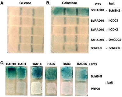
Figure 2.
Specific interaction of MSH2 with RAD10. The indicated baits and preys were cotransformed into EGY48 along with the lacZ reporter, pSH18–34. The preys are expressed only in the presence of galactose. (A and B) Three different transformants were patched on selective plates and then replica-plated onto 5-bromo-4-chloro-indolyl β-d-galactopyranoside (X-Gal) plates with either glucose or galactose. (C) Patches of one transformant containing each indicated pair of baits and preys replica plated onto X-Gal plates containing galactose. Interaction of a bait and a prey induces β-galactosidase activity producing blue coloration.
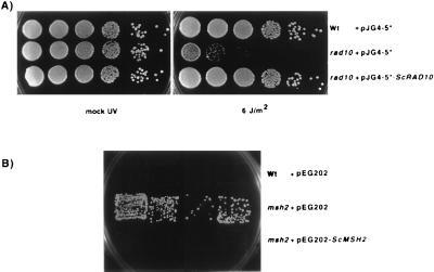
To further analyze whether NER proteins could interact with MSH2, we constructed prey vectors containing ORFs of five additional NER proteins (RAD1, RAD2, RAD3, RAD10, and RAD25) and tested their ability to interact with a LexA-MSH2 bait and activate a lacZ reporter gene. We first confirmed that the LexA-MSH2 and RAD10 fusion proteins were functional and could complement msh2 and rad10 mutations, respectively (Fig. 1). Similar functional complementation was observed for the RAD14 and RAD2 prey constructs (data not shown); RAD1, RAD3, and RAD25 were not tested. In a typical experiment (Fig. 2), RAD10 was found to interact with MSH2. The observed interaction depended on MSH2 and induction of expression of RAD10 by the addition of galactose (compare galactose induction vs. glucose in Fig. 2). MSH2 did not interact with ScNPL3 control prey protein, and RAD10 did not interact with hCDC2, hCDK2, or DmCDC2 control bait proteins, suggesting the activation of the reporter gene was caused by a specific interaction between MSH2 and RAD10. Pairwise interactions between MSH2 bait and RAD1, RAD2, RAD3, RAD10, RAD14, and RAD25 preys were examined in a separate experiment; significant interaction was observed for all of these pairs of proteins under conditions where none of the preys interacted with a nonspecific control bait PRP20 (Fig. 2).
Figure 1.
Functional properties of RAD10 prey and LexA-MSH2 bait. (A) Complementation of the UV sensitivity of a rad10 strain by expression of the RAD10 prey protein. The wild-type and rad10 strains were transformed with indicated plasmids and expression of 12CA5-RAD10 protein in the strains was induced by transferring cells into synthetic drop-out (SD) Trp− medium containing galactose followed by shaking for 4 hr. Then 10-fold serial dilutions were prepared and 10 μl of different dilutions were spotted onto SD Trp− plates containing galactose, and UV sensitivity was determined. (B) Complementation of the mutator phenotype of a msh2 strain by expression of the LexA-MSH2 protein. Patches of the indicated strains on a yeast extract/peptone/dextrose plate were replica-plated to a canavanine plate to allow visualization of Canr papilliae after incubation of the plates at 30° for 2 days.
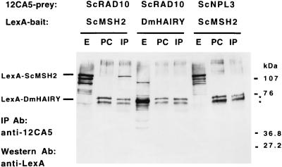
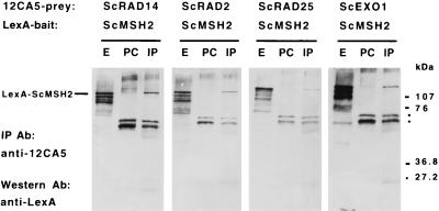
To verify the interaction between MSH2 and different NER proteins, various combinations of LexA-MSH2 and LexA-DmHAIRY were coexpressed with 12CA5-tagged RAD2, RAD10, RAD14, RAD25, and NPL3. Expression of proteins was monitored by Western blotting using anti-LexA and anti-12CA5 antibodies (data not shown). Prey proteins were immunoprecipitated with 12CA5 antibody and detected by Western blotting with LexA antibody. The results showed that LexA-MSH2 coimmunoprecipitated with RAD10 (Fig. 3). In contrast, RAD10 did not interact with the LexA-DmHAIRY control protein and the NPL3 control protein did not interact with LexA-MSH2 (Fig. 3). LexA-MSH2 also coimmunoprecipitated with RAD2, RAD14, and RAD25 proteins (Fig. 4). An EXO1 prey was used as positive control for coimmunoprecipitation (47); the level of precipitation of RAD10 and RAD14 was similar to that of EXO1, whereas the levels of precipitation of RAD2 and RAD25 were somewhat lower (Figs. 3 and 4). We ensured appropriate immunoprecipitation of all preys by stripping the membrane and Western blotting with anti-12CA5 antibodies (data not shown). RAD proteins did not coimmunoprecipitate nonspecifically with a LexA-DmHAIRY control, and 12CA5-NPL3 did not coimmunoprecipitate with LexA-MSH2. This finding indicates that neither LexA-MSH2 nor 12CA5-tagged NER proteins are nonspecifically “sticky” proteins in coimmunoprecipitation experiments.
Figure 3.
Coimmunoprecipitation of MSH2 and RAD10. Whole-cell extracts (200 μg) were prepared from EGY48 strains expressing LexA-tagged baits (MSH2, or DmHAIRY as a negative control) and 12CA5-tagged preys (RAD10, or ScNPL3 as a negative control). The extracts (E) then were analyzed by immunoprecipitation (IP) and Western blotting. PC, proteins eluted from protein G-Sepharose used to preclear; Ab, antibody; *, proteins nonspecifically precipitated by protein-G Sepharose.
Figure 4.
Coimmunoprecipitation of MSH2 and different NER proteins. Whole-cell extracts (200 μg) were prepared from EGY48 expressing LexA-tagged bait (MSH2) and 12CA5-tagged preys (RAD10, RAD14, RAD2, RAD25, or ScEXO1 as positive control). The extracts (E) then were analyzed by immunoprecipitation (IP) and Western blotting. PC, proteins eluted from protein G-Sepharose used to preclear; Ab, antibody; *, proteins nonspecifically precipitated by protein-G Sepharose.
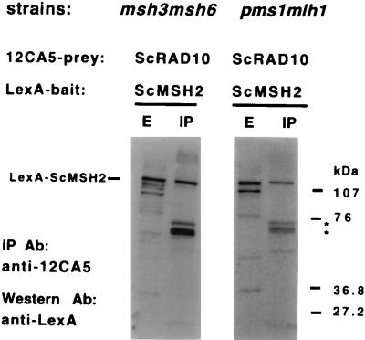
The interactions observed could involve a direct interaction between MSH2 and one or more of the NER proteins tested or they could involve an indirect interaction, presumably requiring either MSH3, MSH6, MLH1, or PMS1. To investigate this possibility, coimmunoprecipitation of LexA-MSH2 and RAD10 was studied in msh3 msh6 and mlh1 pms1 double mutants. The results show that LexA-MSH2 and RAD10 interacted in the absence of MSH3, MSH6, MLH1, or PMS1 (Fig. 5), consistent with the view that the observed interactions directly involve MSH2.
Figure 5.
Interaction of MSH2 and RAD10 in the absence of MSH3, MSH6, MLH1, and PMS1. Whole-cell extracts (200 μg) were prepared from RKY2567 and RKY2752 expressing LexA-tagged bait (MSH2) and 12CA5-tagged prey (RAD10). The extracts (E) then were analyzed by immunoprecipitation (IP) and Western blotting. PC, proteins eluted from protein G-Sepharose used to preclear; Ab, antibody; *, proteins nonspecifically precipitated by protein-G Sepharose.
Overlapping Roles of MMR and NER Proteins in DNA Repair.
The observed interactions between MMR proteins and NER proteins suggest that these proteins could function in the same pathway(s) to some extent or that each of these repair pathways may recognize and repair some of the same types of DNA damage. To test these possibilities, we investigated the effects of mutations in NER and MMR genes on the rate of accumulation of mutations in target genes and on the sensitivity to UV irradiation. Mutations in RAD2, RAD10, and RAD14 caused small increases in the rate of accumulation of Canr mutations (2- to 3-fold) and reversion of the hom3–10 (up to 2-fold) and lys2-Bgl alleles (up to 2-fold) (Table 3), consistent with previously published results on the mutator phenotypes caused by mutations in other NER genes like RAD1 (53) and RAD3 (54). When mutations in these genes were combined with a msh2 mutation, the resulting mutation rates were the same as observed with the msh2 single mutant. It is unlikely that additivity could have been observed because msh2 mutations cause a much greater mutator phenotype than mutations in RAD2, RAD10, or RAD14 but clearly a multiplicative effect was not seen. These results suggest that either RAD2, RAD10, and RAD14 function in a MSH2-dependent repair pathway or they function in a different pathway that can suppress the accumulation of some types of mutations, most likely base substitution mutations (ref. 53 and C. Chen and R.D.K., unpublished results). The observed results are inconsistent with the idea that mutations in RAD2, RAD10, or RAD14 result in the accumulation of DNA damage that is repaired by an MSH2-dependent pathway.
Table 3.
Mutation rates in MMR- and NER-defective strains
| Relevant genotype | Canr | Hom+ | Lys+ |
|---|---|---|---|
| Wild type | 2.4 × 10⁻⁷ (1) | 8.7 × 10⁻⁹ (1) | 1.0 × 10⁻⁸ (1) |
| rad10 | 4.7 × 10⁻⁷ (1.9) | 11.2 × 10⁻⁹ (1.3) | 1.4 × 10⁻⁸ (1.4) |
| rad2 | 8.0 × 10⁻⁷ (3.2) | 7.7 × 10⁻⁹ (0.9) | 1.6 × 10⁻⁸ (1.6) |
| rad14 | 5.6 × 10⁻⁷ (2.2) | 10.5 × 10⁻⁹ (1.2) | 2.0 × 10⁻⁸ (2) |
| msh2 | 2.9 × 10⁻⁶ (12) | 4.5 × 10⁻⁶ (517) | 7.3 × 10⁻⁷ (73) |
| msh2 rad10 | 3.7 × 10⁻⁶ (15) | 5.7 × 10⁻⁶ (655) | ND |
| msh2 rad2 | 3.3 × 10⁻⁶ (13) | 4.5 × 10⁻⁶ (528) | 6.4 × 10⁻⁷ (64) |
| msh2 rad14 | 2.8 × 10⁻⁶ (11.4) | 5.0 × 10⁻⁶ (580) | 5.9 × 10⁻⁷ (59) |
Mutation rates (inactivation of CAN1 and reversion of hom3-10 or lys2-Bgl) were determined and the mean of 3-5 independent experiments is presented. The numbers in parentheses are the increase in mutation rate observed relative to wild type. The strains used are the strains listed in the legend to Fig. 6. Also see Table 2. ND, not determined.
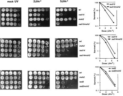
Previous studies of UV-induced recombination in NER-defective E. coli strains suggested that MMR can recognize or interact with UV damage, resulting in increased recombination (40, 41). To determine whether similar effects could be seen in S. cerevisiae, we tested the effect of a mutation in MSH2 on the UV sensitivity of strains containing rad14, rad10, and rad2 deletion mutations. As expected, the rad14, rad10, and rad2 single mutants were highly UV sensitive (Fig. 6). A msh2 mutation did not cause any detectable UV sensitivity alone but did cause an increase in the UV sensitivity of rad10 and rad14 mutants (a 5- to 10-fold range of increased UV sensitivity was seen in different experiments). These data suggest that MSH2 can function in a minor pathway for the repair of UV damage distinct from RAD10, RAD14-dependent NER. The msh2 mutation caused little increase in the UV sensitivity caused by the rad2 mutation, suggesting that RAD2 may function in both pathways.
Figure 6.
Effect of an msh2 mutation on UV sensitivity of NER mutants. The indicated mutant strains were analyzed for UV sensitivity. The graphs shown indicate the % survival observed relative to no UV irradiation. No significant killing of either the wild type or the msh2 mutant strain was observed at the indicated UV dose. Three to 10 independent experiments were performed with each set of strains and one representative example is shown. The strains used were RKY2672, RKY2706 msh2, RKY2350 rad10, RKY2351 msh2 rad10, RKY2343 rad14, RKY2344 msh2 rad14, RKY2352 rad2, and RKY2353 msh2 rad2.
DISCUSSION
The experiments presented here document in vivo interaction between S. cerevisiae MSH2 and six different NER proteins, RAD1, RAD2, RAD3, RAD10, RAD14, and RAD25, which are components of an NER complex (55). We have not yet determined which NER protein(s) interacts directly with MSH2 or which other MMR proteins such as MSH3, MSH6, MLH1, and PMS1 are present in the observed MMR-NER complex(es). These interactions are consistent with genetic experiments implicating MMR and NER proteins in the same repair and recombination reactions.
There is a growing body of evidence that MMR and NER may functionally overlap and that MMR and NER proteins may act together in some types of repair. Previous studies have shown the in vivo requirement of MMR in transcription-coupled repair in E. coli, human, and for some types of damage, S. cerevisiae cells (23–27). NER can repair base-base mispairs in vitro (30), and MSH2 and MSH2-MSH6 complex are known to recognize UV damage, damage by platinum compounds and other adducts in DNA that are thought to be repaired by NER (31–34, 37). However, no clear picture has emerged as to whether the ability of one pathway to recognize damage normally repaired by the other pathway reflects significant repair reactions in vivo (35–37). The results presented here provide insight into these previous observations. For instance, the interaction of MSH2 with essentially all of the NER components required for transcription-coupled repair suggests it is the disruption of this complex in MMR mutants that causes a defect in transcribed strand repair. Second, our observation of increased UV sensitivity of MMR-NER double mutants compared with NER single mutants suggests that an MSH2-dependent process can serve as an alternate, albeit minor, pathway for the repair of UV damage that is independent of RAD1-RAD10 and RAD14 function. Our data showing that rad2 mutants and rad2 msh2 double mutants have similar UV sensitivity suggest that MSH2, and possibly other MMR proteins, recognize UV damage and target the RAD2 endonuclease to this substrate. It should be noted that it has been suggested that XPG, the human RAD2 homolog, plays additional roles in repair of oxidative damage to DNA besides its role in NER (56). Previous studies on the role of MMR in UV-stimulated recombination in E. coli NER-defective mutants and on the role of MMR in lethality of E. coli dam mutants caused by treatment with alkylating agents (38–41) are consistent with the view that MMR proteins can functionally recognize damage that usually is repaired by NER. Finally, our analysis of the mutator phenotype of NER mutants along with the results of previous studies of the mutator phenotype of rad1 mutants and certain rad3 mutants alleles (53, 54) suggests NER proteins could play a direct role in the repair of replication errors. Studies on gene conversion in MMR and NER mutants in S. cerevisiae and in vitro studies indicating the Drosophila MEI9 plays a role in MMR support the view that such repair could involve both NER and MMR proteins in some cases (28, 29), a result that is consistent with the results presented here.
The biological relevance of physical interaction between MMR proteins and NER proteins is even more clear for recombination. RAD1, RAD10, MSH2, and MSH3 are required for recombination between duplicated sequences and double-strand-break-induced single-strand annealing recombination, and RAD1 and RAD10 are required to remove nonhomologous DNA from the 3′ ends of recombining DNA during these processes (20, 57). RAD1, RAD10, and MSH2, but not RAD2 or RAD14, are required for gene conversion of large insertion/deletion mutations that would form large loop mispairs in heteroduplex recombination intermediates (28). Consistent with this requirement, the RAD1-RAD10 complex is a weak endonuclease in vitro that can cleave branched DNA structures and presumably large loop mispairs (57). The RAD1-RAD10 endonuclease does not recognize base damage such as pyrimidine dimers in duplex DNA (58). It has been suggested that the specificity and reactivity of the RAD1-RAD10 endonuclease is modulated by further protein–protein interactions with proteins such as RAD14 that may target the RAD1-RAD10 complex and other NER proteins to the site of DNA damage (for reviews see refs. 55 and 59). Thus, factor(s) likely exist to target this complex to other substrates (i.e., nonhomologous tails and loop mispairs), during recombination events. The genetic data documenting the involvement of MSH2 and MSH3 in the RAD1 RAD10 recombination pathway (17, 18, 28) and the physical interaction we observed suggest that this factor could be MSH2 or the MSH2-MSH3 complex. Indeed, MSH2 by itself and in conjunction with MSH3 has been shown to recognize loop mispairs (2, 5, 6, 60, 61) and MSH2 and MSH2-MSH6 complex recognize branched DNA structures such as Holliday junctions and Y structures in addition to different mispairs (refs. 22 and 60; G. T. Marsischky, S. Lee, J. Griffith, and R.D.K., unpublished results). It is interesting to note that in mammalian cells, the ERCC1 gene product (RAD10 homologue) has been suggested to be involved in the processing of heteroduplex intermediates during recombination (62). Thus, it is possible that MSH2, MSH2-MSH3, and/or MSH2-MSH6 complexes bind to such structures as branched DNAs and loop mispairs and then recruit the RAD1-RAD10 endonuclease by the direct interaction demonstrated here. This interaction could explain the requirement of RAD1, RAD10, MSH2, and MSH3 for recombination reactions and repair of large loops. Similarly, MSH2 and MSH2 containing complexes could target Holliday junction resolution enzymes to Holliday junctions.
The results presented here provide a physical basis for results suggesting functional interactions and overlap between MMR and NER. There are several possibilities for how this overlap could occur. One possibility is that there is a MMR-NER complex containing both the NER proteins described here and MSH2 along with other components of MMR like MSH3, MSH6, MLH1, and PMS1. This complex would contain two different types of DNA damage recognition components, each of which has overlapping damage recognition specificity. Damage recognition by either component then could target other components of both MMR and NER to the site of damage where either could act in repair. Alternately, there could be different complexes containing subsets of MMR and NER proteins, each of which would likely have different DNA damage/structure recognition specificity and this differential specificity would target different combinations NER and/or MMR proteins to the repair of specific DNA structures. The available data suggest that there may exist at least two different types of complexes or reactions, a MSH2-dependent reaction that targets RAD1-RAD10 endonuclease to branched structures and large loop mispairs and a MSH2-dependent reaction that targets RAD2 endonuclease to UV damage. Additional biochemical and genetic studies will be required to distinguish among these possibilities.
Acknowledgments
We thank Pam Silver, Pierre Colas, and Roger Brent for reagent and control plasmids used for two-hybrid experiments, E. Friedberg for pG12-RAD1 and pG12-RAD2 plasmids, and Bernard Lopez and the Kolodner laboratory, especially Gerry Marsischky, Abhijit Datta, Philippe Noirot, Hernan Flores-Rozas, and Michael Kane, for helpful discussions and comments on the manuscript. All of the oligonucleotides and the DNA sequencing facilities used in this study were provided by the Dana-Farber Cancer Institute Molecular Biology Core Facility. This work was supported by National Institutes of Health Grant GM50006 to R.D.K.
ABBREVIATIONS
- NER
nucleotide excision repair
- MMR
DNA mismatch repair
Footnotes
This paper was submitted directly (Track II) to the Proceedings Office.
References
- 1.Kolodner R. Genes Dev. 1996;10:1433–1442. doi: 10.1101/gad.10.12.1433. [DOI] [PubMed] [Google Scholar]
- 2.Acharya S, Wilson T, Gradia S, Kane M F, Guerette S, Marsischky G T, Kolodner R, Fishel R. Proc Natl Acad Sci USA. 1996;93:13629–13634. doi: 10.1073/pnas.93.24.13629. [DOI] [PMC free article] [PubMed] [Google Scholar]
- 3.Drummond J T, Li G-M, Longley M J, Modrich P. Science. 1995;268:1909–1912. doi: 10.1126/science.7604264. [DOI] [PubMed] [Google Scholar]
- 4.Johnson R E, Kovvali G K, Prakash L, Prakash S. J Biol Chem. 1996;271:7285–7288. doi: 10.1074/jbc.271.13.7285. [DOI] [PubMed] [Google Scholar]
- 5.Marsischky G T, Filosi N, Kane M F, Kolodner R. Genes Dev. 1996;10:407–420. doi: 10.1101/gad.10.4.407. [DOI] [PubMed] [Google Scholar]
- 6.Palombo F, Iaccarino I, Najajima E, Ikejima M, Shimada T, Jiricny J. Curr Biol. 1996;6:1181–1184. doi: 10.1016/s0960-9822(02)70685-4. [DOI] [PubMed] [Google Scholar]
- 7.Strand M, Earley M C, Crouse G F, Petes T D. Proc Natl Acad Sci USA. 1995;92:10418–10421. doi: 10.1073/pnas.92.22.10418. [DOI] [PMC free article] [PubMed] [Google Scholar]
- 8.Prolla T A, Pang Q, Alani E, Kolodner R D, Liskay R M. Science. 1994;265:1091–1093. doi: 10.1126/science.8066446. [DOI] [PubMed] [Google Scholar]
- 9.Li G-M, Modrich P. Proc Natl Acad Sci USA. 1995;92:1950–1954. doi: 10.1073/pnas.92.6.1950. [DOI] [PMC free article] [PubMed] [Google Scholar]
- 10.Reenan R A G, Kolodner R D. Genetics. 1992;132:975–985. doi: 10.1093/genetics/132.4.975. [DOI] [PMC free article] [PubMed] [Google Scholar]
- 11.Alani E, Reenan R A, Kolodner R D. Genetics. 1994;137:19–39. doi: 10.1093/genetics/137.1.19. [DOI] [PMC free article] [PubMed] [Google Scholar]
- 12.Detloff P, White M A, Petes T D. Genetics. 1992;132:113–123. doi: 10.1093/genetics/132.1.113. [DOI] [PMC free article] [PubMed] [Google Scholar]
- 13.Datta A, Adjiri A, New L, Crouse G F, Jinks-Robertson S. Mol Cell Biol. 1996;16:1085–1093. doi: 10.1128/mcb.16.3.1085. [DOI] [PMC free article] [PubMed] [Google Scholar]
- 14.Rayssiguier C D, Thaler D S, Radman M. Nature (London) 1989;342:396–401. doi: 10.1038/342396a0. [DOI] [PubMed] [Google Scholar]
- 15.Petit M A, Dimpfli J, Radman M, Echols H. Genetics. 1991;129:327–332. doi: 10.1093/genetics/129.2.327. [DOI] [PMC free article] [PubMed] [Google Scholar]
- 16.Selva E M, New L, Crouse G F, Lahue R S. Genetics. 1995;139:1175–1188. doi: 10.1093/genetics/139.3.1175. [DOI] [PMC free article] [PubMed] [Google Scholar]
- 17.Saparbaev M, Prakash L, Prakash S. Genetics. 1996;142:727–736. doi: 10.1093/genetics/142.3.727. [DOI] [PMC free article] [PubMed] [Google Scholar]
- 18.Sugawara N, Pâques F, Colaiàcovo M, Haber J E. Proc Natl Acad Sci USA. 1997;94:9214–9219. doi: 10.1073/pnas.94.17.9214. [DOI] [PMC free article] [PubMed] [Google Scholar]
- 19.Pâques F, Haber J E. Mol Cell Biol. 1997;17:6765–6771. doi: 10.1128/mcb.17.11.6765. [DOI] [PMC free article] [PubMed] [Google Scholar]
- 20.Fishman-Lobell J, Haber J E. Science. 1992;258:480–484. doi: 10.1126/science.1411547. [DOI] [PubMed] [Google Scholar]
- 21.Ivanov E L, Haber J E. Mol Cell Biol. 1995;15:2245–2251. doi: 10.1128/mcb.15.4.2245. [DOI] [PMC free article] [PubMed] [Google Scholar]
- 22.Alani E, Lee S, Kane M F, Griffith J, Kolodner R D. J Mol Biol. 1997;265:289–301. doi: 10.1006/jmbi.1996.0743. [DOI] [PubMed] [Google Scholar]
- 23.Mellon I, Rajpal D K, Koi M, Boland C R, Champe G N. Science. 1996;272:557–560. doi: 10.1126/science.272.5261.557. [DOI] [PubMed] [Google Scholar]
- 24.Mellon I, Champe G N. Proc Natl Acad Sci USA. 1996;93:1292–1297. doi: 10.1073/pnas.93.3.1292. [DOI] [PMC free article] [PubMed] [Google Scholar]
- 25.Leadon S A, Avrutskaya A V. Cancer Res. 1997;57:3784–3791. [PubMed] [Google Scholar]
- 26.Sweder K S, Verhage R A, Crowley D J, Crouse G F, Brouwer J, Hanawalt P C. Genetics. 1996;143:1127–1135. doi: 10.1093/genetics/143.3.1127. [DOI] [PMC free article] [PubMed] [Google Scholar]
- 27.Leadon S A, Avrutskaya A V. Mutat Res. 1998;407:177–187. doi: 10.1016/s0921-8777(98)00007-x. [DOI] [PubMed] [Google Scholar]
- 28.Kirkpatrick D T, Petes T D. Nature (London) 1997;387:929–931. doi: 10.1038/43225. [DOI] [PubMed] [Google Scholar]
- 29.Bhui-Kaur A, Goodman M F, Tower J. Mol Cell Biol. 1998;18:1436–1443. doi: 10.1128/mcb.18.3.1436. [DOI] [PMC free article] [PubMed] [Google Scholar]
- 30.Huang J, Hsu D S, Kazantsev A, Sancar A. Proc Natl Acad Sci USA. 1994;91:12213–12217. doi: 10.1073/pnas.91.25.12213. [DOI] [PMC free article] [PubMed] [Google Scholar]
- 31.Duckett D R, Drummond J T, Murchie A I H, Reardon J T, Sancar A, Lilley D M, Modrich P. Proc Natl Acad Sci USA. 1996;93:6443–6447. doi: 10.1073/pnas.93.13.6443. [DOI] [PMC free article] [PubMed] [Google Scholar]
- 32.Li G-M, Wang H, Romano L J. J Biol Chem. 1996;271:24084–24088. [PubMed] [Google Scholar]
- 33.Mello J A, Acharya S, Fishel R, Essigmann J M. Chem Biol. 1996;3:579–589. doi: 10.1016/s1074-5521(96)90149-0. [DOI] [PubMed] [Google Scholar]
- 34.Yamada M, O’Regan E, Brown R, Karran P. Nucleic Acids Res. 1997;25:491–496. doi: 10.1093/nar/25.3.491. [DOI] [PMC free article] [PubMed] [Google Scholar]
- 35.Ceccotti S, Aquilina G, Macpherson P, Yamada M, Karran P, Bignami M. Curr Biol. 1996;6:1528–1531. doi: 10.1016/s0960-9822(96)00758-0. [DOI] [PubMed] [Google Scholar]
- 36.Moggs J G, Szymkowski D E, Yamada M, Karran P, Wood R D. Nucleic Acids Res. 1997;25:480–491. doi: 10.1093/nar/25.3.480. [DOI] [PMC free article] [PubMed] [Google Scholar]
- 37.Mu D, Tursun M, Duckett D R, Drummond J T, Modrich P, Sancar A. Mol Cell Biol. 1997;17:760–769. doi: 10.1128/mcb.17.2.760. [DOI] [PMC free article] [PubMed] [Google Scholar]
- 38.Fram R J, Cusick P S, Wilson J M, Marinus M G. Mol Pharmacol. 1985;28:51–55. [PubMed] [Google Scholar]
- 39.Karran P, Marinus M G. Nature (London) 1982;296:868–869. doi: 10.1038/296868a0. [DOI] [PubMed] [Google Scholar]
- 40.Feng W, Lee E, Hays J B. Genetics. 1991;129:1007–1020. doi: 10.1093/genetics/129.4.1007. [DOI] [PMC free article] [PubMed] [Google Scholar]
- 41.Feng W, Hays J B. Genetics. 1995;140:1175–1186. doi: 10.1093/genetics/140.4.1175. [DOI] [PMC free article] [PubMed] [Google Scholar]
- 42.Tishkoff D X, Filosi N, Gaida G M, Kolodner R D. Cell. 1997;88:253–263. doi: 10.1016/s0092-8674(00)81846-2. [DOI] [PubMed] [Google Scholar]
- 43.Finley R L, Brent R. Proc Natl Acad Sci USA. 1994;91:12980–12984. doi: 10.1073/pnas.91.26.12980. [DOI] [PMC free article] [PubMed] [Google Scholar]
- 44.Geitz D, St. Jean A, Woods R A, Schiestl R H. Nucleic Acids Res. 1992;20:1425. doi: 10.1093/nar/20.6.1425. [DOI] [PMC free article] [PubMed] [Google Scholar]
- 45.Baudin A, Ozier-Kalogeropoulos O, Denouel A, Lacroute F, Cullin C. Nucleic Acids Res. 1993;21:3329–3330. doi: 10.1093/nar/21.14.3329. [DOI] [PMC free article] [PubMed] [Google Scholar]
- 46.Rothstein R. Methods Enzymol. 1983;101:202–211. doi: 10.1016/0076-6879(83)01015-0. [DOI] [PubMed] [Google Scholar]
- 47.Tishkoff D X, Boerger A L, Bertrand P, Filosi N, Gaida G M, Kane M F, Kolodner R D. Proc Natl Acad Sci USA. 1997;94:7487–7492. doi: 10.1073/pnas.94.14.7487. [DOI] [PMC free article] [PubMed] [Google Scholar]
- 48.Gyuris J, Golemis E, Chertkov H, Brent R. Cell. 1993;75:791–803. doi: 10.1016/0092-8674(93)90498-f. [DOI] [PubMed] [Google Scholar]
- 49.Golemis E A, Brent R. Mol Cell Biol. 1992;12:3006–3014. doi: 10.1128/mcb.12.7.3006. [DOI] [PMC free article] [PubMed] [Google Scholar]
- 50.Zervos A S, Gyuris J, Brent R. Cell. 1993;72:223–232. doi: 10.1016/0092-8674(93)90662-a. [DOI] [PubMed] [Google Scholar]
- 51.Elion E A, Satterberg B, Kranz J E. Mol Biol Cell. 1993;4:495–510. doi: 10.1091/mbc.4.5.495. [DOI] [PMC free article] [PubMed] [Google Scholar]
- 52.Bankmann M, Prakash L, Prakash S. Nature (London) 1992;355:555–558. doi: 10.1038/355555a0. [DOI] [PubMed] [Google Scholar]
- 53.Kunz B A, Kohalmi L, Kang X, Magnusson K. J Bacteriol. 1990;172:3009–3014. doi: 10.1128/jb.172.6.3009-3014.1990. [DOI] [PMC free article] [PubMed] [Google Scholar]
- 54.Montelone B A, Gilbertson L A, Nassar R, Giroux C, Malone R E. Mutat Res. 1992;267:55–66. doi: 10.1016/0027-5107(92)90110-n. [DOI] [PubMed] [Google Scholar]
- 55.Friedberg E C, Walker G C, Siede W. DNA Repair and Mutagenesis. Washington, DC: Am. Soc. Microbiol.; 1995. pp. 233–281. [Google Scholar]
- 56.Cooper P K, Nouspikel T, Clarkson S G, Leadon S A. Science. 1997;275:990–993. doi: 10.1126/science.275.5302.990. [DOI] [PubMed] [Google Scholar]
- 57.Bardwell A J, Bardwell L, Tomkinson A E, Friedberg E C. Science. 1994;265:2082–2085. doi: 10.1126/science.8091230. [DOI] [PubMed] [Google Scholar]
- 58.Tomkinson A E, Bardwell A J, Bardwell N J, Tappe N J, Friedberg E C. Nature (London) 1993;362:860–862. doi: 10.1038/362860a0. [DOI] [PubMed] [Google Scholar]
- 59.Wood R D. Methods Enzymol. 1996;65:135–167. [Google Scholar]
- 60.Alani E, Chi N-W, Kolodner R. Genes Dev. 1995;9:234–247. doi: 10.1101/gad.9.2.234. [DOI] [PubMed] [Google Scholar]
- 61.Habraken Y, Sung P, Prakash L, Prakash S. Curr Biol. 1996;6:1185–1187. doi: 10.1016/s0960-9822(02)70686-6. [DOI] [PubMed] [Google Scholar]
- 62.Sargent R G, Rolig R L, Kilburn A E, Adair G M, Wilson J H, Nairn R S. Proc Natl Acad Sci USA. 1997;94:13122–13127. doi: 10.1073/pnas.94.24.13122. [DOI] [PMC free article] [PubMed] [Google Scholar]


