Abstract
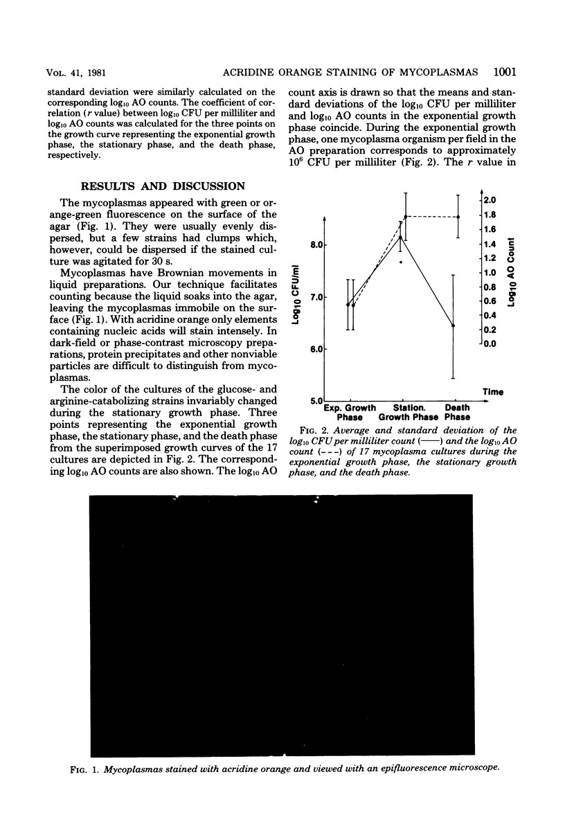
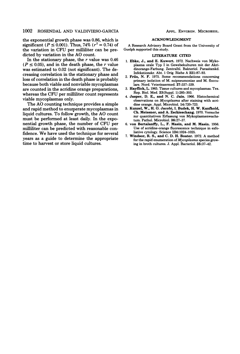
Samples of liquid mycoplasma cultures were mixed with equal part of a 0.01% solution of acridine orange and placed on agar plates. The number of fluorescing organisms per field was counted in an epifluorescence microscope at an X 1,000 magnification. When the number of fluorescing organisms per field was related to the number of colony-forming units per milliliter during the growth cycle, highly significant correlation was found in cultures with greater than or equal to 10(6) colony-forming units per ml during the exponential growth phase. The counts were weakly correlated during the stationary phase and not correlated during the death phase. This technique provides a mean to enumerate mycoplasmas in liquid cultures.
Full text
PDF


Images in this article
Selected References
These references are in PubMed. This may not be the complete list of references from this article.
- Ebke J., Kuwert E. Nachweis von Mykoplasma orale Typ I in Gewebekulturen mit der Akridinorange-Färbung. Zentralbl Bakteriol Orig A. 1972 Jul;221(1):87–93. [PubMed] [Google Scholar]
- Friis N. F. Some recommendations concerning primary isolation of Mycoplasma suipneumoniae and Mycoplasma flocculare a survey. Nord Vet Med. 1975 Jun;27(6):337–339. [PubMed] [Google Scholar]
- Hayflick L. Tissue cultures and mycoplasmas. Tex Rep Biol Med. 1965 Jun;23(Suppl):285+–285+. [PubMed] [Google Scholar]
- Jasper D. E., Jain N. C. Histochemical observations on Mycoplasma after staining with acridine orange. Appl Microbiol. 1966 Sep;14(5):720–723. doi: 10.1128/am.14.5.720-723.1966. [DOI] [PMC free article] [PubMed] [Google Scholar]
- Künzel W., Jacobi H. D., Budek I., Kaufhold H. W., Meissner C., Zschieschang A. Versuche zur quantitativen Erfassung von Mykoplasmenwachstum. Pathol Microbiol (Basel) 1970;36(1):27–37. [PubMed] [Google Scholar]
- MASIN F., MASIN M., VON BERTALANFFY L. Use of acridine-orange fluorescence technique in exfoliative cytology. Science. 1956 Nov 23;124(3230):1024–1025. doi: 10.1126/science.124.3230.1024. [DOI] [PubMed] [Google Scholar]
- Windsor R. S., Boarer C. D. A method for the rapid enumeration of Mycoplasma species growing in broth culture. J Appl Bacteriol. 1972 Mar;35(1):37–42. doi: 10.1111/j.1365-2672.1972.tb03671.x. [DOI] [PubMed] [Google Scholar]


