Abstract
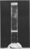
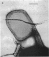
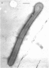
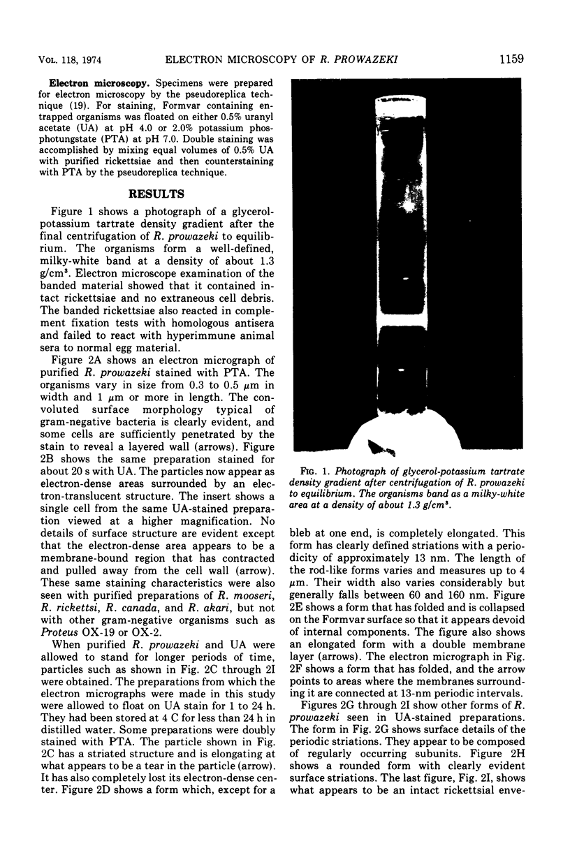
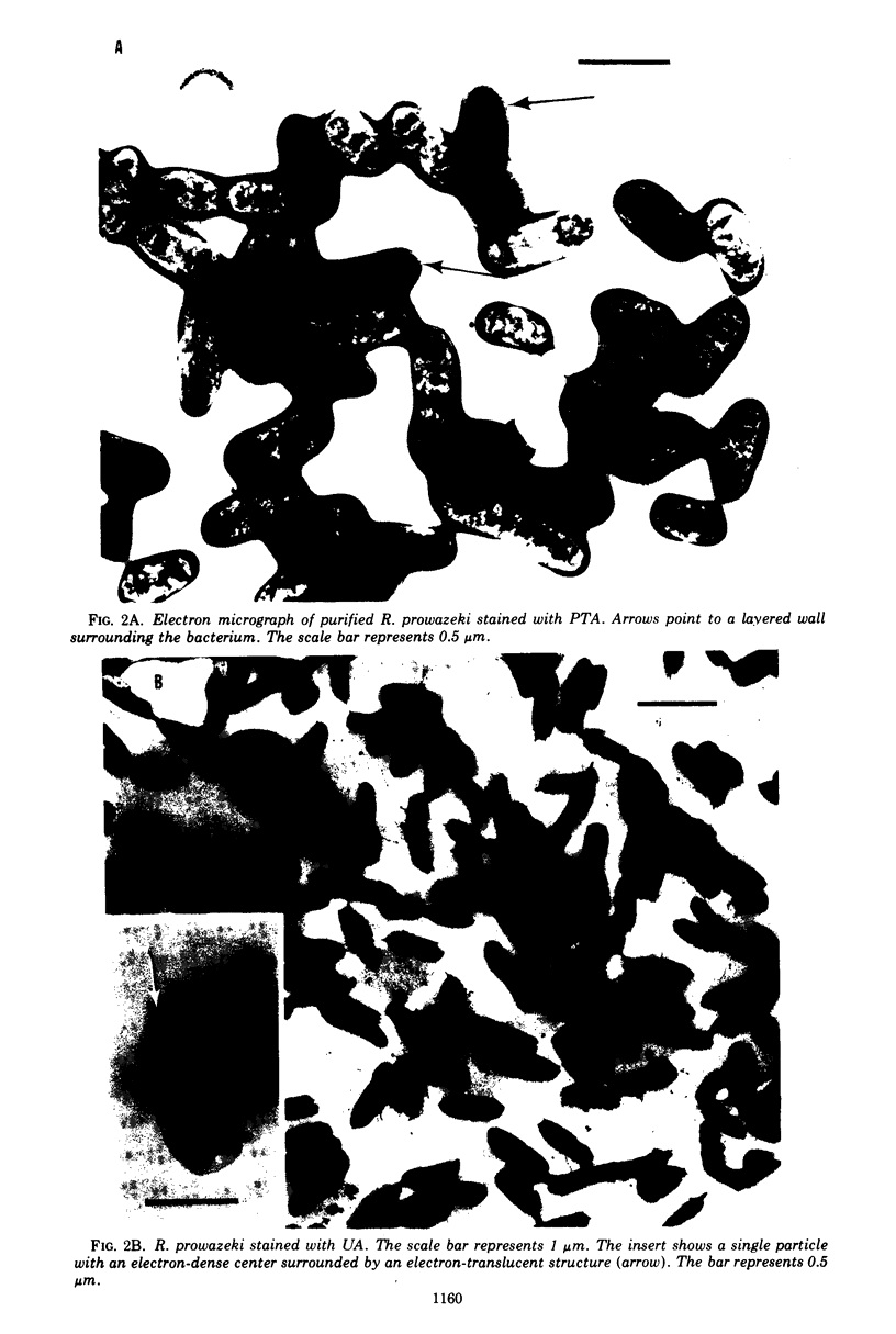
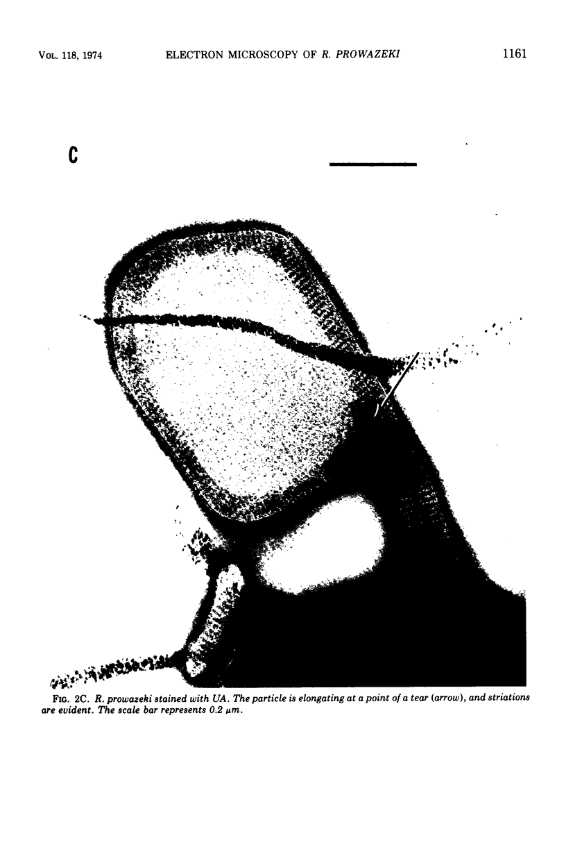
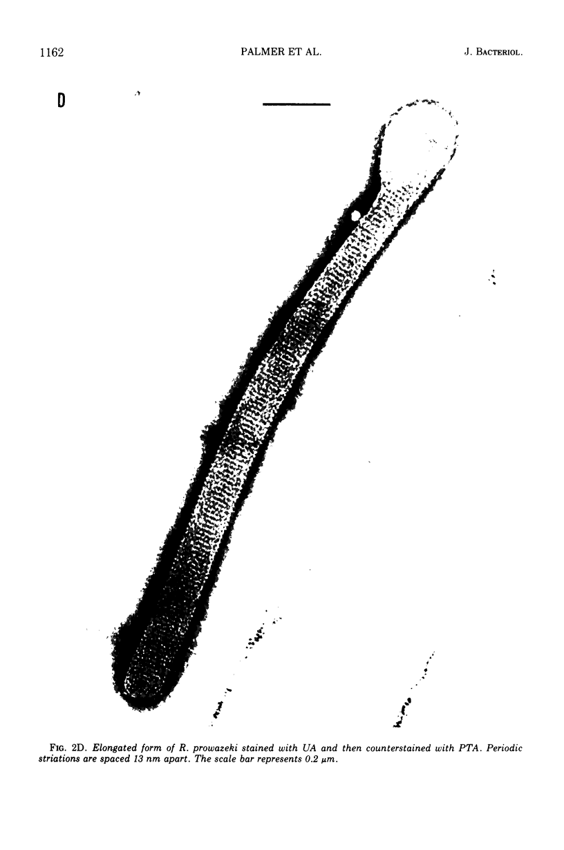
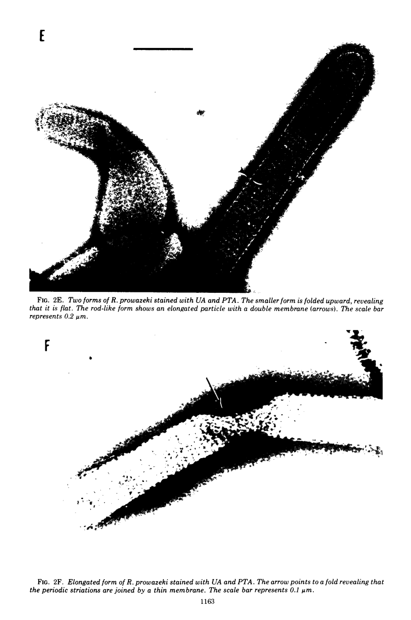
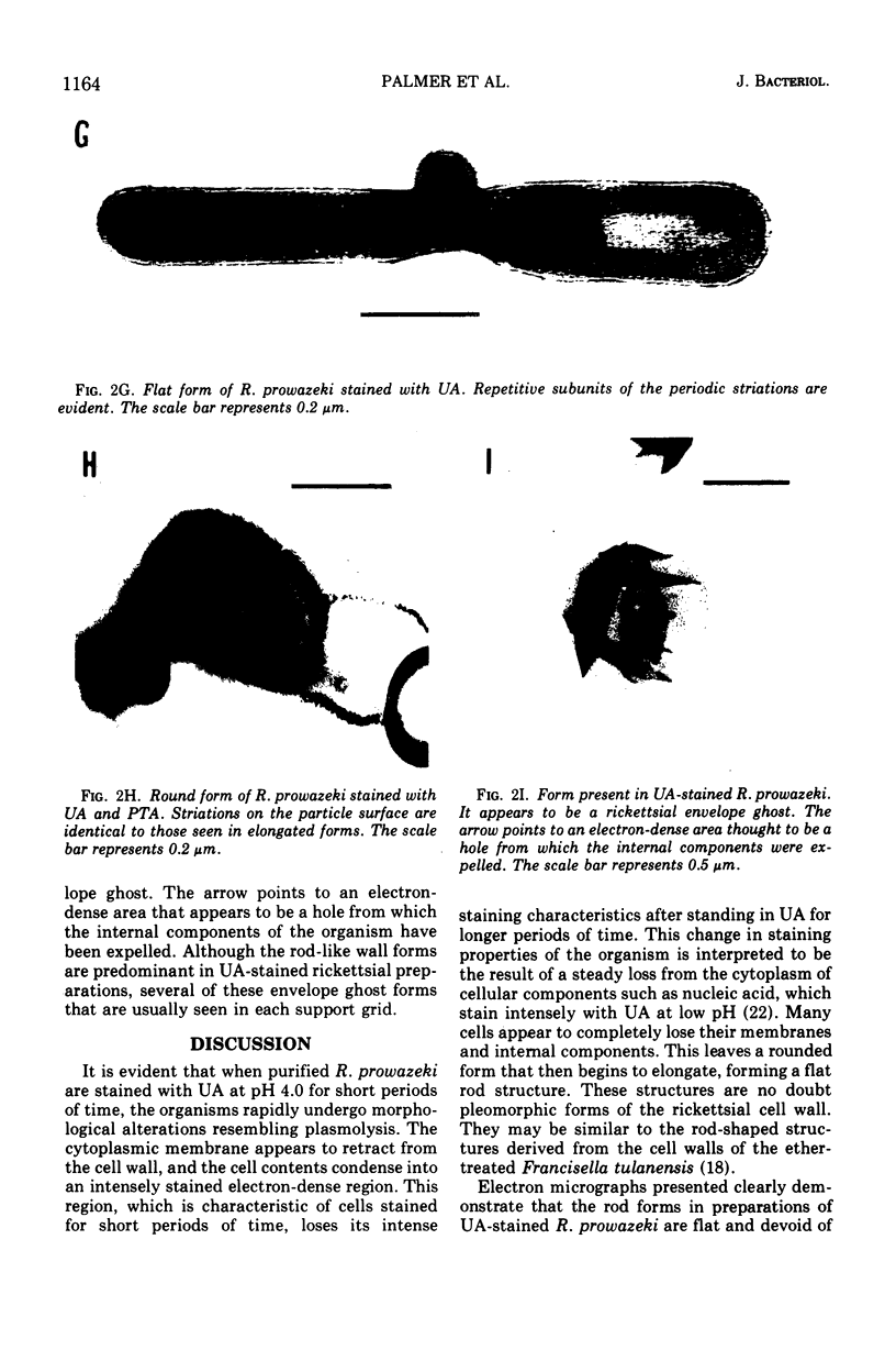
Purified Rickettsia prowazeki were found to undergo morphological changes resembling plasmolysis when stained with uranyl acetate, resulting in rod-like forms. Sequential electron micrographs of disintegrating organisms provide evidence for the cell wall origin of these rod-like forms. The substructure of the cell wall was discerned by using negative-contrast electron microscopy. The wall was found to be composed of repetitive subunits with a periodicity of 13 nm and was surrounded by a thin membrane.
Full text
PDF
Images in this article
Selected References
These references are in PubMed. This may not be the complete list of references from this article.
- Almeida J. D., Waterson A. P., Berry D. M., Turner L. H. Structures associated with leptospires possibly relevant to the Marburg agent. Lancet. 1969 Feb 1;1(7588):235–237. doi: 10.1016/s0140-6736(69)91243-4. [DOI] [PubMed] [Google Scholar]
- Anacker R. L., Pickens E. G., Lackman D. B. Details of the ultrastructure of Rickettsia prowazekii grown in the chick yolk sac. J Bacteriol. 1967 Jul;94(1):260–262. doi: 10.1128/jb.94.1.260-262.1967. [DOI] [PMC free article] [PubMed] [Google Scholar]
- Anderson D. L., Johnson R. C. Electron microscopy of immune disruption of leptospires: action of complement and lysozyme. J Bacteriol. 1968 Jun;95(6):2293–2309. doi: 10.1128/jb.95.6.2293-2309.1968. [DOI] [PMC free article] [PubMed] [Google Scholar]
- Anderson D. R., Hopps H. E., Barile M. F., Bernheim B. C. Comparison of the ultrastructure of several rickettsiae, ornithosis virus, and Mycoplasma in tissue culture. J Bacteriol. 1965 Nov;90(5):1387–1404. doi: 10.1128/jb.90.5.1387-1404.1965. [DOI] [PMC free article] [PubMed] [Google Scholar]
- BOVARNICK M. R., ALLEN E. G. Reversible inactivation of typhus rickettsiae at O C. J Bacteriol. 1957 Jan;73(1):56–62. doi: 10.1128/jb.73.1.56-62.1957. [DOI] [PMC free article] [PubMed] [Google Scholar]
- Buckmire F. L., Murray R. G. Studies on the cell wall of Spirillum serpens. 1. Isolation and partial purification of the outermost cell wall layer. Can J Microbiol. 1970 Oct;16(10):1011–1022. doi: 10.1139/m70-171. [DOI] [PubMed] [Google Scholar]
- COHN Z. A., HAHN F. E., CEGLOWSKI W., BOZEMAN F. M. Unstable nucleic acids of Rickettsia mooseri. Science. 1958 Feb 7;127(3293):282–283. doi: 10.1126/science.127.3293.282. [DOI] [PubMed] [Google Scholar]
- Fischman D. A., Weinbaum G. Hexagonal pattern in cell walls of Escherichia coli B. Science. 1967 Jan 27;155(3761):472–474. doi: 10.1126/science.155.3761.472. [DOI] [PubMed] [Google Scholar]
- GIMENEZ D. F. STAINING RICKETTSIAE IN YOLK-SAC CULTURES. Stain Technol. 1964 May;39:135–140. doi: 10.3109/10520296409061219. [DOI] [PubMed] [Google Scholar]
- Higashi N. Recent advances in electron microscope studies on ultrastructure of rickettsiae. Zentralbl Bakteriol Orig. 1968 Apr;206(3):277–283. [PubMed] [Google Scholar]
- Ormsbee R. A. Rickettsiae (as organisms). Annu Rev Microbiol. 1969;23:275–292. doi: 10.1146/annurev.mi.23.100169.001423. [DOI] [PubMed] [Google Scholar]
- PERKINS H. R., ALLISON A. C. Cell-wall constituents of rickettsiae and psittacosis-lymphogranuloma organisms. J Gen Microbiol. 1963 Mar;30:469–480. doi: 10.1099/00221287-30-3-469. [DOI] [PubMed] [Google Scholar]
- Peters D., Muller G. The Marburg agent and structures associated with leptospira. Lancet. 1969 May 3;1(7601):923–925. doi: 10.1016/s0140-6736(69)92551-3. [DOI] [PubMed] [Google Scholar]
- Reynolds J. A. Red cell membranes: fact and fancy. Fed Proc. 1973 Oct;32(10):2034–2038. [PubMed] [Google Scholar]
- SHEPARD C. C., RIBI E., LARSON C. Electron microscopically revealed structural elements of Bacterium tularense and their in vitro and in vivo role in immunologic reactions. J Immunol. 1955 Jul;75(1):7–14. [PubMed] [Google Scholar]
- Thorne K. J., Thornley M. J., Glauert A. M. Chemical analysis of the outer membrane and other layers of the cell envelope of Acinetobacter sp. J Bacteriol. 1973 Oct;116(1):410–417. doi: 10.1128/jb.116.1.410-417.1973. [DOI] [PMC free article] [PubMed] [Google Scholar]
- WISSEMAN C. L., Jr, JACKSON E. B., HAHN F. E., LEY A. C., SMADEL J. E. Metabolic studies of rickettsiae. I. The effects of antimicrobial substances and enzyme inhibitors on the oxidation of glutamate by purified rickettsiae. J Immunol. 1951 Aug;67(2):123–136. [PubMed] [Google Scholar]
- Weiss E. Growth and physiology of rickettsiae. Bacteriol Rev. 1973 Sep;37(3):259–283. doi: 10.1128/br.37.3.259-283.1973. [DOI] [PMC free article] [PubMed] [Google Scholar]
- Wisseman C. L., Jr Some biological properties of rickettsiae pathogenic for man. Zentralbl Bakteriol Orig. 1968 Apr;206(3):299–313. [PubMed] [Google Scholar]
- Wood W. H., Jr, Wisseman C. L., Jr The cell wall of Rickettsia mooseri. I. Morphology and chemical composition. J Bacteriol. 1967 Mar;93(3):1113–1118. doi: 10.1128/jb.93.3.1113-1118.1967. [DOI] [PMC free article] [PubMed] [Google Scholar]


