Abstract
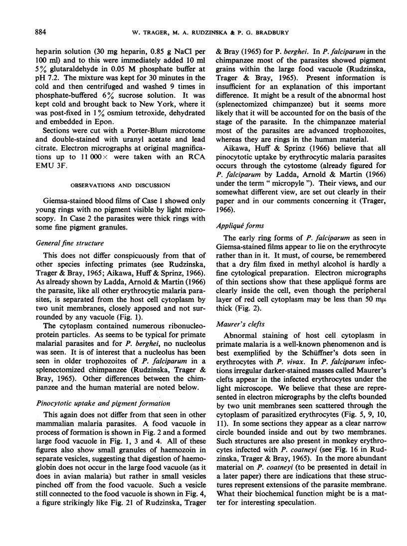
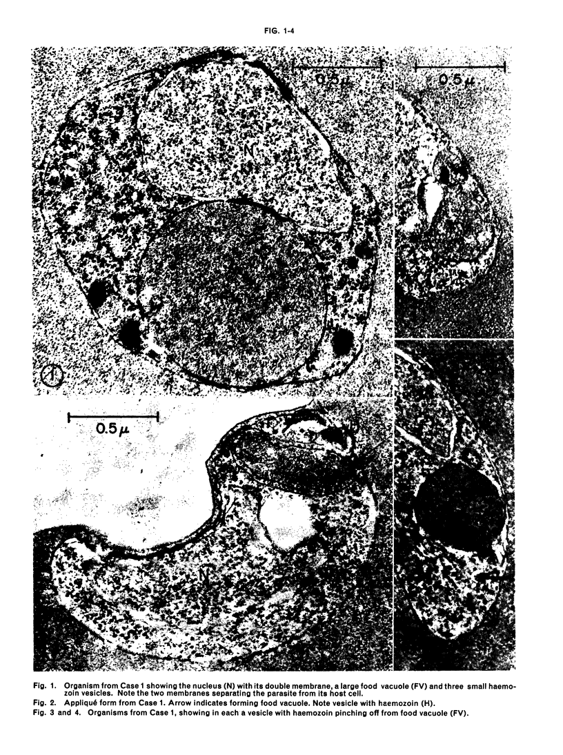
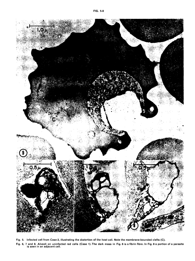
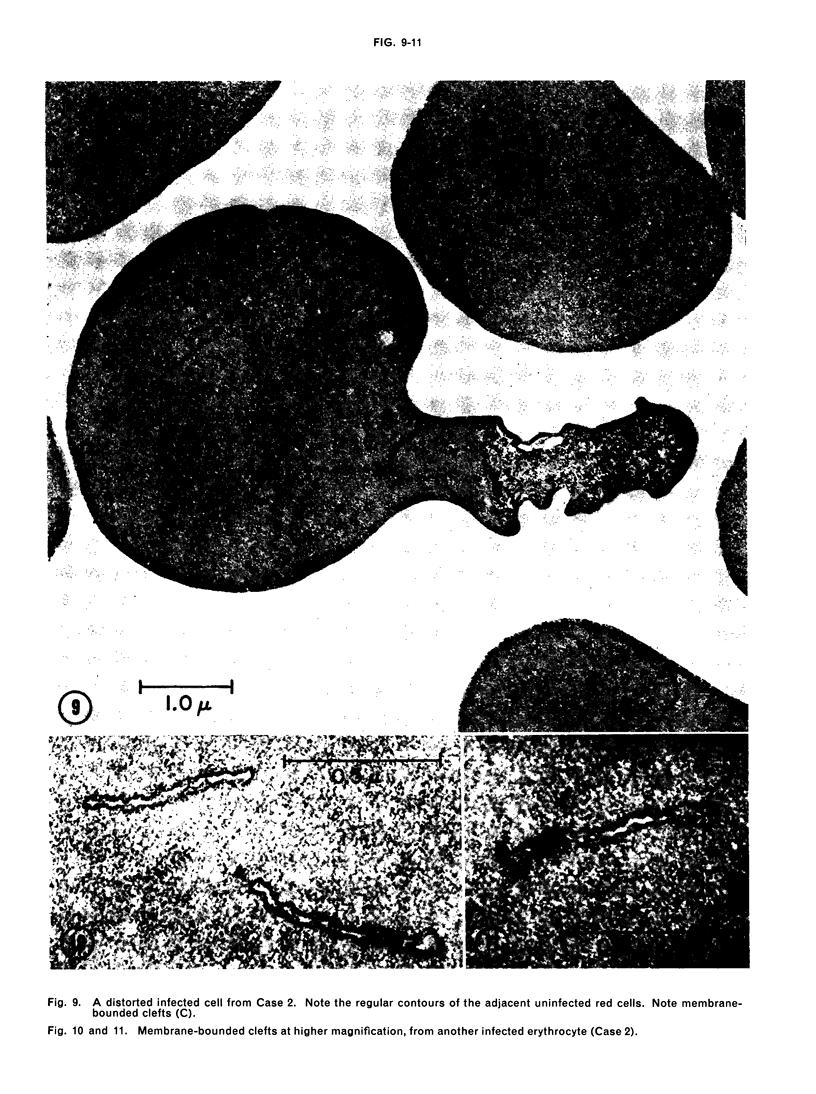
Electron micrographs of ultra-thin sections of erythrocytes taken from two Liberian children ill with Plasmodium falciparum malaria show that the appliqué forms of this parasite are clearly within the host cell. The general fine structure of the parasites resembles that of other mammalian malaria parasites, as does the mode of ingestion of host cell material by pinocytosis. Granules of haemozoin were usually found in small vesicles pinched off from the large food vacuole. Scattered through the cytoplasm of infected red cells were narrow clear clefts bounded on each side by two unit membranes. These probably represent the Maurer's clefts seen in light microscopy. The surface of infected erythrocytes was notably distorted, a phenomenon which may have a bearing on the stickiness of the infected red cells in human falciparum malaria and the segregation of these cells in the capillaries. Many uninfected erythrocytes showed a multiple alveolar, blister-like abnormality of a portion of the cell membrane; this was not seen in otherwise comparable blood from a case of P. ovale infection.
Full text
PDF
Images in this article
Selected References
These references are in PubMed. This may not be the complete list of references from this article.
- Aikawa M., Hepler P. K., Huff C. G., Sprinz H. The feeding mechanism of avian malarial parasites. J Cell Biol. 1966 Feb;28(2):355–373. doi: 10.1083/jcb.28.2.355. [DOI] [PMC free article] [PubMed] [Google Scholar]
- FULTON J. D., FLEWETT T. H. The relation of Plasmodium berghei and Plasmodium knowlesi to their respective red-cell hosts. Trans R Soc Trop Med Hyg. 1956 Mar;50(2):150–156. doi: 10.1016/0035-9203(56)90076-1. [DOI] [PubMed] [Google Scholar]
- Ladda R., Arnold J., Martin D. Electron microscopy of Plasmodium falciparum . 1. The structure of trophozoites in erythrocytes of human volunteers. Trans R Soc Trop Med Hyg. 1966;60(3):369–375. doi: 10.1016/0035-9203(66)90302-6. [DOI] [PubMed] [Google Scholar]
- Lewis S. M., Danon D., Marikovsky Y. Electron-microscope studies of the red cell in paroxysmal nocturnal haemoglobinuria. Br J Haematol. 1965 Nov;11(6):689–695. doi: 10.1111/j.1365-2141.1965.tb00118.x. [DOI] [PubMed] [Google Scholar]
- RUDZINSKA M. A., TRAGER W. Phagotrophy and two new structures in the malaria parasite Plasmodium berghei. J Biophys Biochem Cytol. 1959 Aug;6(1):103–112. doi: 10.1083/jcb.6.1.103. [DOI] [PMC free article] [PubMed] [Google Scholar]
- Rudzinska M. A., Trager W., Bray R. S. Pinocytotic uptake and the digestion of hemoglobin in malaria parasites. J Protozool. 1965 Nov;12(4):563–576. doi: 10.1111/j.1550-7408.1965.tb03256.x. [DOI] [PubMed] [Google Scholar]


