

Abstract
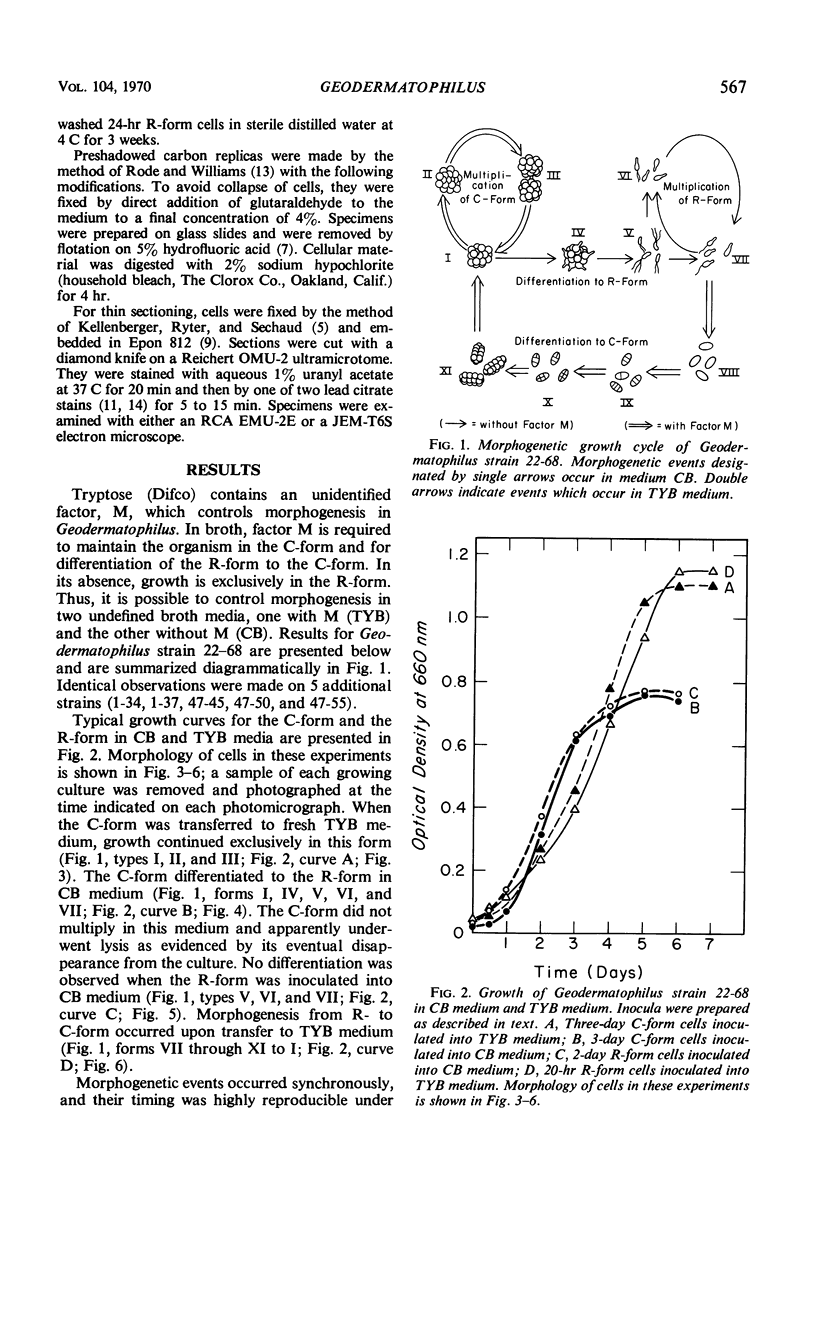
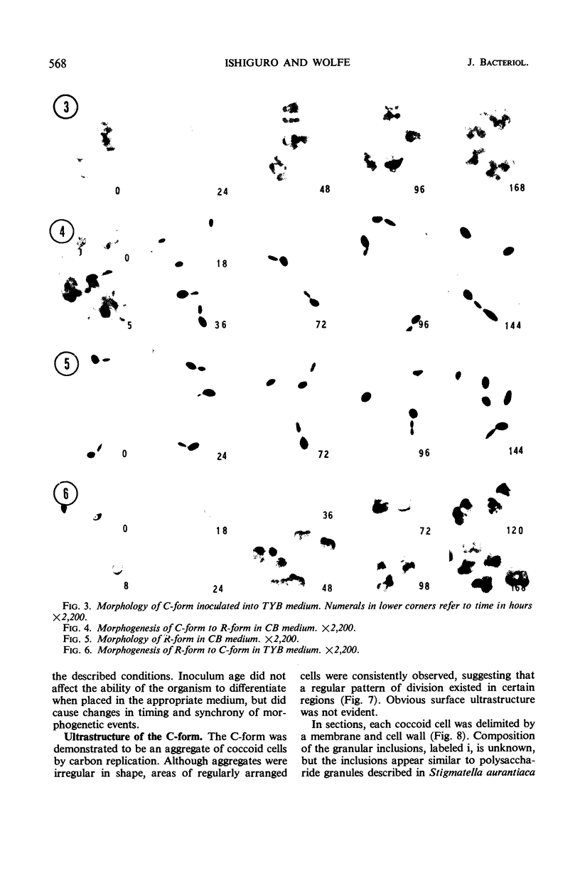
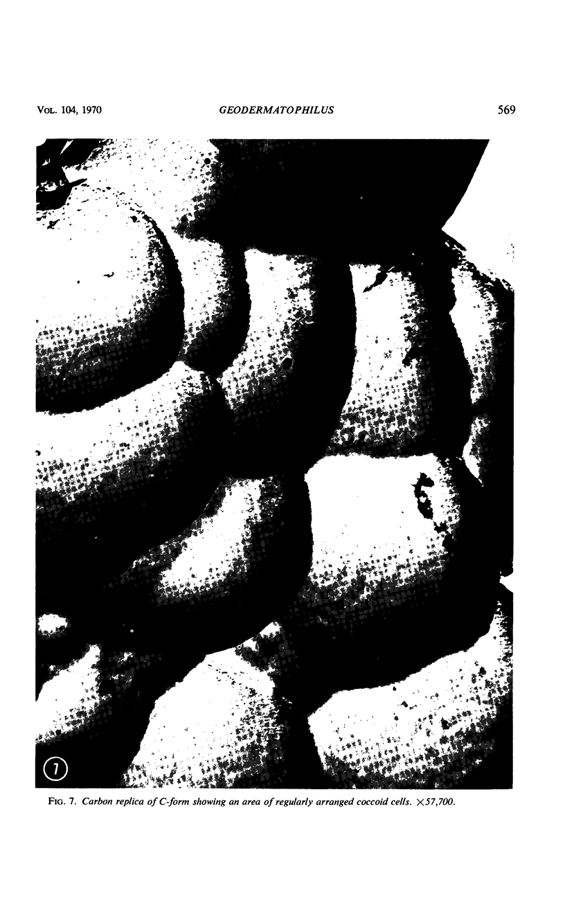
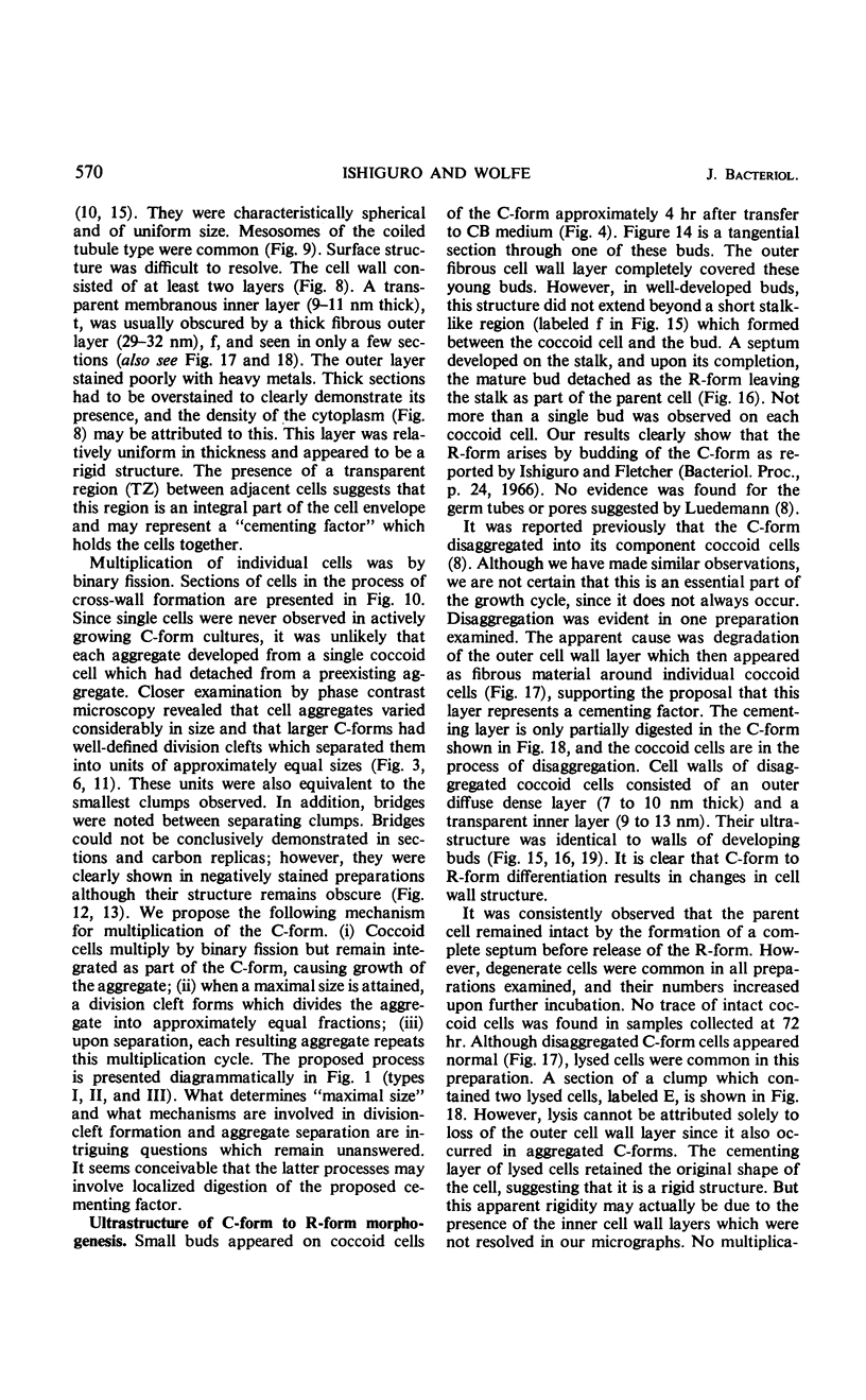
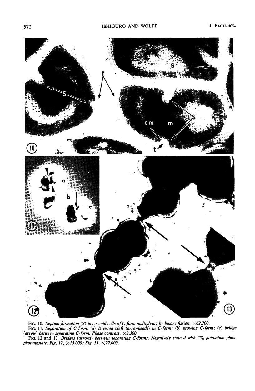
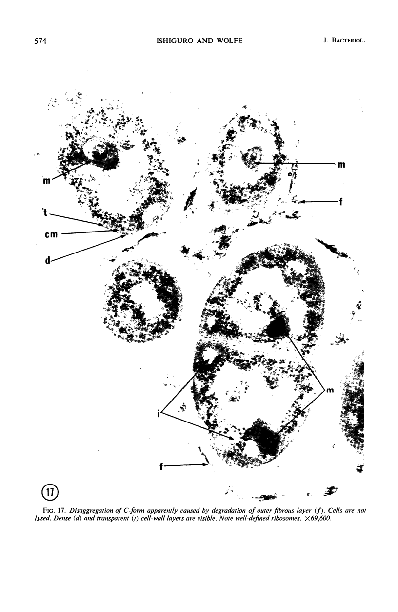
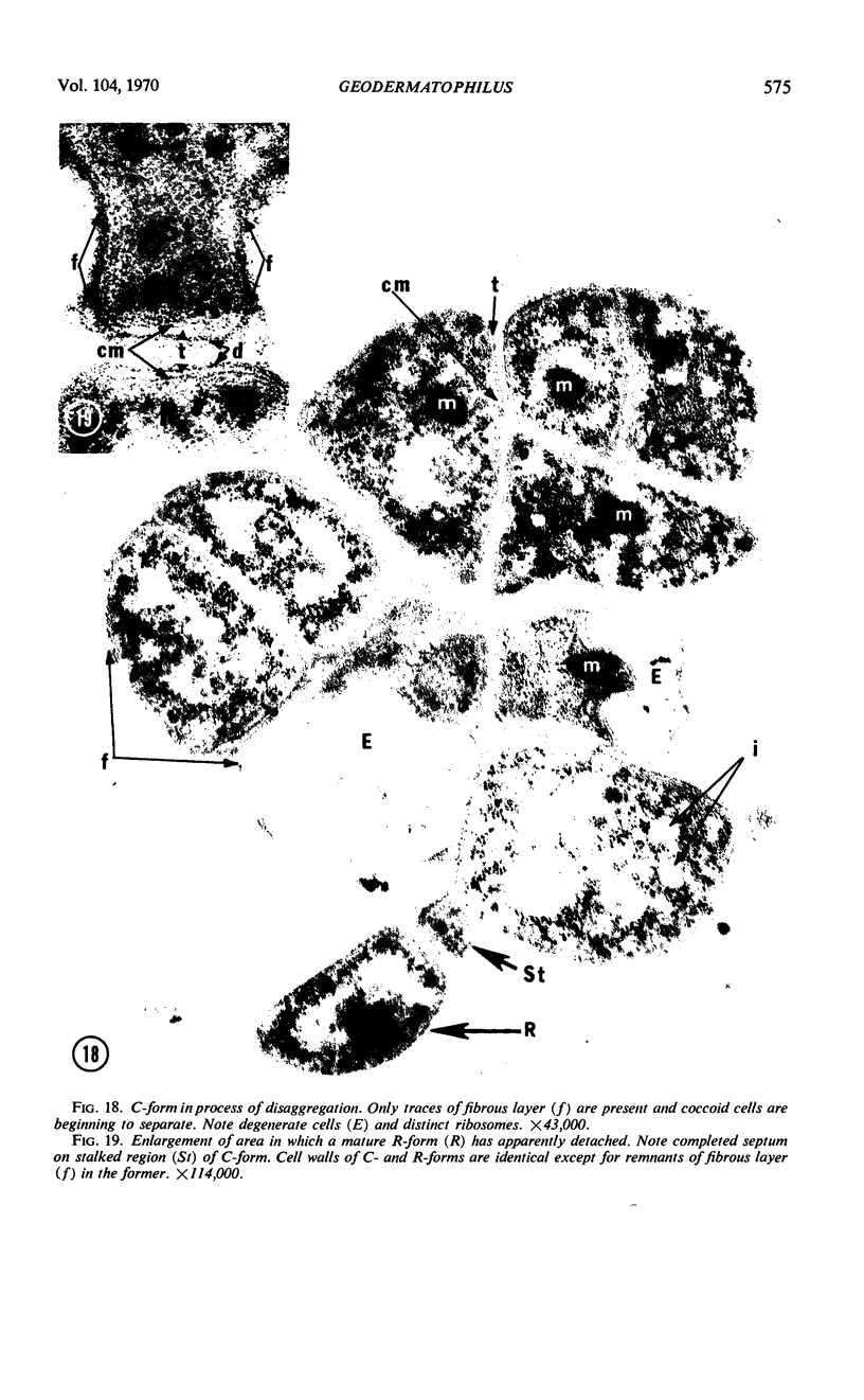
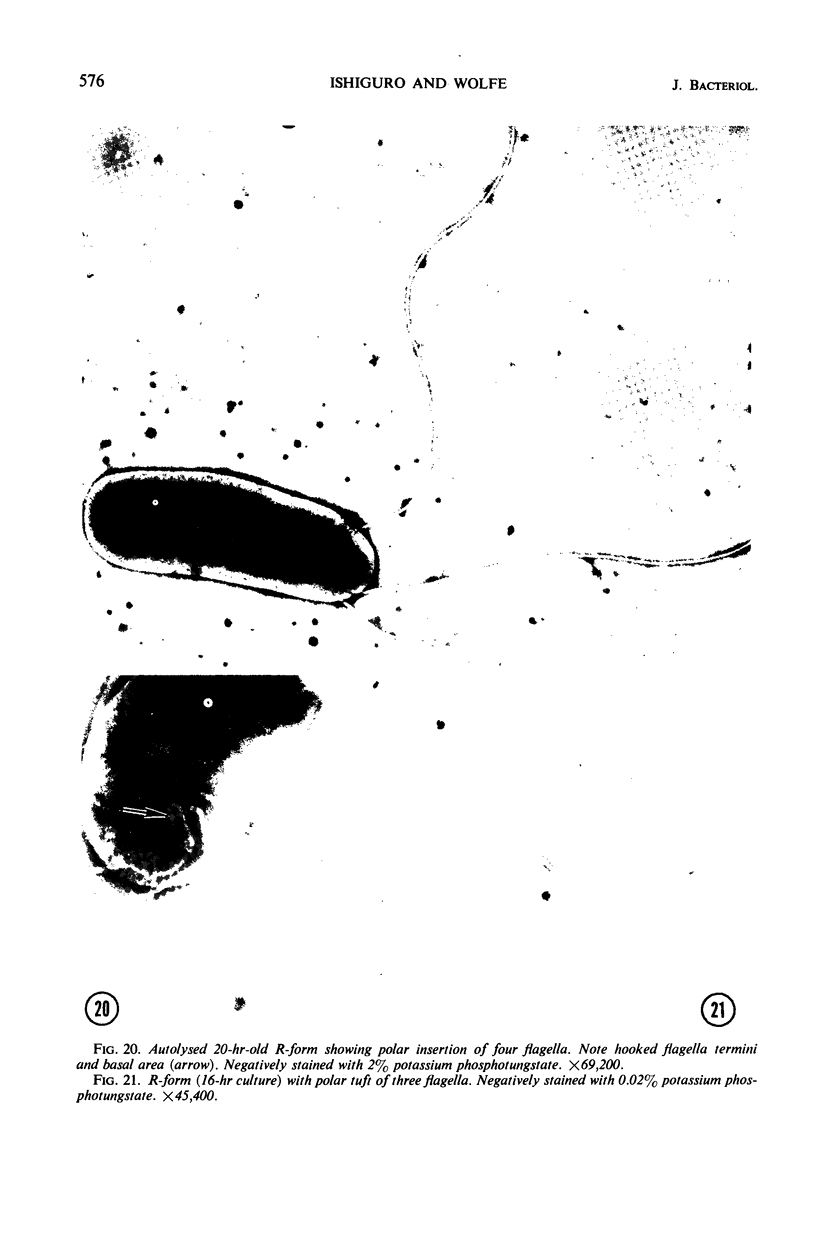
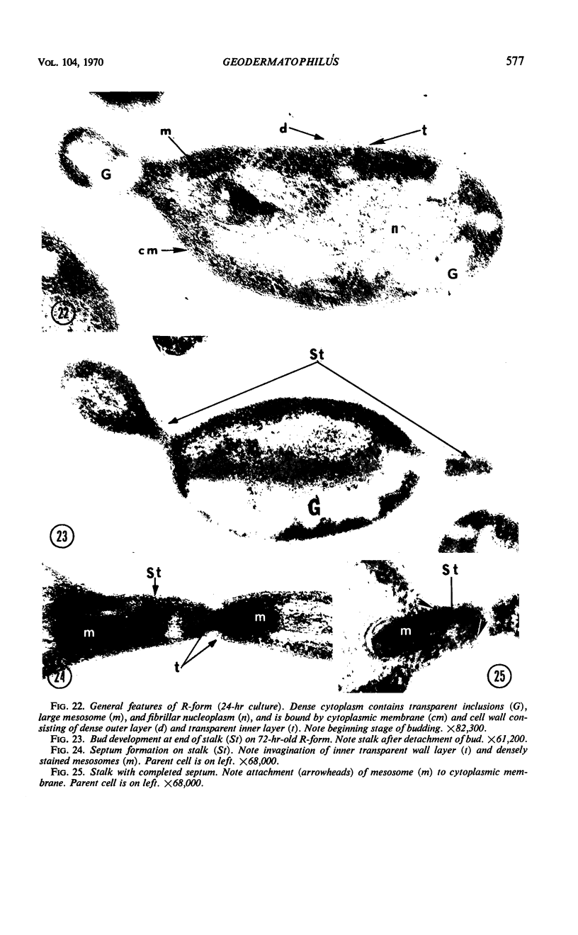
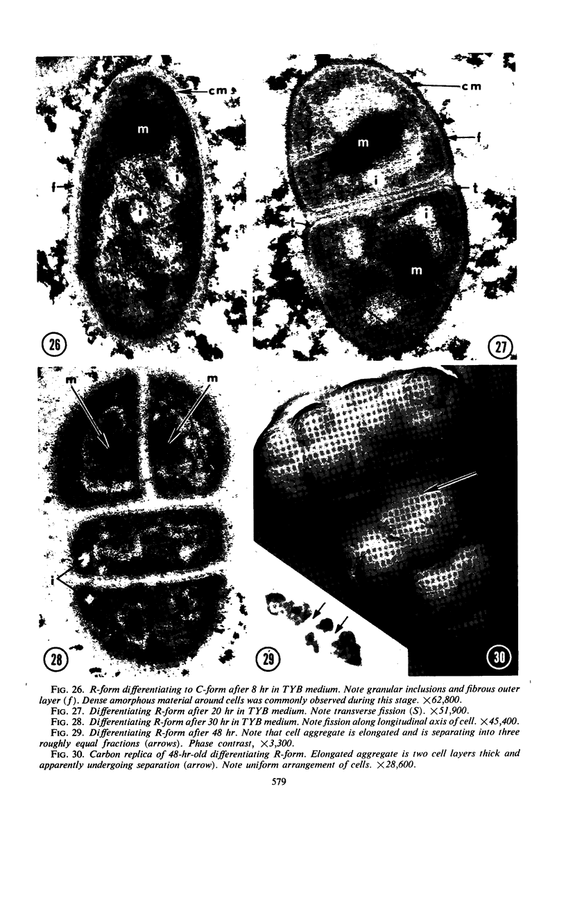
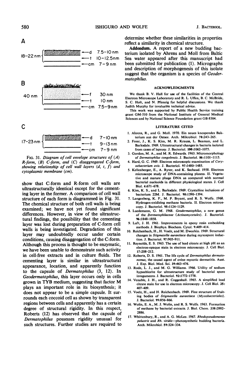
Geodermatophilus grows in two major forms, a nonmotile irregularly shaped aggregate of coccoid cells (C-form) and a motile budding rod (R-form). Morphogenesis can be controlled by an unidentified factor in Tryptose which is required for maintenance of the organism in the C-form and for differentiation of the R-form to the C-form. Morphogenetic events occur synchronously in the described system. Ultrastructural studies show that the major difference between C- and R-forms is in cells envelope structure. R-form cell walls consist of two layers, an inner transparent membranous layer (10 to 12.5 nm thick) and an outer dense diffuse layer (7.5 to 10 nm). In addition to these layers, the C-form has a thick fibrous layer (30 nm) over the dense layer. This layer appears to be a cementing substance which holds the coccoid cells together.
Full text
PDF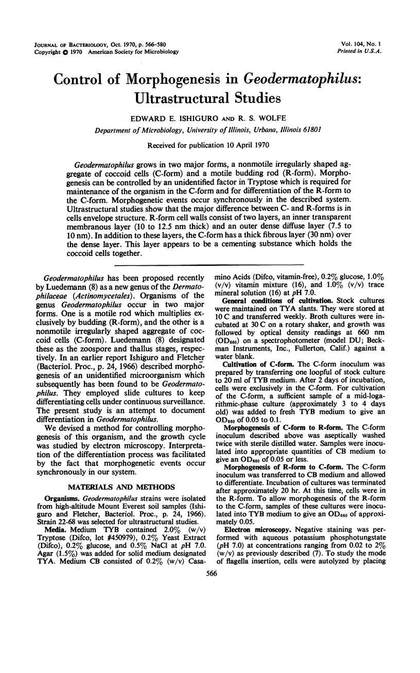
Images in this article

Selected References
These references are in PubMed. This may not be the complete list of references from this article.
- Ahrens R., Moll G. Ein neues knospendes Bakterium aus der Ostee. Arch Mikrobiol. 1970;70(3):243–265. [PubMed] [Google Scholar]
- Freer J., Kim K. S., Krauss M. R., Beaman L., Barksdale L. Ultrastructural changes in bacteria isolated from cases of leprosy. J Bacteriol. 1969 Nov;100(2):1062–1075. doi: 10.1128/jb.100.2.1062-1075.1969. [DOI] [PMC free article] [PubMed] [Google Scholar]
- GORDON M. A., EDWARDS M. R. MICROMORPHOLOGY OF DERMATOPHILUS CONGOLENSIS. J Bacteriol. 1963 Nov;86:1101–1115. doi: 10.1128/jb.86.5.1101-1115.1963. [DOI] [PMC free article] [PubMed] [Google Scholar]
- Hard G. C. Electron microscopic examination of Corynebacterium ovis. J Bacteriol. 1969 Mar;97(3):1480–1485. doi: 10.1128/jb.97.3.1480-1485.1969. [DOI] [PMC free article] [PubMed] [Google Scholar]
- KELLENBERGER E., RYTER A., SECHAUD J. Electron microscope study of DNA-containing plasms. II. Vegetative and mature phage DNA as compared with normal bacterial nucleoids in different physiological states. J Biophys Biochem Cytol. 1958 Nov 25;4(6):671–678. doi: 10.1083/jcb.4.6.671. [DOI] [PMC free article] [PubMed] [Google Scholar]
- Kim K. S., Barksdale L. Crystalline inclusions of bacterium 22M. J Bacteriol. 1969 Jun;98(3):1390–1394. doi: 10.1128/jb.98.3.1390-1394.1969. [DOI] [PMC free article] [PubMed] [Google Scholar]
- LUFT J. H. Improvements in epoxy resin embedding methods. J Biophys Biochem Cytol. 1961 Feb;9:409–414. doi: 10.1083/jcb.9.2.409. [DOI] [PMC free article] [PubMed] [Google Scholar]
- Langenberg K. F., Bryant M. P., Wolfe R. S. Hydrogen-oxidizing methane bacteria. II. Electron microscopy. J Bacteriol. 1968 Mar;95(3):1124–1129. doi: 10.1128/jb.95.3.1124-1129.1968. [DOI] [PMC free article] [PubMed] [Google Scholar]
- Luedemann G. M. Geodermatophilus, a new genus of the Dermatophilaceae (Actinomycetales). J Bacteriol. 1968 Nov;96(5):1848–1858. doi: 10.1128/jb.96.5.1848-1858.1968. [DOI] [PMC free article] [PubMed] [Google Scholar]
- REYNOLDS E. S. The use of lead citrate at high pH as an electron-opaque stain in electron microscopy. J Cell Biol. 1963 Apr;17:208–212. doi: 10.1083/jcb.17.1.208. [DOI] [PMC free article] [PubMed] [Google Scholar]
- ROBERT D. S. The life cycle of Dermatophilus dermatonomus, the causal agent of ovine mycotic dermatitis. Aust J Exp Biol Med Sci. 1961 Oct;39:463–476. [PubMed] [Google Scholar]
- Reichenbach H., Voelz H., Dworkin M. Structural changes in Stigmatella aurantiaca during myxospore induction. J Bacteriol. 1969 Feb;97(2):905–911. doi: 10.1128/jb.97.2.905-911.1969. [DOI] [PMC free article] [PubMed] [Google Scholar]
- Rode L. J., Williams M. G. Utility of sodium hypochlorite for ultrastructure study of bacterial spore integuments. J Bacteriol. 1966 Dec;92(6):1772–1778. doi: 10.1128/jb.92.6.1772-1778.1966. [DOI] [PMC free article] [PubMed] [Google Scholar]
- VENABLE J. H., COGGESHALL R. A SIMPLIFIED LEAD CITRATE STAIN FOR USE IN ELECTRON MICROSCOPY. J Cell Biol. 1965 May;25:407–408. doi: 10.1083/jcb.25.2.407. [DOI] [PMC free article] [PubMed] [Google Scholar]
- Voelz H., Reichenbach H. Fine structure of fruiting bodies of Stigmatella aurantiaca (Myxobacterales). J Bacteriol. 1969 Sep;99(3):856–866. doi: 10.1128/jb.99.3.856-866.1969. [DOI] [PMC free article] [PubMed] [Google Scholar]
- WOLIN E. A., WOLIN M. J., WOLFE R. S. FORMATION OF METHANE BY BACTERIAL EXTRACTS. J Biol Chem. 1963 Aug;238:2882–2886. [PubMed] [Google Scholar]
- Whittenbury R., McLee A. G. Rhodopseudomonas palustris and Rh. viridis--photosynthetic budding bacteria. Arch Mikrobiol. 1967;59(1):324–334. doi: 10.1007/BF00406346. [DOI] [PubMed] [Google Scholar]