Abstract
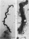
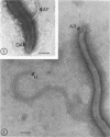
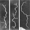
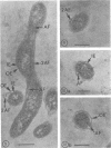
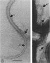
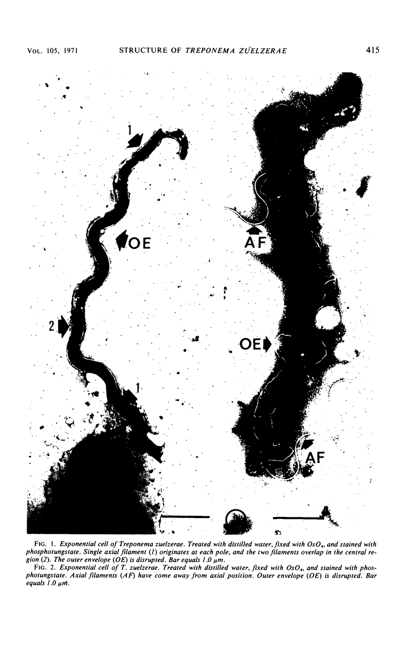
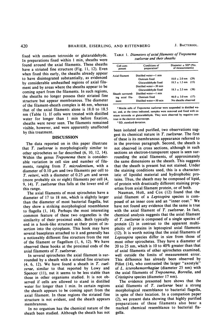
The fine structure of the spirochete Treponema zuelzerae, and particularly of its axial filaments, was investigated by using the electron microscope. The cell consists of a protoplasmic core surrounded by two concentric envelopes, each approximately 12 nm in width. Between these envelopes are two axial filaments, one originating at each pole of the cell, which overlap and lie side by side in the central region of the cell. The diameter of the axial filaments is 18.0 to 18.5 nm. The terminal region of each filament at its proximal end consists of a hook-like structure, very similar in appearance to the proximal end of a bacterial flagellum. The outer envelope of the cell is readily disrupted with distilled water, and this treatment often results in the release of the filaments from their axial position. A sheath is seen surrounding the filaments when cells are treated with distilled water for no more than 1 min and fixed immediately with osmium tetroxide or glutaraldehyde. This sheath has a striated fine structure and a diameter of 46 nm.
Full text
PDF
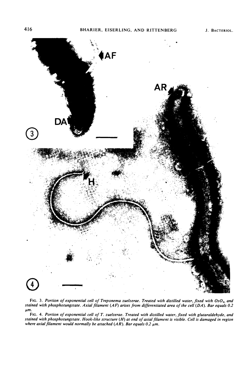
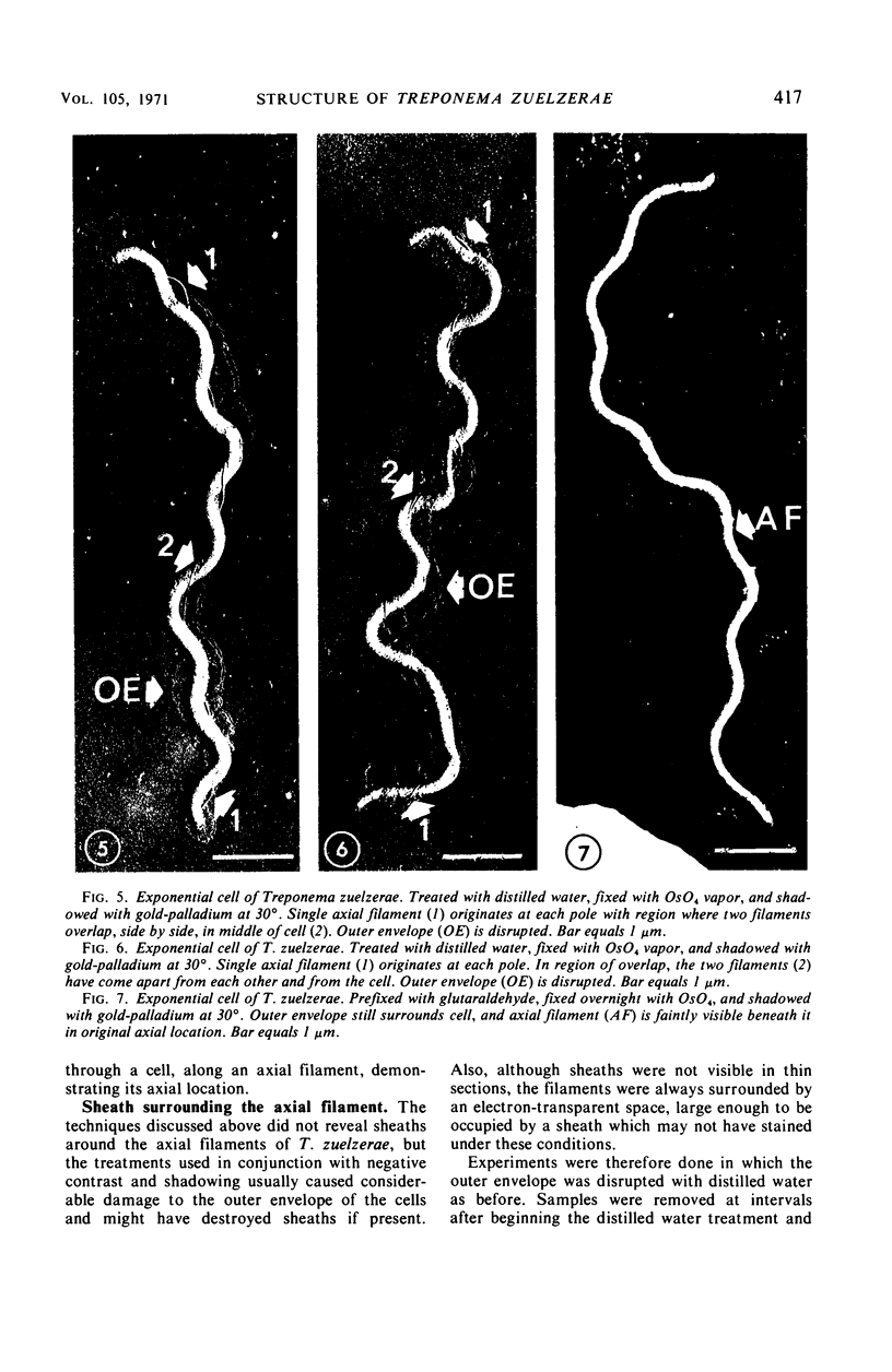
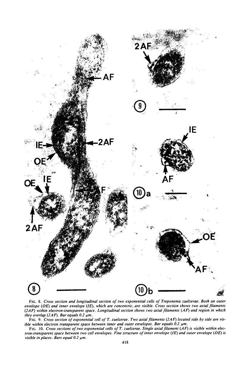
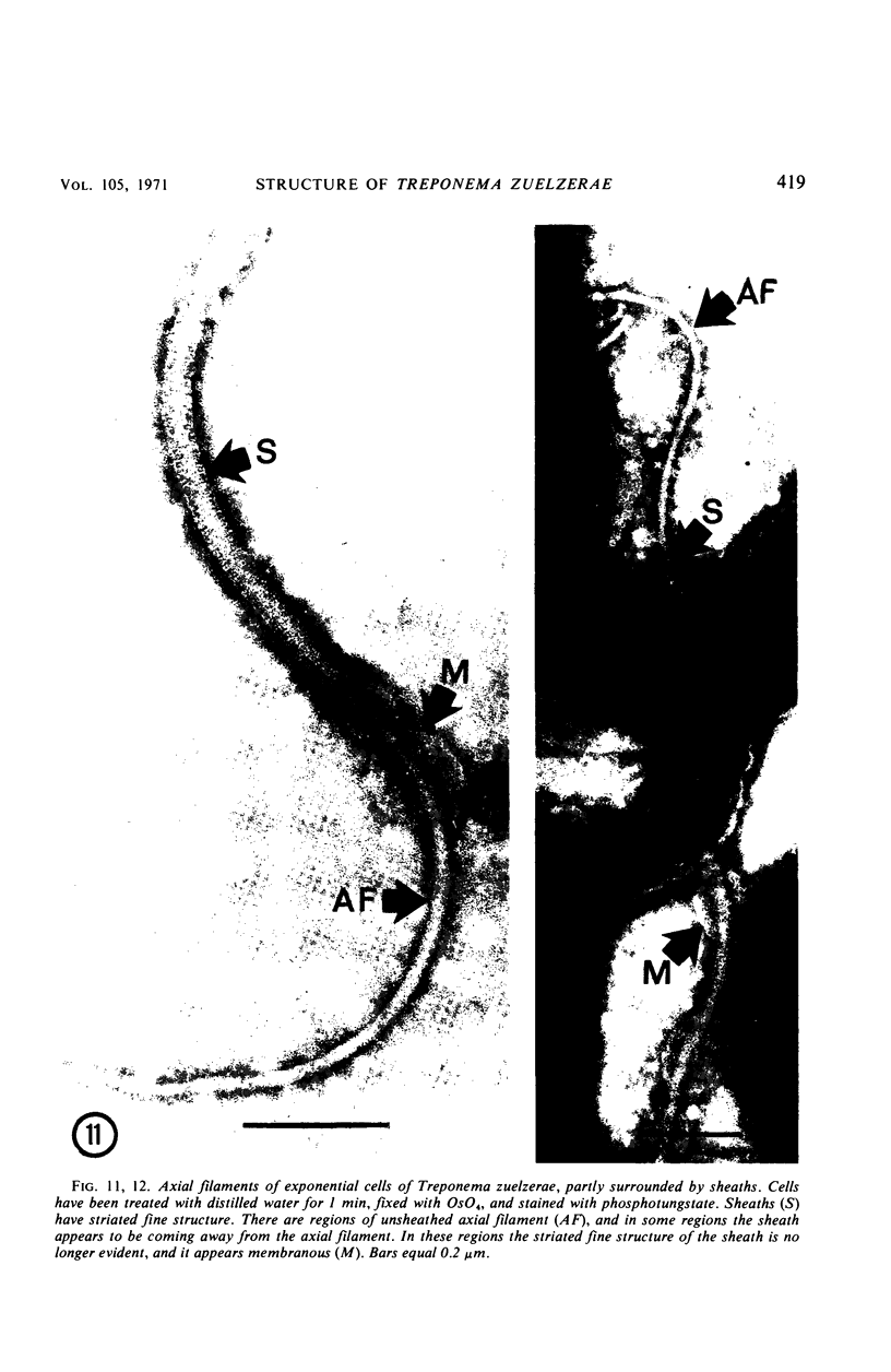
Images in this article
Selected References
These references are in PubMed. This may not be the complete list of references from this article.
- Abram D., Vatter A. E., Koffler H. Attachment and structural features of flagella of certain bacilli. J Bacteriol. 1966 May;91(5):2045–2068. doi: 10.1128/jb.91.5.2045-2068.1966. [DOI] [PMC free article] [PubMed] [Google Scholar]
- BRADFIELD J. R. G., CATER D. B. Electron-microscopic evidence on the structure of spirochaetes. Nature. 1952 Jun 7;169(4310):944–946. doi: 10.1038/169944a0. [DOI] [PubMed] [Google Scholar]
- Bharier M. A., Rittenberg S. C. Chemistry of axial filaments of Treponema zuezerae. J Bacteriol. 1971 Jan;105(1):422–429. doi: 10.1128/jb.105.1.422-429.1971. [DOI] [PMC free article] [PubMed] [Google Scholar]
- Bharier M. A., Rittenberg S. C. Immobilization effects of anticell and antiaxial filament sera on Treponema zuelzerae. J Bacteriol. 1971 Jan;105(1):430–437. doi: 10.1128/jb.105.1.430-437.1971. [DOI] [PMC free article] [PubMed] [Google Scholar]
- Bladen H. A., Hampp E. G. Ultrastructure of Treponema microdentium and Borrelia vincentii. J Bacteriol. 1964 May;87(5):1180–1191. doi: 10.1128/jb.87.5.1180-1191.1964. [DOI] [PMC free article] [PubMed] [Google Scholar]
- Holt S. C., Canale-Parola E. Fine structure of Spirochaeta stenostrepta, a free-living, anaerobic spirochete. J Bacteriol. 1968 Sep;96(3):822–835. doi: 10.1128/jb.96.3.822-835.1968. [DOI] [PMC free article] [PubMed] [Google Scholar]
- JAHN T. L., LANDMAN M. D. LOCOMOTION OF SPIROCHETES. Trans Am Microsc Soc. 1965 Jul;84:395–406. [PubMed] [Google Scholar]
- KELLENBERGER E., RYTER A., SECHAUD J. Electron microscope study of DNA-containing plasms. II. Vegetative and mature phage DNA as compared with normal bacterial nucleoids in different physiological states. J Biophys Biochem Cytol. 1958 Nov 25;4(6):671–678. doi: 10.1083/jcb.4.6.671. [DOI] [PMC free article] [PubMed] [Google Scholar]
- LISTGARTEN M. A., LOESCHE W. J., SOCRANSKY S. S. MORPHOLOGY OF TREPONEMA MICRODENTIUM AS REVEALED BY ELECTRON MICROSCOPY OF ULTRATHIN SECTIONS. J Bacteriol. 1963 Apr;85:932–939. doi: 10.1128/jb.85.4.932-939.1963. [DOI] [PMC free article] [PubMed] [Google Scholar]
- LISTGARTEN M. A., SOCRANSKY S. S. ELECTRON MICROSCOPY OF AXIAL FIBRILS, OUTER ENVELOPE, AND CELL DIVISION OF CERTAIN ORAL SPIROCHETES. J Bacteriol. 1964 Oct;88:1087–1103. doi: 10.1128/jb.88.4.1087-1103.1964. [DOI] [PMC free article] [PubMed] [Google Scholar]
- Lowy J., Spencer M. Structure and function of bacterial flagella. Symp Soc Exp Biol. 1968;22:215–236. [PubMed] [Google Scholar]
- Nauman R. K., Holt S. C., Cox C. D. Purification, ultrastructure, and composition of axial filaments from Leptospira. J Bacteriol. 1969 Apr;98(1):264–280. doi: 10.1128/jb.98.1.264-280.1969. [DOI] [PMC free article] [PubMed] [Google Scholar]
- REYNOLDS E. S. The use of lead citrate at high pH as an electron-opaque stain in electron microscopy. J Cell Biol. 1963 Apr;17:208–212. doi: 10.1083/jcb.17.1.208. [DOI] [PMC free article] [PubMed] [Google Scholar]
- VELDKAMP H. Isolation and characteristics of Treponema zuelzerae nov. spec., and anaerobic, free-living spirochete. Antonie Van Leeuwenhoek. 1960;26:103–125. doi: 10.1007/BF02538999. [DOI] [PubMed] [Google Scholar]


