Abstract
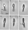
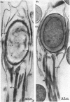
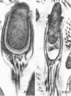
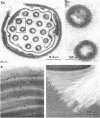
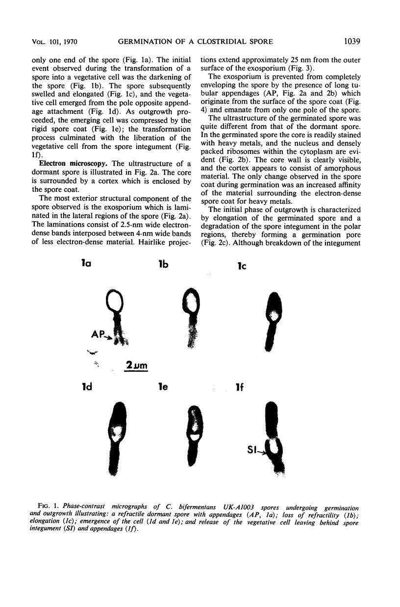
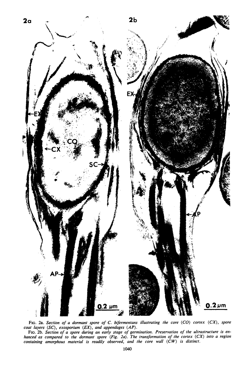
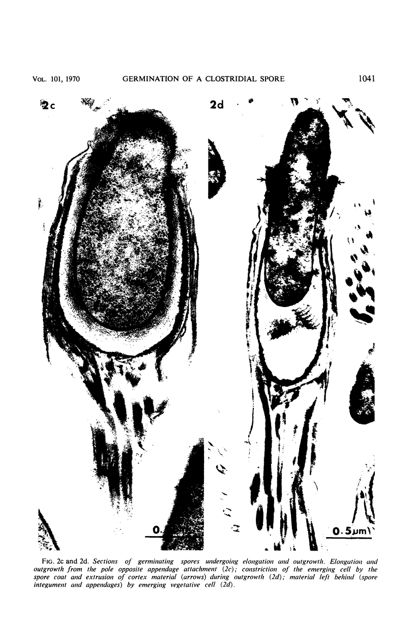
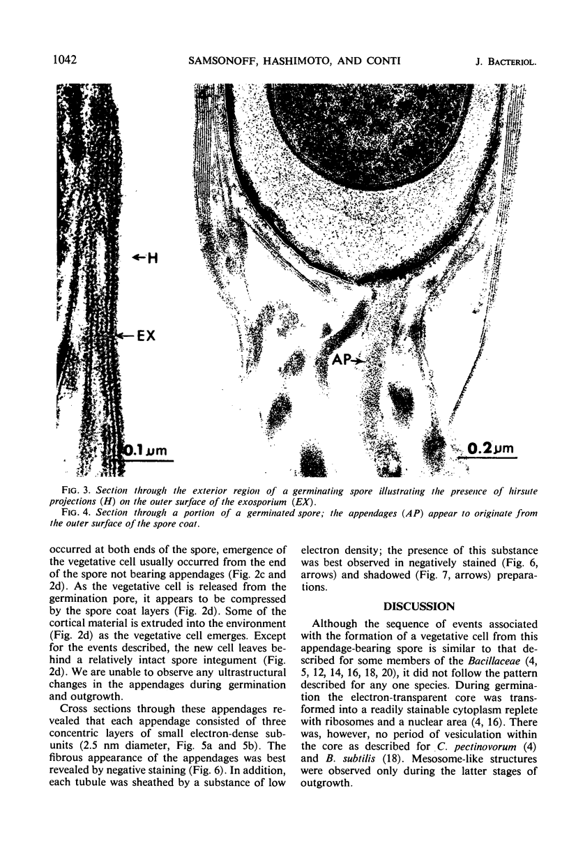
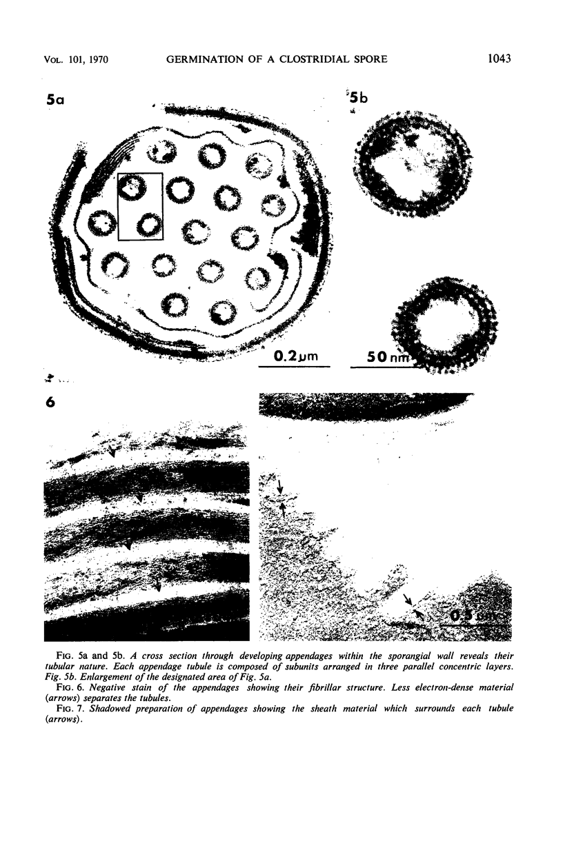
The sequence of events occurring during the germination and outgrowth of appendage-bearing spores of Clostridium bifermentans was studied by phase-contrast and electron microscopy. The mature spore was characterized ultrastructurally as having the normal spore components as well as long tubular appendages which orginated from the surface of the spore coat. Spores were incompletely enclosed by a distinctly laminated exosporium which possessed hairlike projections on its outermost layer. During germination, structural changes were observed in the core, core wall, cortex, and spore coat layers. Cortical material was extruded from the spore during outgrowth, which usually occurred from the pole opposite the appendages. The subunits comprising the structure of the appendages and the morphology of the mature appendages were observed. No discernible changes could be observed in the spore appendages during germination and outgrowth.
Full text
PDF
Images in this article
Selected References
These references are in PubMed. This may not be the complete list of references from this article.
- Hodgkiss W., Ordal Z. J., Cann D. C. The morphology and ultrastructure of the spore and exosporium of some Clostridium species. J Gen Microbiol. 1967 May;47(2):213–225. doi: 10.1099/00221287-47-2-213. [DOI] [PubMed] [Google Scholar]
- Hodgkiss W., Ordal Z. J. Morphology of the spore of some strains of Clostridium botulinum type E. J Bacteriol. 1966 May;91(5):2031–2036. doi: 10.1128/jb.91.5.2031-2036.1966. [DOI] [PMC free article] [PubMed] [Google Scholar]
- Hoeniger J. F., Headley C. L. Cytology of spore germination in Clostridium pectinovorum. J Bacteriol. 1968 Nov;96(5):1835–1847. doi: 10.1128/jb.96.5.1835-1847.1968. [DOI] [PMC free article] [PubMed] [Google Scholar]
- Hoeniger J. F., Headley C. L. Ultrastructural aspects of spore germination and outgrowth in Clostridium sporogenes. Can J Microbiol. 1969 Sep;15(9):1061–1065. doi: 10.1139/m69-189. [DOI] [PubMed] [Google Scholar]
- KELLENBERGER E., RYTER A., SECHAUD J. Electron microscope study of DNA-containing plasms. II. Vegetative and mature phage DNA as compared with normal bacterial nucleoids in different physiological states. J Biophys Biochem Cytol. 1958 Nov 25;4(6):671–678. doi: 10.1083/jcb.4.6.671. [DOI] [PMC free article] [PubMed] [Google Scholar]
- Knaysi G., Baker R. F., Hillier J. A Study, with the High-Voltage Electron Microscope, of the Endospore and Life Cycle of Bacillus mycoides. J Bacteriol. 1947 May;53(5):525–537. doi: 10.1128/jb.53.5.525-537.1947. [DOI] [PMC free article] [PubMed] [Google Scholar]
- Knaysi G., Hillier J. PRELIMINARY OBSERVATIONS ON THE GERMINATION OF THE ENDOSPORE IN BACILLUS MEGATHERIUM AND THE STRUCTURE OF THE SPORE COAT. J Bacteriol. 1949 Jan;57(1):23–29. doi: 10.1128/jb.57.1.23-29.1949. [DOI] [PMC free article] [PubMed] [Google Scholar]
- Moberly B. J., Shafa F., Gerhardt P. Structural details of anthrax spores during stages of transformation into vegetative cells. J Bacteriol. 1966 Jul;92(1):220–228. doi: 10.1128/jb.92.1.220-228.1966. [DOI] [PMC free article] [PubMed] [Google Scholar]
- Pope L., Yolton D. P., Rode L. J. Appendages of Clostridium bifermentans spores. J Bacteriol. 1967 Oct;94(4):1206–1215. doi: 10.1128/jb.94.4.1206-1215.1967. [DOI] [PMC free article] [PubMed] [Google Scholar]
- Propst A., Möse J. R. Sporulation und Germination beim Clostridium butyricum M 55. Elektronenmikroskopische Untersuchung. Zentralbl Bakteriol Orig. 1966 Nov;201(3):373–395. [PubMed] [Google Scholar]
- REYNOLDS E. S. The use of lead citrate at high pH as an electron-opaque stain in electron microscopy. J Cell Biol. 1963 Apr;17:208–212. doi: 10.1083/jcb.17.1.208. [DOI] [PMC free article] [PubMed] [Google Scholar]
- Rode L. J., Crawford M. A., Williams M. G. Clostridium spores with ribbon-like appendages. J Bacteriol. 1967 Mar;93(3):1160–1173. doi: 10.1128/jb.93.3.1160-1173.1967. [DOI] [PMC free article] [PubMed] [Google Scholar]
- Rousseau M., Fléchon J., Hermier J. Etude au microscope électronique de la germination de la spore chex Bacillus subtilis. Ann Inst Pasteur (Paris) 1966 Aug;111(2):149–160. [PubMed] [Google Scholar]
- Santo L. M., Hohl H. R., Frank H. A. Ultrastructure of putrefactive anaerobe 3679h during sporulation. J Bacteriol. 1969 Sep;99(3):824–833. doi: 10.1128/jb.99.3.824-833.1969. [DOI] [PMC free article] [PubMed] [Google Scholar]
- TAKAGI A., KAWATA T., YAMAMOTO S. Electron microscope studies on ultrathin sections of spores of the Clostridium group, with special reference to the sporulation and germination process. J Bacteriol. 1960 Jul;80:37–46. doi: 10.1128/jb.80.1.37-46.1960. [DOI] [PMC free article] [PubMed] [Google Scholar]
- Yolton D. P., Pope L., Williams M. G., Rode L. J. Further electron microscope characterization of spore appendages of Clostridium bifermentans. J Bacteriol. 1968 Jan;95(1):231–238. doi: 10.1128/jb.95.1.231-238.1968. [DOI] [PMC free article] [PubMed] [Google Scholar]


