

Abstract
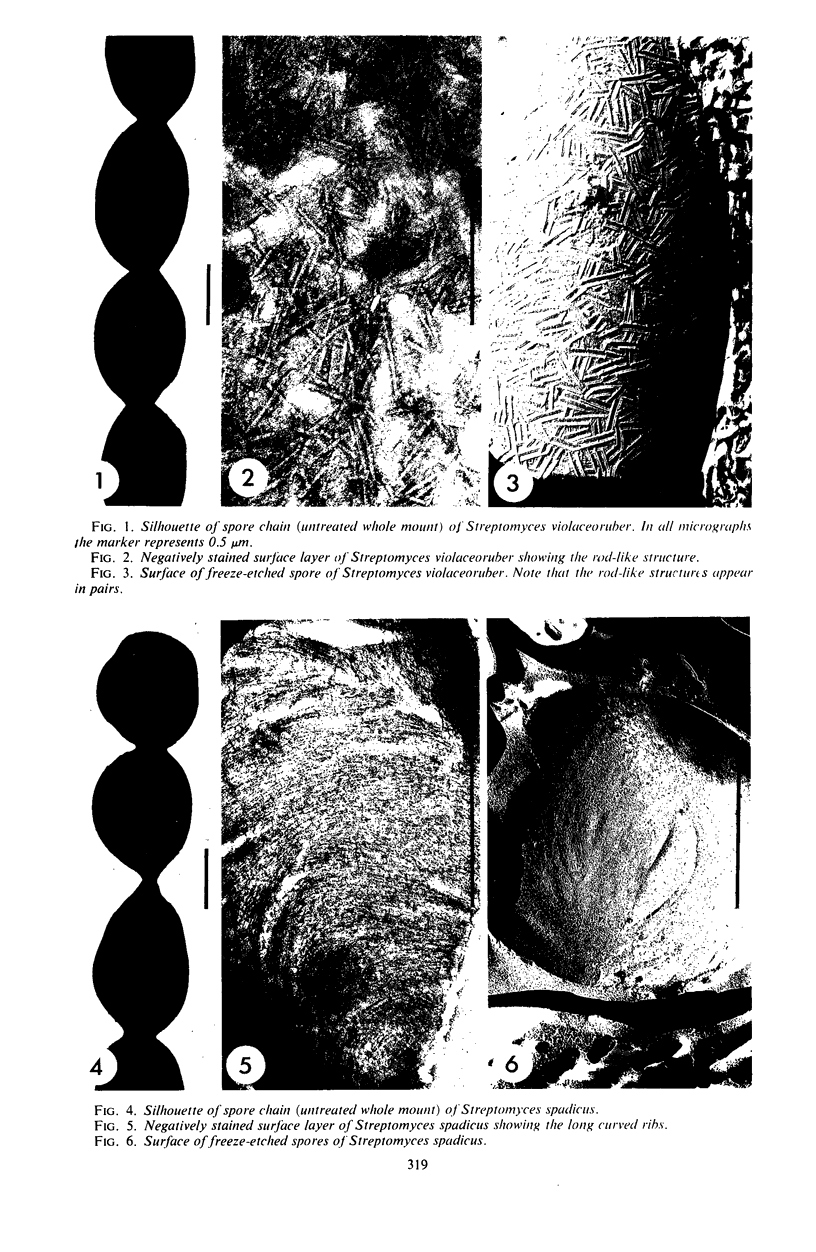
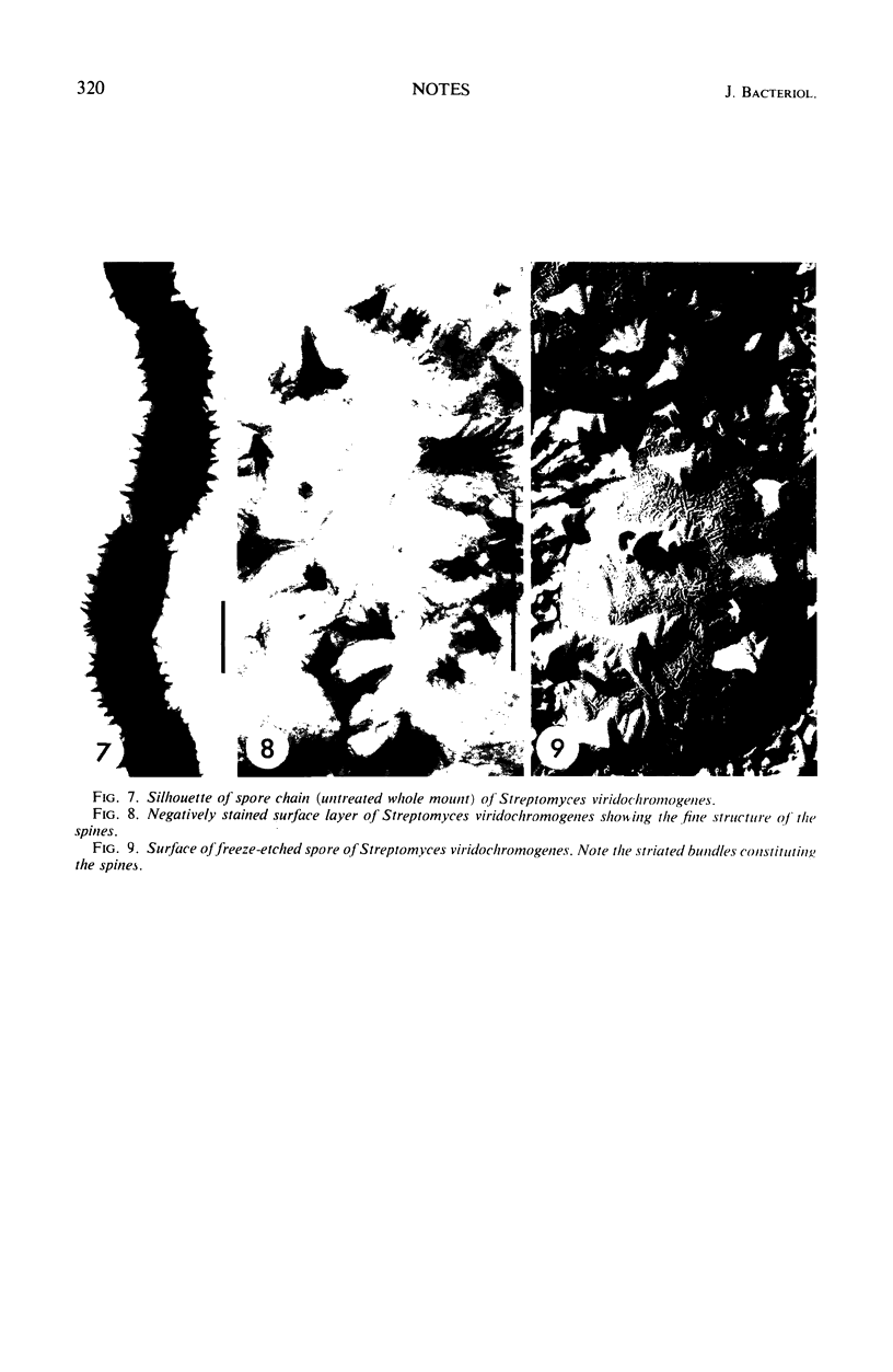
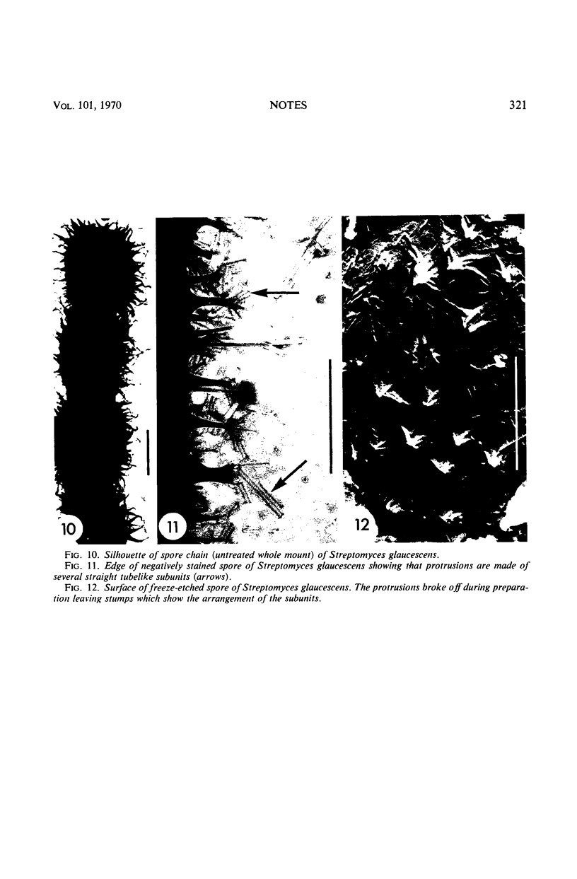
The application of a simple negative staining technique and freeze-etching has revealed detailed information on the species-specific surface structure of spores and aerial hyphae of streptomycetes.
Full text
PDF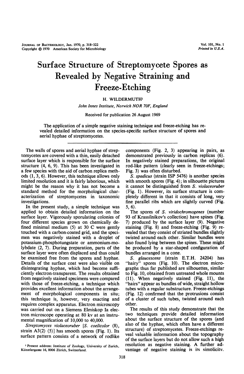
Images in this article

Selected References
These references are in PubMed. This may not be the complete list of references from this article.
- BRENNER S., HORNE R. W. A negative staining method for high resolution electron microscopy of viruses. Biochim Biophys Acta. 1959 Jul;34:103–110. doi: 10.1016/0006-3002(59)90237-9. [DOI] [PubMed] [Google Scholar]
- Bradley S. G., Ritzi D. Composition and ultrastructure of Streptomyces venezuelae. J Bacteriol. 1968 Jun;95(6):2358–2364. doi: 10.1128/jb.95.6.2358-2364.1968. [DOI] [PMC free article] [PubMed] [Google Scholar]
- DIETZ A., MATHEWS J. Taxonomy by carbon replication. I. An examination of Streptomyces hygroscopicus. Appl Microbiol. 1962 May;10:258–263. doi: 10.1128/am.10.3.258-263.1962. [DOI] [PMC free article] [PubMed] [Google Scholar]
- GLAUERT A. M., HOPWOOD D. A. The fine structure of Streptomyces violaceoruber (S. coelicolor). III. The walls of the mycelium and spores. J Biophys Biochem Cytol. 1961 Aug;10:505–516. doi: 10.1083/jcb.10.4.505. [DOI] [PMC free article] [PubMed] [Google Scholar]
- HOPWOOD D. A., GLAUERT A. M. Electron microscope observations on the surface structures of Streptomyces violaceoruber. J Gen Microbiol. 1961 Oct;26:325–330. doi: 10.1099/00221287-26-2-325. [DOI] [PubMed] [Google Scholar]
- HORNE R. W. THE APPLICATION OF NEGATIVE STAINING METHODS TO QUANTITATIVE ELECTRON MICROSCOPY. Lab Invest. 1965 Jun;14:1054–1068. [PubMed] [Google Scholar]
- Hopwood D. A. Genetic analysis and genome structure in Streptomyces coelicolor. Bacteriol Rev. 1967 Dec;31(4):373–403. doi: 10.1128/br.31.4.373-403.1967. [DOI] [PMC free article] [PubMed] [Google Scholar]
- KUTZNER H. J., WAKSMAN S. A. Streptomyces coelicolor Mueller and Streptomyces violaceoruber Waksman and Curtis, two distinctly different organisms. J Bacteriol. 1959 Oct;78:528–538. doi: 10.1128/jb.78.4.528-538.1959. [DOI] [PMC free article] [PubMed] [Google Scholar]
- RANCOURT M. W., LECHEVALIER H. A. ELECTRON MICROSCOPIC STUDY OF THE FORMATION OF SPINY CONIDIA IN SPECIES OF STREPTOMYCES. Can J Microbiol. 1964 Jun;10:311–316. doi: 10.1139/m64-042. [DOI] [PubMed] [Google Scholar]