Abstract
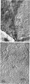
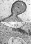
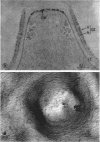
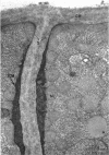
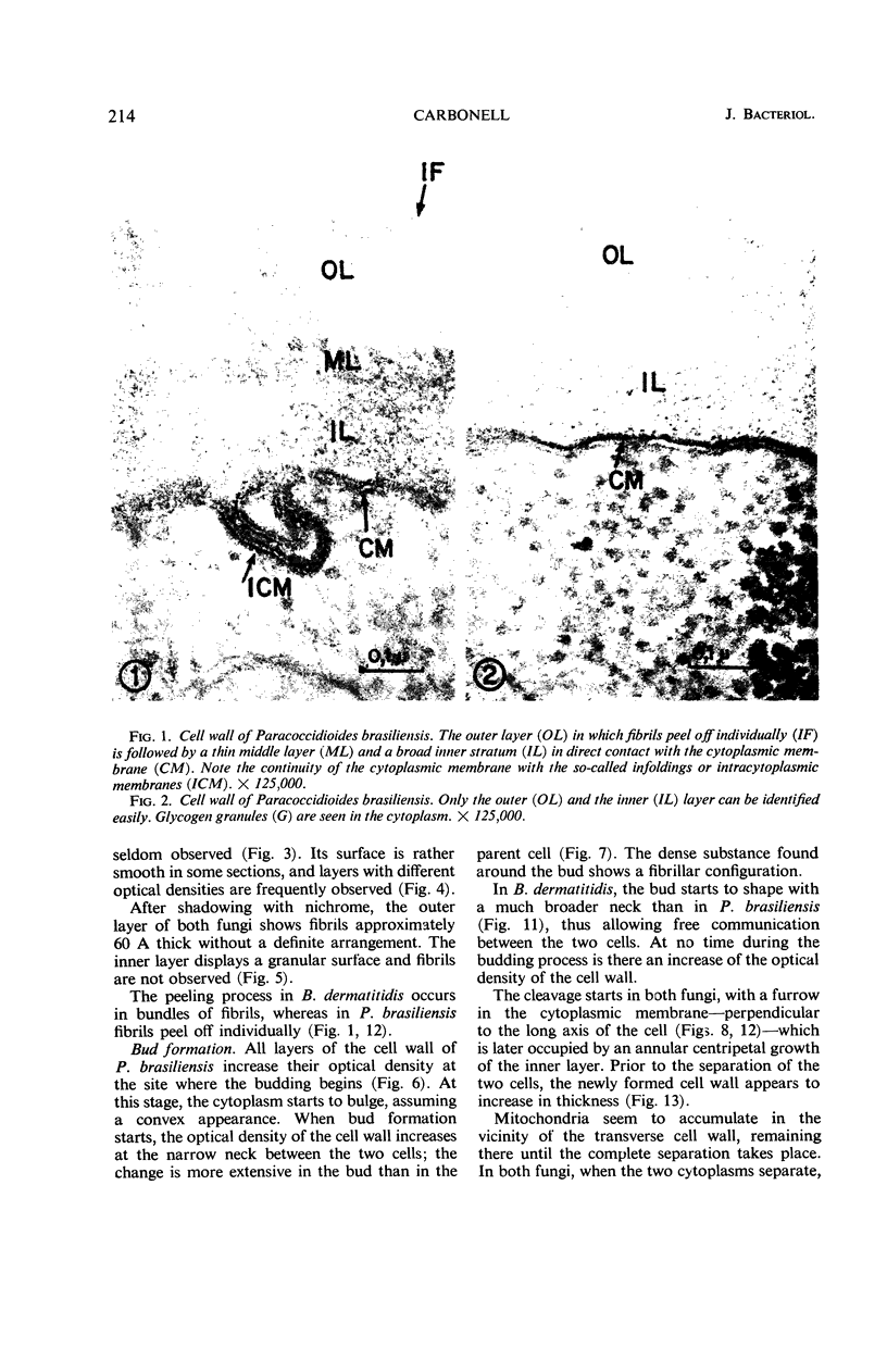
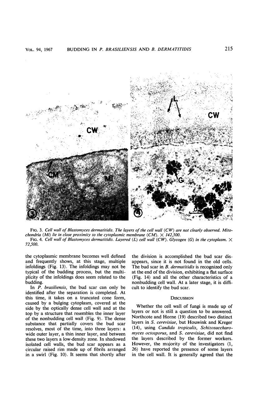
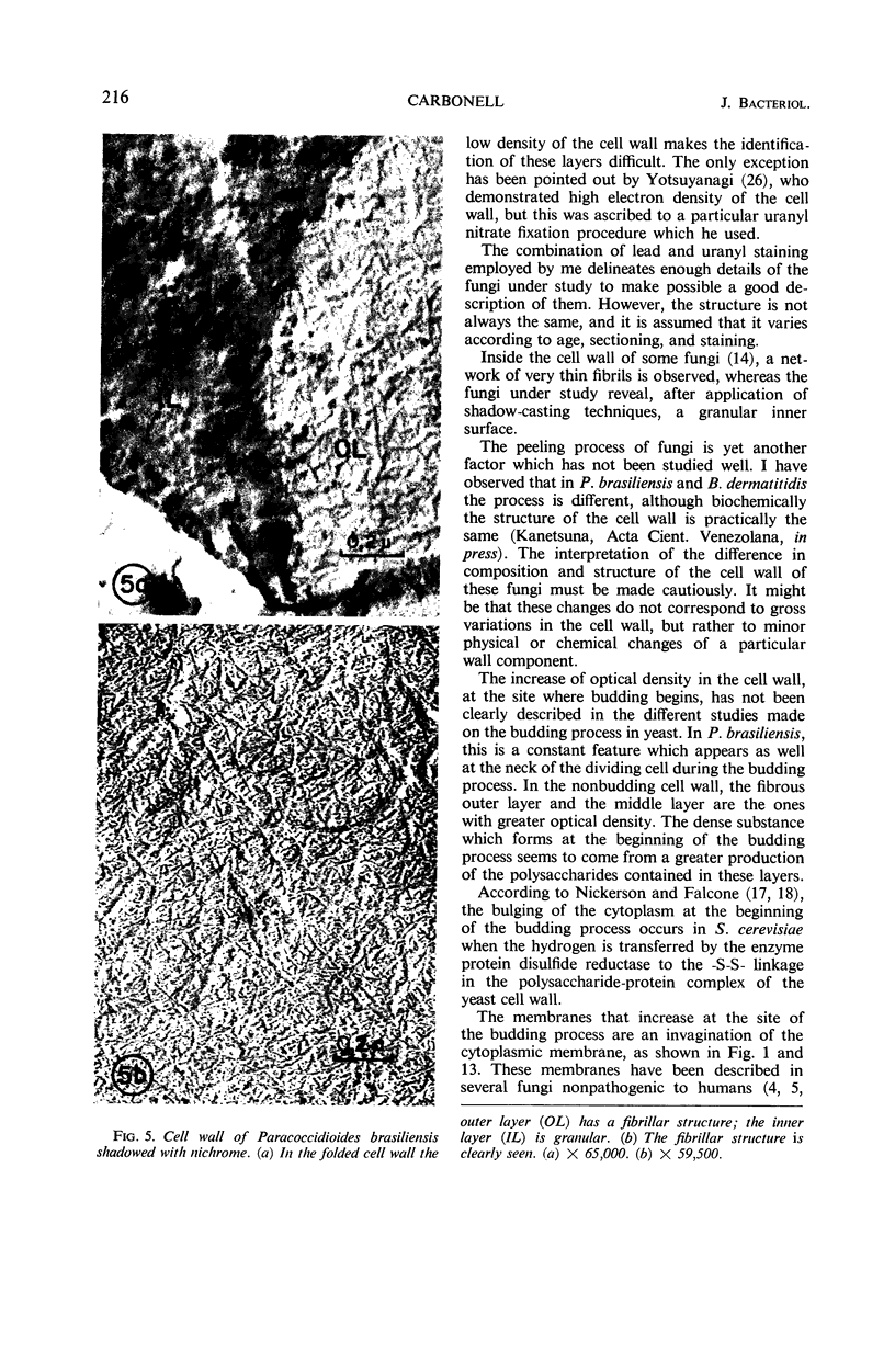
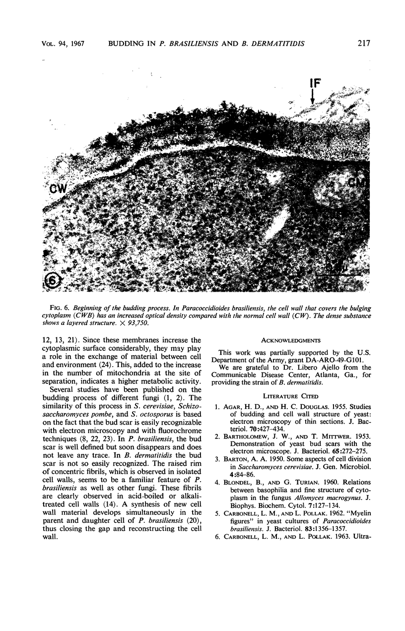
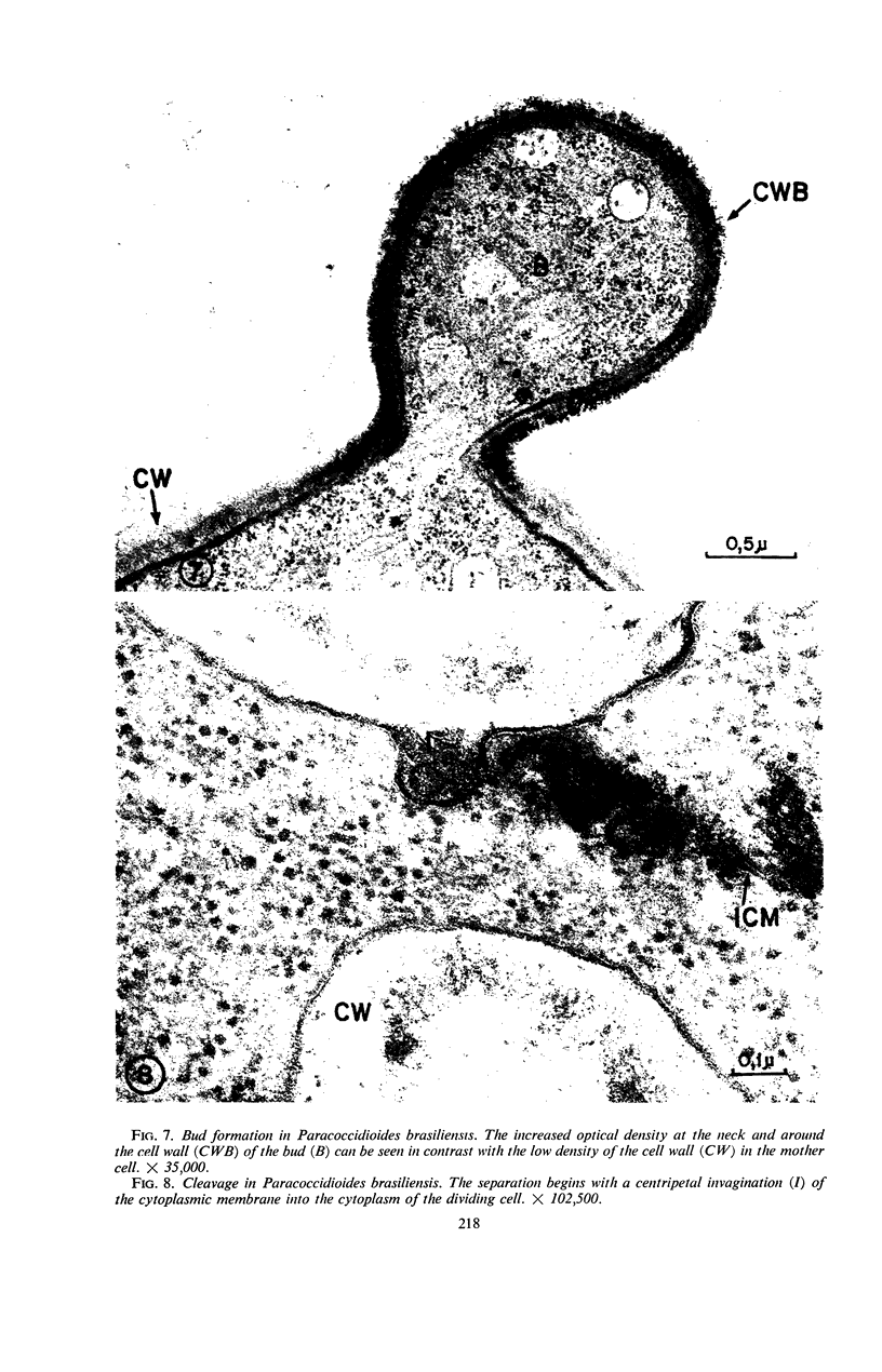
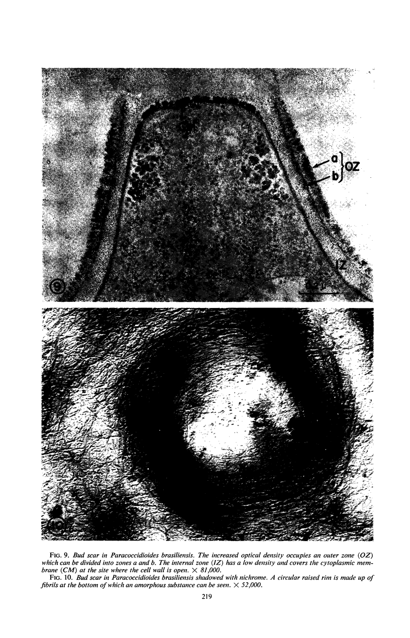
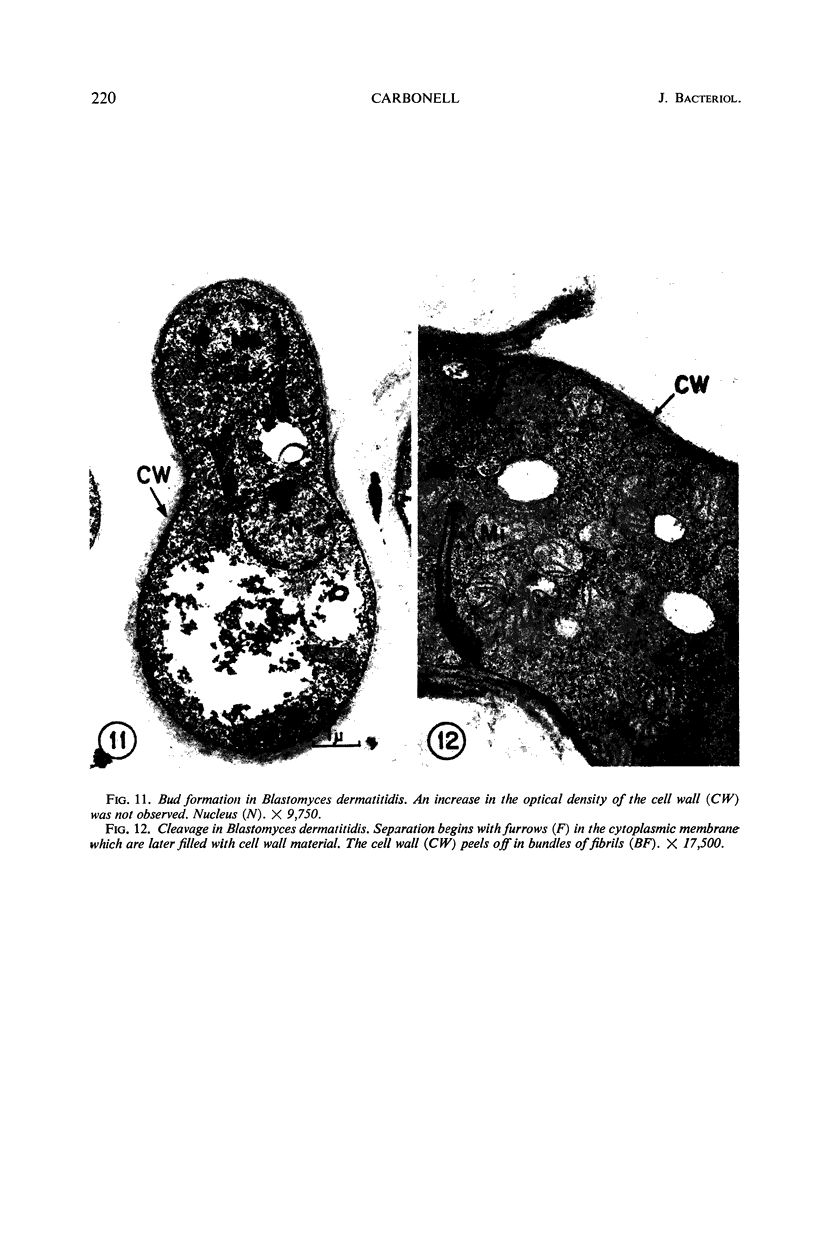
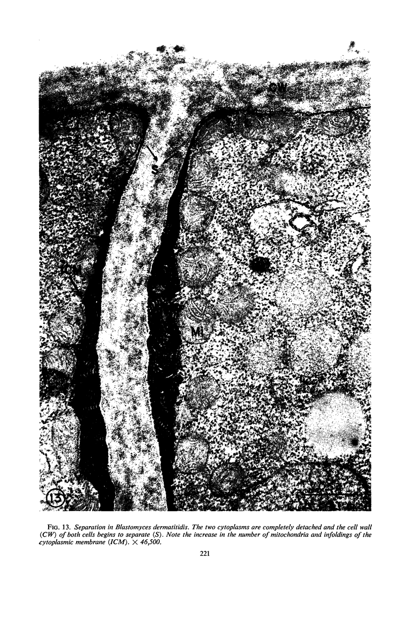
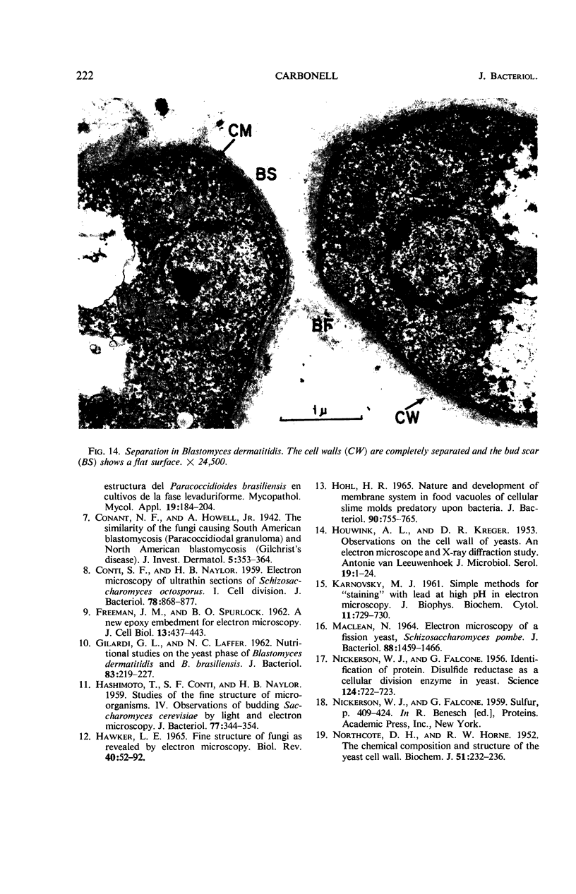
The difference between the budding process of Paracoccidioides brasiliensis and Blastomyces dermatitidis is reported herein. A characteristic feature in P. brasiliensis is that the optical density of the cell wall increases at the site where budding begins and at the neck of the dividing cell, whereas B. dermatitidis does not undergo this alteration. The neck which is formed between the mother and daughter cell at the site of division is much wider in B. dermatitidis than in P. brasiliensis. The bud scar in P. brasiliensis appears as a truncated cone, the top of which is covered only by the inner layer of the cell wall; in comparison, in B. dermatitidis the bud scar exhibits a flattened surface covered by the cell wall. Both fungi show an increase in the number of mitochondria and infoldings of the cytoplasmic membrane at the site of separation, which indicates that at this site there is an increase of metabolic activity.
Full text
PDF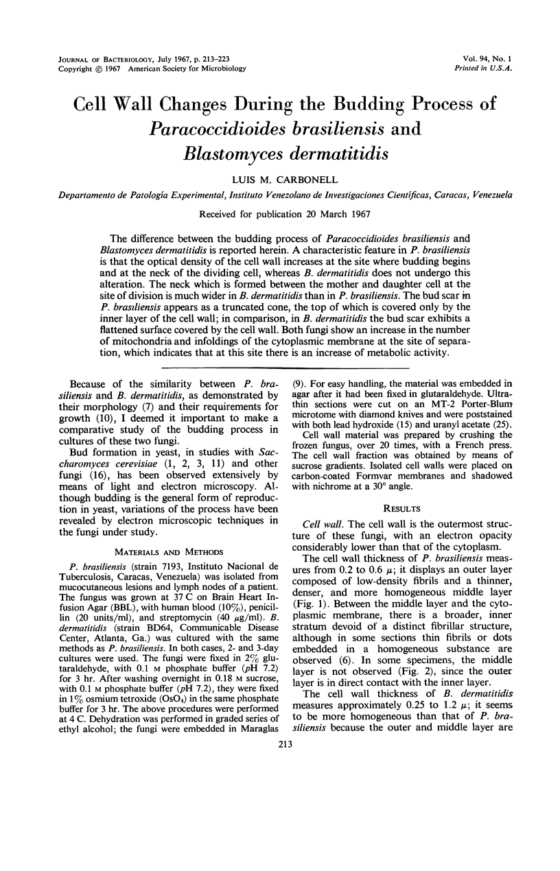
Images in this article
Selected References
These references are in PubMed. This may not be the complete list of references from this article.
- AGAR H. D., DOUGLAS H. C. Studies of budding and cell wall structure of yeast; electron microscopy of the sections. J Bacteriol. 1955 Oct;70(4):427–434. doi: 10.1128/jb.70.4.427-434.1955. [DOI] [PMC free article] [PubMed] [Google Scholar]
- BARTHOLOMEW J. W., MITTWER T. Demonstration of yeast bud scars with the electron microscope. J Bacteriol. 1953 Mar;65(3):272–275. doi: 10.1128/jb.65.3.272-275.1953. [DOI] [PMC free article] [PubMed] [Google Scholar]
- BARTON A. A. Some aspects of cell division in saccharomyces cerevisiae. J Gen Microbiol. 1950 Jan;4(1):84–86. doi: 10.1099/00221287-4-1-84. [DOI] [PubMed] [Google Scholar]
- BLONDEL B., TURIAN G. Relation between basophilia and fine structure of cytoplasm in the fungus Allomyces macrogynus Em. J Biophys Biochem Cytol. 1960 Feb;7:127–134. doi: 10.1083/jcb.7.1.127. [DOI] [PMC free article] [PubMed] [Google Scholar]
- CARBONELL L. M., POLLAK L. "Myelin figures" in yeast cultures of Paracoccidioides brasiliensis. J Bacteriol. 1962 Jun;83:1356–1357. doi: 10.1128/jb.83.6.1356-1357.1962. [DOI] [PMC free article] [PubMed] [Google Scholar]
- CONTI S. F., NAYLOR H. B. Electron microscopy of ultrathin sections of Schizosaccharomyces octosporus. I. Cell division. J Bacteriol. 1959 Dec;78:868–877. doi: 10.1128/jb.78.6.868-877.1959. [DOI] [PMC free article] [PubMed] [Google Scholar]
- FALCONE G., NICKERSON W. J. Identification of protein disulfide reductase as a cellular division enzyme in yeasts. Science. 1956 Oct 19;124(3225):722–723. doi: 10.1126/science.124.3225.722. [DOI] [PubMed] [Google Scholar]
- FREEMAN J. A., SPURLOCK B. O. A new epoxy embedment for electron microscopy. J Cell Biol. 1962 Jun;13:437–443. doi: 10.1083/jcb.13.3.437. [DOI] [PMC free article] [PubMed] [Google Scholar]
- GILARDI G. L., LAFFER N. C. Nutritional studies on the yeast phase of Blastomyces dermatitidis and B. brasiliensis. J Bacteriol. 1962 Feb;83:219–227. doi: 10.1128/jb.83.2.219-227.1962. [DOI] [PMC free article] [PubMed] [Google Scholar]
- HASHIMOTO T., CONTI S. F., NAYLOR H. B. Studies of the fine structure of microorganisms. IV. Observations on budding Saccharomyces cerevisiae by light and electron microscopy. J Bacteriol. 1959 Mar;77(3):344–354. doi: 10.1128/jb.77.3.344-354.1959. [DOI] [PMC free article] [PubMed] [Google Scholar]
- HAWKER L. E. FINE STRUCTURE OF FUNGI AS REVEALED BY ELECTRON MICROSCOPY. Biol Rev Camb Philos Soc. 1965 Feb;40:52–92. doi: 10.1111/j.1469-185x.1965.tb00795.x. [DOI] [PubMed] [Google Scholar]
- HOUWINK A. L., KREGER D. R. Observations on the cell wall of yeasts; an electron microscope and x-ray diffraction study. Antonie Van Leeuwenhoek. 1953;19(1):1–24. doi: 10.1007/BF02594830. [DOI] [PubMed] [Google Scholar]
- Hohl H. R. Nature and Development of Membrane Systems in Food Vacuoles of Cellular Slime Molds Predatory upon Bacteria. J Bacteriol. 1965 Sep;90(3):755–765. doi: 10.1128/jb.90.3.755-765.1965. [DOI] [PMC free article] [PubMed] [Google Scholar]
- KARNOVSKY M. J. Simple methods for "staining with lead" at high pH in electron microscopy. J Biophys Biochem Cytol. 1961 Dec;11:729–732. doi: 10.1083/jcb.11.3.729. [DOI] [PMC free article] [PubMed] [Google Scholar]
- MACLEAN N. ELECTRON MICROSCOPY OF A FISSION YEAST, SCHIZOSACCHAROMYCES POMBE. J Bacteriol. 1964 Nov;88:1459–1466. doi: 10.1128/jb.88.5.1459-1466.1964. [DOI] [PMC free article] [PubMed] [Google Scholar]
- NORTHCOTE D. H., HORNE R. W. The chemical composition and structure of the yeast cell wall. Biochem J. 1952 May;51(2):232–236. doi: 10.1042/bj0510232. [DOI] [PMC free article] [PubMed] [Google Scholar]
- PHAFF H. J. CELL WALL OF YEASTS. Annu Rev Microbiol. 1963;17:15–30. doi: 10.1146/annurev.mi.17.100163.000311. [DOI] [PubMed] [Google Scholar]
- SHATKIN A. J., TATUM E. L. Electron microscopy of Neurospora crassa mycelia. J Biophys Biochem Cytol. 1959 Dec;6:423–426. doi: 10.1083/jcb.6.3.423. [DOI] [PMC free article] [PubMed] [Google Scholar]
- Streiblová E., Málek I., Beran K. Structural changes in the cell wall of Schizosaccharomyces pombe during cell division. J Bacteriol. 1966 Jan;91(1):428–435. doi: 10.1128/jb.91.1.428-435.1966. [DOI] [PMC free article] [PubMed] [Google Scholar]
- TOMASZ A., JAMIESON J. D., OTTOLENGHI E. THE FINE STRUCTURE OF DIPLOCOCCUS PNEUMONIAE. J Cell Biol. 1964 Aug;22:453–467. doi: 10.1083/jcb.22.2.453. [DOI] [PMC free article] [PubMed] [Google Scholar]
- WATSON M. L. Staining of tissue sections for electron microscopy with heavy metals. J Biophys Biochem Cytol. 1958 Jul 25;4(4):475–478. doi: 10.1083/jcb.4.4.475. [DOI] [PMC free article] [PubMed] [Google Scholar]
- YOTSUYANAGI Y. [Study of yeast mitochondria. I. Variations in mitochondrial ultrastructure during the aerobic growth cycle]. J Ultrastruct Res. 1962 Aug;7:121–140. doi: 10.1016/s0022-5320(62)80031-8. [DOI] [PubMed] [Google Scholar]


