Abstract
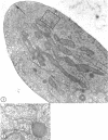
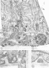
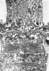
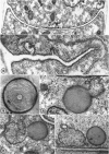
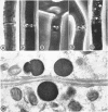
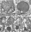
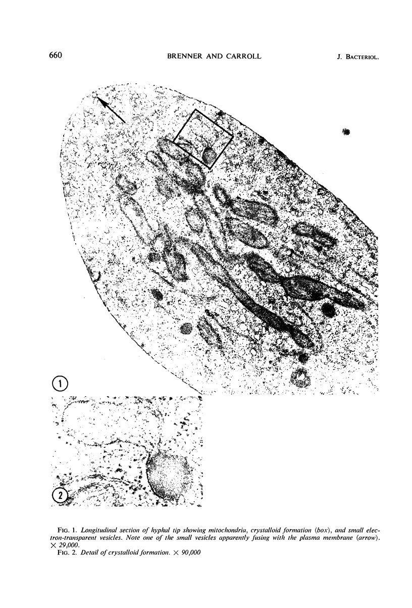
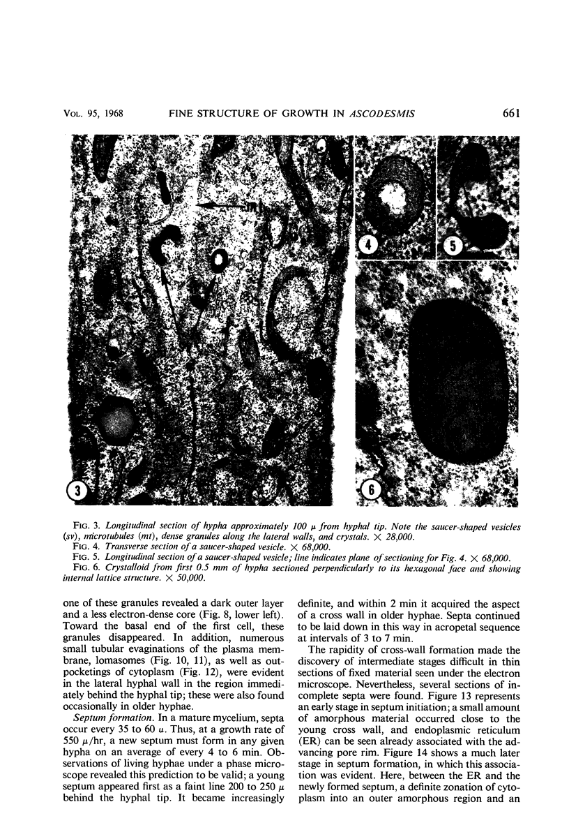
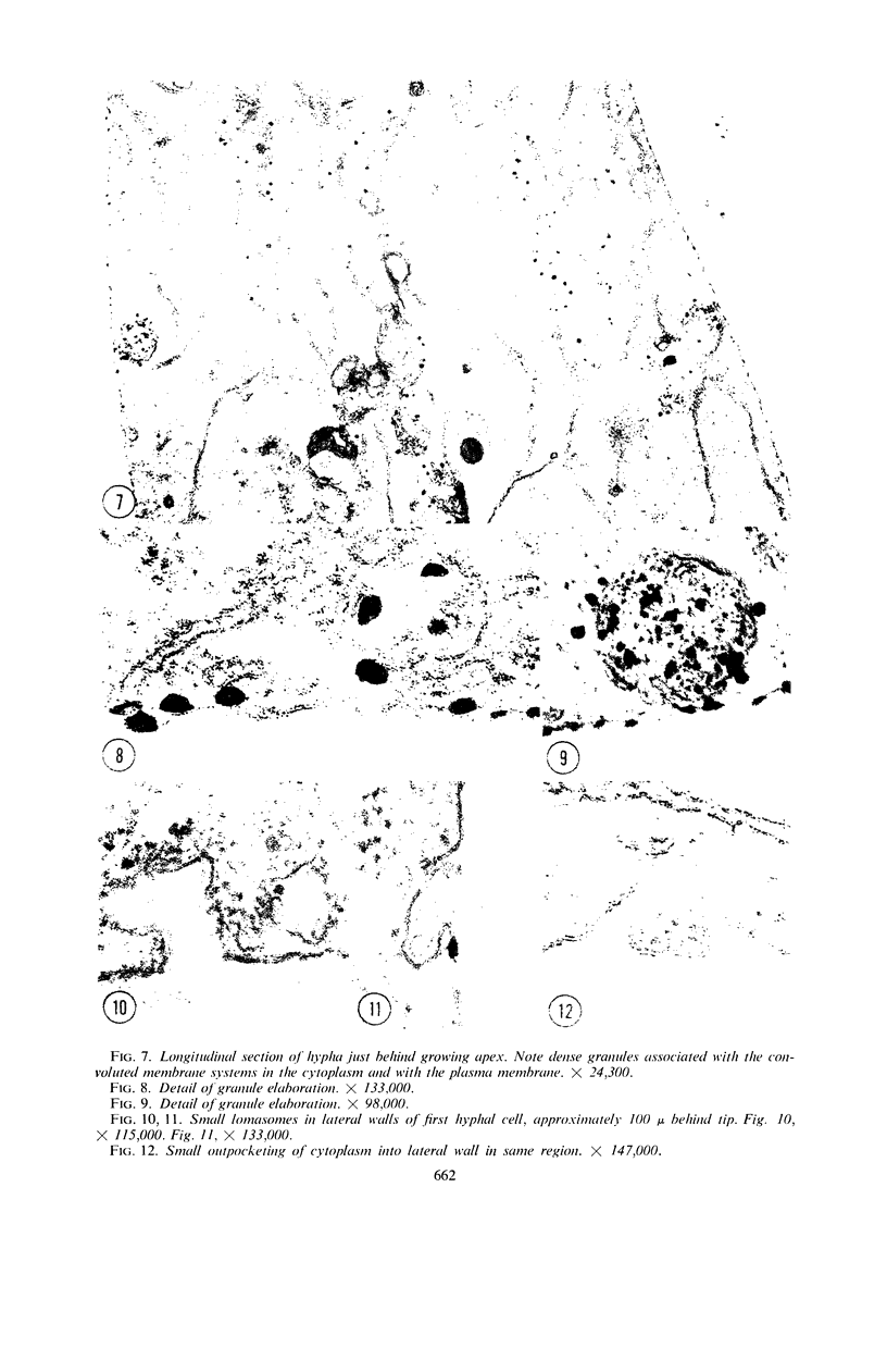
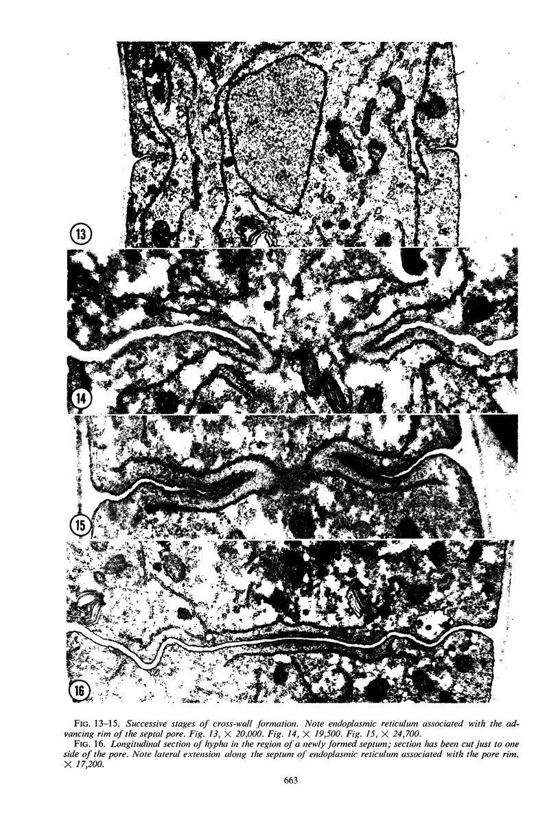
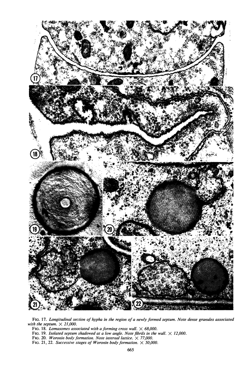
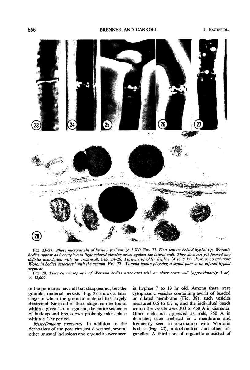
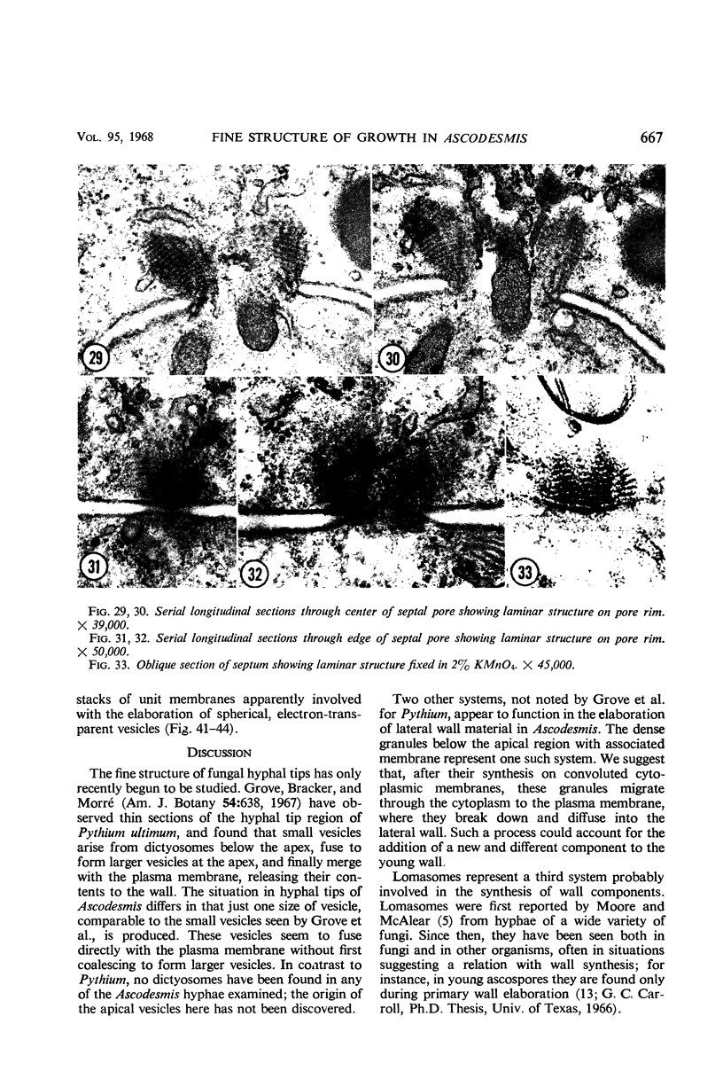
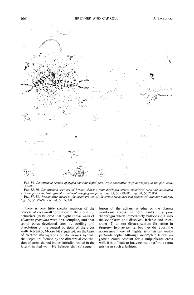
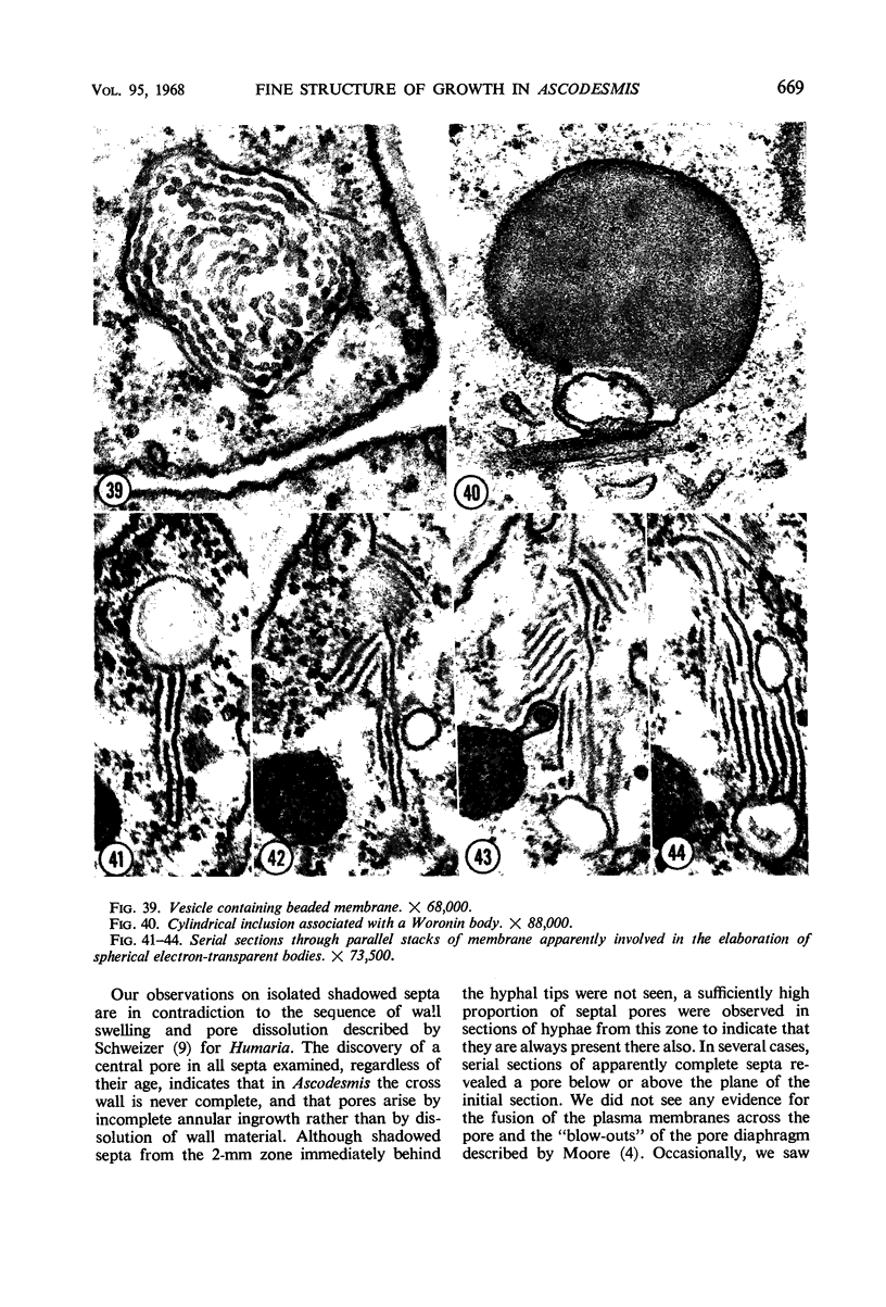
Mycelial mats of Ascodesmis sphaerospora were fixed and embedded for electron microscopy, and thin sections of 1-mm blocks, taken from the 1st to the 7th mm behind the hyphal tips, were cut parallel to the long axis of the hyphae. The hyphal tip region is characterized by an outer zone of electron-transparent vesicles, 500 to 1,000 A in diameter, and is apparently associated with wall elaboration. Immediately behind this region, dense granules become evident along convoluted membrane systems and along the plasma membrane; in the same region are numerous small lomasomes in the lateral wall. As the hypha grows, septa are laid down at 3- to 7-min intervals at a distance of 200 to 250 μ behind the hyphal tip. A cylinder of endoplasmic reticulum is intimately involved in cross-wall deposition from its earliest stages; as the wall grows in, it becomes increasingly constricted in the pore region, finally assuming a torus-like configuration. Woronin bodies are shown to have a crystalline substructure and to originate in pouch-like membrane systems. Cross-walls from a 7- to 13-hr-old mycelium frequently show highly ordered structures in the vicinity of the pore. These structures may appear either as laminar stacks of discs to one side of the pore or as series of stubby concentric rings within the pore area itself. In the latter case, a mass of granular material is frequently seen plugging the pore. Other unusual organelles and inclusions in 7- to 13-hr hyphae are vesicles containing swirls of beaded or dilated membrane, membrane-enclosed rods, and stacks of unit membranes associated with spherical, electron-transparent vesicles.
Full text
PDF
Images in this article
Selected References
These references are in PubMed. This may not be the complete list of references from this article.
- SHATKIN A. J., TATUM E. L. Electron microscopy of Neurospora crassa mycelia. J Biophys Biochem Cytol. 1959 Dec;6:423–426. doi: 10.1083/jcb.6.3.423. [DOI] [PMC free article] [PubMed] [Google Scholar]
- TSUDA S., TATUM E. L. Intracellular crystalline ergosterol in Neurospora. J Biophys Biochem Cytol. 1961 Oct;11:171–177. doi: 10.1083/jcb.11.1.171. [DOI] [PMC free article] [PubMed] [Google Scholar]
- Wilsenach R., Kessel M. The role of lomasomes in wall formation in Penicillium vermiculatum. J Gen Microbiol. 1965 Sep;40(3):401–404. doi: 10.1099/00221287-40-3-401. [DOI] [PubMed] [Google Scholar]


