Abstract
Humans express a unique subset of high-density lipoproteins (HDLs) called trypanosome lytic factors (TLFs) that kill many Trypanosoma parasite species. The proteins apolipoprotein (apo) A-I, apoL-I, and haptoglobin-related protein, which are involved in TLF structure and function, were expressed through the introduction of transgenes in mice to explore their physiological roles in vivo. Transgenic expression of human apolipoprotein L-I alone conferred trypanolytic activity in vivo. Coexpression of human apolipoprotein A-I and haptoglobin-related protein (Hpr) had an effect on the integration of apolipoprotein L-I into HDL, and both proteins were required to increase the specific activity of TLF, which was measurable in vitro. Unexpectedly, truncated apolipoprotein L-I devoid of the serum resistance gene interacting domain, which was previously shown to kill human infective trypanosomes, was not trypanolytic in transgenic mice despite being coexpressed with human apolipoprotein A-I and Hpr and incorporated into HDLs. We conclude that all three human apolipoproteins act cooperatively to achieve maximal killing capacity and that truncated apolipoprotein L-I does not function in transgenic animals.
There are many molecular subsets of human high-density lipoproteins (HDLs) that are distinguished by differences in their lipid and protein composition. Apolipoprotein A-I (apoA-I) is the defining component of all HDLs, which, along with different combinations of proteins, confer many functions to HDLs, the best known being atheroprotection (1). Trypanosome lytic factors (TLFs 1 and 2) are a subset of HDLs that contain two unique protein components, haptoglobin-related protein (Hpr) (2) and apolipoprotein L-I (apoL-I) (3). TLF1 is a 500-kD lipid-rich HDL particle and TLF2 is a lipid-poor HDL that circulates in blood as an immunocomplex with polyclonal IgM (4, 5).
TLF is the major innate defense mechanism by which humans and certain primates kill the majority of trypanosome species; the two subspecies of trypanosomes that are resistant to lysis by TLF infect humans (6). Trypanosome subspecies expressing the serum resistance–associated protein (SRA), either naturally (Trypanosoma brucei rhodesiense) or via genetic modification (Trypanosoma brucei brucei-SRA), are resistant to lysis by TLF (3) because SRA purportedly binds apoL-I and neutralizes TLF activity or redirects TLF intracellular trafficking to prevent lysis (3, 7). Tantalizingly, a truncated form of apoL-I (Tr-apoL-I) devoid of the SRA-interacting domain, which was expressed in bacteria or conjugated to nanobodies that recognize a common carbohydrate epitope on the variant surface glycoprotein (VSG) coat of trypanosomes, was reported to escape binding of SRA and kill TLF-resistant trypanosomes (3, 8). Although it is possible to imagine that transgenic cattle expressing this Tr-apoL-I could resist infection by all trypanosome subspecies, this activity has not been examined.
Although not infective to humans, T. b. brucei infects economically important African cattle species because they do not make TLFs. This parasite can be killed in vitro by human plasma, purified TLF, or recombinant apoL-I by a pore-forming mechanism (9, 10). Early reports indicated that purified Hpr was trypanolytic in vitro (11), but more recent data indicate that Hpr cooperates to increase the lytic activity of TLF1 (12) or to increase the uptake of TLF1 (13) via an Hp/Hpr receptor on trypanosomes (11, 14). Both mechanisms are currently proposed to be instrumental in the level of TLF activity in vitro, with the finding that hemoglobin must be bound to Hpr to generate an increase in both lytic capacity and the receptor-mediated uptake of TLF (15).
RESULTS AND DISCUSSION
Creation of TLF mice using hydrodynamic-based gene delivery
To elaborate on the role of Hpr, apoL-I, and Tr-apoL-I and to explore reconstitution of TLF activity in vivo, we created a physiologically relevant system that allows transgenic expression of individual or multiple TLF components in a mouse. Using a hydrodynamic gene-based delivery approach (16), we generated Tg mice that express native human Hpr, apoL-I or Tr-apoL-I, or both components predominantly from the liver (17), which is the normal site where TLF components are synthesized and assembled. Expression plasmids that encode Hpr and/or apoL-I or Tr-apo L-I are expressed and proteins are secreted into the circulation within 24 h.
Human TLF has several well-characterized properties that define it as a specific subset of HDL. We used the following criteria as a benchmark for comparing the activity of reconstituted TLFs from Tg mice: (a) coimmunoprecipitation experiments have revealed that Hpr and apoL-I are coexpressed in the same particle despite constituting only 1% of HDL molecules, suggesting that their distribution in HDL particles is nonrandom (12, 15). (b) Trypanolytic activity of human plasma or purified TLF against T. b. brucei can be demonstrated in an in vitro killing assay during which the trypanosomes swell and burst. Alternatively, human plasma can be i.v. injected into a mouse to provide protection against T. b. brucei infection (18). (c) Trypanosomes that express SRA either naturally (T. b. rhodesiense) or artificially (T. brucei-SRA) are resistant to trypanolysis by TLF in vitro (3) or in vivo after transfer of human plasma or purified TLF i.v. to mice (18). (d) Trypanolytic activity in vitro can be blocked by addition of weak bases to prevent acidification of internal vesicles, which is required to activate TLF after uptake and internalization of the lipoprotein particle by the parasite (19).
Apolipoprotein L-I protects mice from infection with T. b. brucei, T. evansi, and T. congolense
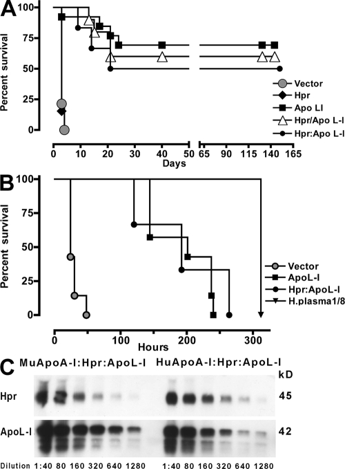
Tg mice expressing genes that encode for Hpr and apoL-I individually reveal that only mice expressing the apoL-I transgene (Tg-apoL-I) are protected from challenge with T. b. brucei. In contrast, mice that express the Hpr transgene (Tg-Hpr) are not protected and succumb to infection with the same kinetics as mice given the empty vector (Fig. 1 A). We then evaluated whether the expression of Hpr and apoL-I together in vivo would give additional protection beyond that seen with apoL-I. There is no increase in protection when the mice express both Hpr and apoL-I either from individual plasmids (Tg-Hpr/apoL-I) or coexpress them from a single plasmid (Tg-Hpr:apoL-I), which allows protein synthesis in the same transfected cell. These results suggest that in vivo apoL-I is the main lytic component of TLF.
Figure 1.
Survival kinetics of mice infected with T. b. brucei ILTat 1.25, relative lytic units, and protein expression in Tg mice. (A) Mice expressing Hpr (black diamond; n = 13), apoL-I (black square; n = 13), or a combination of Hpr and apoL-I in individual plasmids (white triangle; Hpr/apoL-I; n = 10) or in a single plasmid (black circle, Hpr:apoL-I; n = 6) were infected i.p. with 1.78 × 10⁶ trypanosomes. Control mice were treated with empty vector (gray circle; n = 14). The parasitemia was monitored and time of death was recorded. (B) Survival kinetics of naive mice infected with T. b. brucei ILTat 1.25 that were administered plasma i.v. from mice transfected with empty vector (gray circle; n = 7), apoL-I (black square; n = 7), or a combination of Hpr and apoL-I in a single plasmid (black circle; Hpr:apoL-I; n = 3). The protection obtained by normal human plasma (dilution 1/8) is indicated by the inverted triangle. (C) Western blot with serial dilutions of plasma from Tg mice expressing murine apoA-I and human Hpr and apoL-I from the same plasmid (Hpr:apoL-I) and Tg mice expressing human apoA-I and human Hpr and apoL-I from the same plasmid (Hpr:apoL-I). Hpr and apoL-I were detected with monoclonal antibodies.
Tg-apoL-I mice mimic humans in their resistance and sensitivity to infection with a variety of trypanosome species (Table I). Nonprimate mammals do not have the genes that encode for Hpr and apoL-I, and therefore they do not produce TLF. They are thus susceptible to a broader range of trypanosome species than humans, such as T. congolense, T. evansi, and T. b. brucei, which limits their survival in Central Africa. T. congolense and T. evansi are both trypanosomes whose course of infection is mild and prolonged because of relapsing but ultimately lethal parasitemias, whereas the T. b. brucei used in these experiments has a short course of infection that is rapidly lethal. We show that Tg-apoL-I mice can ameliorate infections with all these trypanosome species and find that all Tg-apoL-I survivors are parasite-free 20 d after infection (Table I). As expected, Tg-apoL-I mice infected with T. brucei-SRA were unable to control the parasitemia, and all mice succumbed to infection within 3 d with kinetics identical to mice that received the empty vector (Table I).
Table I.
Transgenic mice that express the apoL-I gene ameliorate infection with T. brucei, T. evansi, and T. congolense
| Transgenic DNA |
Trypanosome species (1.78 × 10⁶ injected i.p.) |
Number mice alive/ number mice infected |
||
|---|---|---|---|---|
| Day 3 | Day 20 | Day 50 | ||
| ApoL-I | T. brucei | 5/5 | 3/5b | |
| Vector | 0/5a | |||
| ApoL-I | T. brucei SRA | 0/5a | ||
| Vector | 0/5a | |||
| ApoL-I | T. evansi | 5/5 | 4/5c | |
| Vector | 5/5 | 1/5d | ||
| ApoL-I | T. congolense | 5/5 | 3/5b | |
| Vector | 2/5 | 2/5d | ||
Mice expressing the gene for apoL-I were infected with 1.78 × 10⁶T. b. brucei (Lister 427-derived line), T. b. brucei-SRA (Lister 427-derived line expressing the SRA gene), T. evansi (AnTat 3), or T. congolense (STIB 68Q). n = 5 per group.
Parasitemia ≥ 1 × 10⁹ before mice were killed.
No parasites detectable.
Died of unknown cause, did not have detectable parasites.
Parasitemia = 1 × 10⁶/ ml.
Transgene expression decreases with time in the Tg mice; blood levels of apoL-I begin to decrease around day 14 and Hpr levels begin to decrease around day 7, but low levels of protein can still be detected by day 45 (unpublished data). Consequently, we sometimes observe a recrudescence of parasites at 20–25 d after infection, which can develop into a highly lethal parasitemia (109/ml). If there is no detectable parasitemia by day 25 in the Tg mice, they will remain parasite-free indefinitely. These parasites that recrudesce are not resistant to TLF, and we therefore conclude that some parasites are in tissue spaces to which TLF has no access, or that the TLF titer is inadequate to kill all of the trypanosomes.
Although Tg-apoL-I mice exhibit many of the properties of human TLF, experiments in which killing activity was assessed by plasma transfer to naive mice indicate that expression of apoL-I gives lower trypanolytic activity than that observed for human plasma. Transfer of 300 μl of Tg-apoL-I plasma to naive mice already infected with T. b. brucei delayed the time to peak parasitemia (109/ml) with a median survival of 201 h (Fig. 1 B). Despite the variability in the plasma transfer assay it is clear that plasma from both Tg-apoL-I (median survival 201 h) and Tg-Hpr:apoL-I (median survival 192 h) confer equal protection, which indicates that coexpression of Hpr in the context of murine HDL does not increase lytic activity.
Transfer of 300 μl human plasma completely cleared the trypanosome infection and protected the mice indefinitely. To gauge the relative amount of lytic units within the plasma from different Tg mice, we serially diluted human plasma until survival time was ∼300 h (1:8, 37 μl; Fig. 1 B). In this plasma transfer assay, we find that Tg mouse plasma has ∼10% the lytic capacity of normal human plasma. In contrast to human plasma, Tg-apoL-I plasma or Tg-Hpr:apoL-I plasma showed no trypanolytic activity in vitro within the time frame of our standard assay (unpublished data), which suggests that the Tg plasma is missing some additional human component that augments or stabilizes activity in vitro or in vivo.
Human apoA-I and haptoglobin-related protein increase the specific activity of HDL synthesized by Tg mice
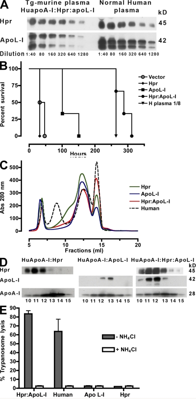
We next evaluated whether human apoA-I could augment or stabilize the Tg-TLF activity in vitro or in vivo. In contrast to mice, humans have three major HDL subfractions designated HDL1 ∼11.4 nm, HDL2 ∼10.2 nm, and HDL3 ∼8.7 nm. Mice containing a stably integrated human apoA-I transgene (HuapoA-I) express ∼2 mg/ml human apoA-I and 0.1 mg/ml murine apoA-I and exhibit a humanlike HDL profile of HDL2 and HDL3, indicating that the amino acid sequence of apoA-I is a determinant of the HDL distribution (20). Plasma from HuapoA-I mice transfected with Hpr and apoL-I (Hpr:apoL-I) display similar levels of protein either compared with plasma from MuapoA-I mice transfected with Hpr and apoL-I (Fig. 1 C) or compared with human plasma (Fig. 2 A), corresponding to ∼5-10 μg/ml of apoL-I (21) and ∼20–30 μg/ml of Hpr (22).
Figure 2.
Increased specific activity of Tg-HDL in the presence of human apoA-I and Hpr. (A) Western blot with serial dilutions of plasma from HuapoA-I mice expressing Hpr and apoL-I from a single plasmid (HuapoA-I:Hpr:apoL-I) and normal human plasma. Hpr and apoL-I were detected with monoclonal antibodies. (B) Survival kinetics of naive mice infected with T. b. brucei ILTat 1.25 and then given 300 μl plasma i.v. from HuapoA-I mice expressing Hpr (black diamond; n = 3), apoL-I (black square; n = 3), or both Hpr and apoL-I from a single plasmid (black circle, Hpr:apoL-I; n = 3). The protection obtained by normal human plasma (dilution 1/8) is indicated by the inverted triangle. (C) A280 profile of KBr-purified lipoproteins from human (dashed line) and HuapoA-I mouse plasma expressing Hpr:apoL-I (red line), Hpr (green line), and apoL-I (blue line) separated by size on a Superdex 200 column; fractions 6–7 are void, fractions 8–9 are human LDL, fractions 10–14 are HDLs, and fraction 15 is albumin. (D) Western blot of different size-fractionated lipoproteins from HuapoA-I mice expressing Hpr only (HuapoA-I:Hpr), apoL-I only (HuapoA-I:apoL-I), or Hpr and apoL-I from the same plasmid (HuapoA-I:Hpr:apoL-I). Hpr and apoL-I were detected with monoclonal antibodies. (E) In vitro lytic activity of size-fractionated HDL (fraction 11) from human and HuapoA-I mice expressing Hpr, apoL-I, or both from a single plasmid (dark gray bars). Open bars represent the lytic activity obtained in the presence of NH4Cl (10 mM). Data show the mean and SEM of five independent measurements.
In contrast to the murine apoA-I plasma transfer experiments, we find that expression of apoL-I in the presence of human apoA-I results in plasma that is less lytic than that obtained when both TLF components (Hpr:apoL-I) are expressed. Plasma transfer from HuapoA-I mice transfected with Hpr to naive mice infected with T. b. brucei show no protection by Hpr (median survival 32 h), equivalent to vector alone (median survival 39.5 h; Fig. 2 B).
Plasma from HuapoA-I:Hpr:apoL-I transferred to naive mice infected with T. b. brucei has an increase in lytic capacity (median survival time 288 h) compared with that observed with HuapoA-I:apoL-I (median survival 101 h; Fig. 2 B). These data reveal that coexpression of Hpr in the context of human apoA-I increases lytic activity, which suggests that human apoA-I may augment or stabilize Tg-TLF (Hpr:apoL-I) activity in vivo. Despite an increase in activity, the triple Tg mouse plasma (HuapoA-I:Hpr:apoL-I) is not as potent as human plasma in transfer experiments. Additional factors present in human plasma, such as TLF2, which is an immune complex of polyclonal IgM and lipid-poor TLF1 that circulates in plasma at ∼10 μg/ml (4), could explain the difference in lytic capacity.
To evaluate the assembly of Hpr and apoL-I into HDLs and the lytic capacity of our various Tg mice, we purified all of the different Tg-HDLs containing human apoA-I by density gradient ultracentrifugation followed by size fractionation and analyzed each fraction by Western blot for localization of Hpr, apoL-I, and apoA-I. Size fractionation of lipoproteins of the HuapoA-I mice show a broad HDL peak (plain lines, #10-14; Fig. 2 C), which matches the distribution of human HDL (dashed line). When mice were transfected with a plasmid that encodes for Hpr, we find that Hpr-HDL is enriched in fractions 11 and 12 (Fig. 2 D). When mice were transfected with a plasmid that encodes for apoL-I, we find that apoL-I-HDL is enriched in fractions 12 and 13 (Fig. 2 D). When we transfected a single plasmid that encodes both genes under the control of individual promoters to drive expression in the same cell, we find that apoL-I has been “redistributed” upon coexpression with Hpr predominantly into fractions 11 and 12 (Fig. 2 D). This suggests that when coexpressed, Hpr and apoL-I localize to the same HDL particle (Fig. S1, available at http://www.jem.org/cgi/content/full/jem.20071463/DC1). We observe a similar redistribution of apoL-I upon coexpression of Hpr in murine apoA-I HDLs (unpublished data).
Purified human TLFs containing either Hpr or apoL-I have low specific activities for in vitro trypanolysis relative to TLFs that contain both Hpr and apoL-I in the same particle (12). Notably, in our experimental system, only the purified triple Tg-HDL, which is composed of HuapoA-I:Hpr:apoL-I, showed trypanolytic activity in vitro within the time frame of our standard assay (Fig. 2 E). The lytic capacity of the triple Tg-HDL was equivalent to that of human TLF-enriched HDL (both isolated by the same method). In addition, trypanosome lysis by both HDLs was inhibited by incubation of trypanosomes with ammonium chloride (10 mM), a lysosomotropic agent that blocks the acidification of internal organelles (Fig. 2 E). This indicates that we have created a functional Tg TLF (HuapoA-I:Hpr:apoL-I) that mimics the properties of human TLF. These data reveal that human apoA-I contributes to an increase in activity of Tg-Hpr:apoL-I HDLs (but not of Tg-apoL-I) that is measurable in plasma transfer experiments in vivo (Fig. 2 B), and more so in lytic assays in vitro (Fig. 2 E). There was no measurable activity in mouse apoA-I:Hpr:apoL-I HDL in vitro (unpublished data). Therefore, we conclude that the increase in specific activity observed previously in vitro (13) is caused not only by the presence of just Hpr and apoL-I, but by an interaction between the three human proteins apoA-I, Hpr, and apoL-I (Fig. 2, B and E).
The difference in the specific activity of the Tg-HDLs could arise from changes in the assembly and stability of the HDL complex or changes in the trafficking of Tg-HDLs in the trypanosome. We do not detect any differences in the assembly of TLFs generated with mouse or human apoA-I based on the following observations. Hpr and apoL-I are exclusively found associated with HDL. We consistently find redistribution, and sometimes an increase, in concentration of apoL-I upon coexpression with Hpr. This suggests that there is an interaction of Hpr with apoL-I. Purification of the Tg-Hpr:apoL-I HDLs by KBr ultracentrifugation and size chromatography gave equivalent yields of Hpr and apoL-I (unpublished data). This suggests that there was no loss of protein components during the purification of TLFs generated with mouse or human apoA-I. Given that no difference in the relative binding to trypanosomes of purified human HDL subclasses containing either Hpr or apoL-I or both Hpr and apoL-I has been reported (12), we hypothesize that human apoA-I in the presence of Hpr and apoL-I effects either the trafficking of the purified triple Tg HDL or the resistance to trypanosomal proteases within the lysosome in agreement with others (23).
Apolipoprotein L-I devoid of the “SRA-interacting domain” does not protect mice from trypanosome infection
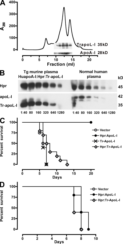
Having established that transient transgenesis provides the first animal model capable of validating reconstituted TLF activity in vivo, we next assessed the trypanolytic potential of a truncated apoL-I devoid of the SRA-interacting domain (Tr-apoL-I). Full-length apoL-I can kill T. brucei, but not T. brucei that expresses SRA. Tr-apoL-I, which is constructed by deletion of the C-terminal α-helix starting at amino acid 340, has been synthesized in bacteria and cell-free systems and is reported to kill SRA-expressing trypanosomes both in vitro (3) and in vivo when conjugated to nanobodies directed against VSG carbohydrate epitopes and injected into T. brucei–SRA–infected mice (8). Transgenic expression of Tr-apoL-I in livestock could therefore conceivably create animals that are resistant to infection by all species of trypanosomes. Tr-apoL-I–transfected mice revealed robust expression of Tr-apoL-I that readily assembled into HDL particles in a manner similar to full-length apoL-I (Fig. 3 A). To our surprise, we were unable to detect any lytic activity in vivo against SRA-expressing trypanosomes, or even against the susceptible T. b. brucei (Table II). To improve the potential activity of Tr-apoL-I, we generated a double plasmid (Hpr:Tr-apoL-I) and transfected Tg-HuapoA-I mice, thereby creating HuapoA-I:Hpr:Tr-apoL-I mice. Serial dilution of Tg mice plasma revealed robust expression of both proteins (Fig. 3 B), which could be coimmunoprecipitated indicating their localization in the same HDL (Fig. S1). However, inoculation of mice with a low dose of 5,000 parasites did not reveal any significant trypanolytic activity (P > 0.05) of HuapoA-I:Hpr:Tr-apoL-I against T. b. brucei (Fig. 3 C) or T. b. brucei-SRA (Fig. 3 D).
Figure 3.
Tr-apoL-I gene expression in Tg-HuapoA-I mice does not protect from infection with T. b. brucei or T. b. brucei-SRA. (A) A280 profile of KBr-purified lipoproteins separated by size on a Superdex 200 column; fractions 6–7 are void, fractions 10–14 are HDLs as indicated by the distribution of apoA-I, and fraction 15 is albumin. (inset) A Western blot of the fractions probed for apoA-I (28 kD) and Tr-apoL-I (∼35 kD) detected with polyclonal anti–apoL-I. (B) Western blot with serial dilutions of plasma from HuapoA-I mice expressing Hpr and Tr-apoL-I from a single plasmid (HuapoA-I:Hpr:Tr-apoL-I) and normal human plasma. Hpr, apoL-I, and Tr-apoL-I were detected with polyclonal antibodies. (C) Mice expressing combination of Hpr and apoL-I in a single plasmid (black circle, Hpr:apoL-I; n = 3), Tr-apoL-I (cross, n = 5), combination of Hpr and Tr-apoL-I in a single plasmid (gray diamond; Hpr:Tr-apoL-I; n = 10) were infected i.p. with 5,000 T. b. brucei trypanosomes. Control mice were treated with empty vector (gray circle; n = 4). The parasitemia was monitored and time of death was recorded. (D) Mice expressing a combination of Hpr and apoL-I in a single plasmid (black circle, Hpr:apoL-I; n = 5) and a combination of Hpr and Tr-apoL-I in a single plasmid (gray diamond; Hpr:Tr-apoL-I; n = 5) were infected i.p. with 5,000 T. b. brucei-SRA trypanosomes. Control mice were treated with empty vector (gray circle; n = 2). The parasitemia was monitored and the time of death was recorded.
Table II.
Transgenic mice that express the truncated apoL-I devoid of the SRA-interacting domain do not protect from infection with T. b. brucei or T. b. brucei-SRA
| Transgenic DNA |
Trypanosome species (1.78 × 10⁶ injected i.p.) |
Number mice alive/ number mice infected |
|
|---|---|---|---|
| Day 3 | Day 50 | ||
| ApoL-I | T. b. brucei | 5/5 | 3/5a |
| Tr-apoL-I | 0/5b | ||
| Vector | 0/5b | ||
| ApoL-I | T. b. brucei SRA | 0/5b | |
| Tr-ApoL-I | 0/5b | ||
| Vector | 0/5b | ||
Tg mice expressing full-length apoL-I or truncated apoL-I infected with 1.78 x10⁶T. b. brucei (Lister 427-derived line) or T. b. brucei-SRA (Lister 427-derived line expressing the SRA gene). n = 5 per group.
No parasites detectable.
Parasitemia ≥ 1 × 10⁹/ml before mice were killed.
The discrepancy between active Tr-apoL-I generated in vitro and inactive Tr-apoL-I generated in vivo could be caused by potential differences in conformation, binding, trafficking, and accumulation within trypanosomes. It is unknown how apoL-I folds in solution when synthesized in bacteria in vitro compared with that present in HDL molecules when synthesized in transgenic mice in vivo, but it is likely that the structures are different. Lipid-poor apolipoproteins, depending on the concentration, will form oligomers in physiological media, whereas apolipoproteins bound to lipid-rich HDL particles do not (24). The recombinant “lipid-free” Tr-apoL-I–nanobody binds to VSG all over the surface of the parasite, whereas HDL and TLF bind to specific receptors in the flagellar pocket (11, 15). TLF is trafficked to lysosomes, wherein it is activated (7). It is unknown where the “lipid free” Tr-apoL-I nanobody traffics, accumulates, or acts.
Despite the lack of activity of Tr-apoL-I, full-length apoL-I bound to HDL is completely effective in vivo (Table I and Fig. 1). Coexpression with human apoA-I and Hpr increases the lytic activity of full-length apoL-I that is measurable in vivo, by plasma transfer (Fig. 2 B), and in vitro (Fig. 2 E). In contrast, there is no significant activity of Tr-apoL-I bound to HDL, even when coexpressed with human apoA-I and Hpr (Fig. 3, C and D). Therefore, the data indicate that the C terminus of apoL-I clearly contributes to lytic activity and is absolutely required when associated with HDL.
An individual from India was diagnosed with T. evansi, which is not normally infective for humans (25). He had two mutated apoL-I alleles that prevented the production of functional apoL-I protein. It was speculated that the lack of the apoL-I pore-forming domain (stop codon at aa 149) or membrane-addressing domain (stop codon at 268) were key to his susceptibility to T. evansi (26). In contrast, our Tg-Tr-apoL-I mice data show that deletion of the last α-helix at the C terminus (stop codon at 342) is sufficient to eliminate TLF activity in vivo, even though Tr-apoL-I is synthesized and incorporated into HDL and contains both the pore-forming and membrane-addressing domains. Therefore, it is unlikely that a transgene that encodes for this Tr-apoL-I will lead to the production of trypanosome-resistant transgenic animals.
Conclusion
Overall, our in vivo data show that apoL-I is necessary and sufficient to kill trypanosomes. Conversely, Tr-apoL-I, which was predicted to be lytic for T. b. brucei–SRA, and therefore T. b. rhodesiense, is unable to kill any trypanosomes in vivo or in vitro, thereby underscoring the importance of the C-terminal α-helix of apoL-I in the context of HDL. Hpr expressed in vivo does not cause the lysis of African trypanosomes. Although Tg-Hpr mice have been previously described, no lytic activity was detected in purified HDL in vitro (27), which is in agreement with our data. We find that the addition of Hpr does not enhance the in vivo protection directly within the Tg mice, over and above apoL-I. Despite the observation that Hpr redistributes apoL-I into higher molecular mass HDLs, Hpr in combination with apoL-I is not sufficient to generate TLF with lytic activity that is measurable in vitro within the time frame of our assay. It is the combination of human apoA-I, Hpr, and apoL-I that recreates a fully functional “human TLF” in vitro, with equivalent lytic capacity to purified human TLF. These data lead us to conclude that all three human proteins are necessary to effect maximal killing and emphasize the importance of Tg mice to validate hypotheses generated through in vitro experimentation. Further use of these Tg mice, coupled with the development of newly engineered lines of mice, should help unravel many questions associated with the in vivo biological properties of TLF.
MATERIALS AND METHODS
Cloning and expression of apolipoprotein L-I and haptoglobin-related protein in individual plasmids and together in one plasmid.
pRG977 plasmid was obtained from Regeneron Pharmaceuticals, Inc.
Human haptoglobin-related protein encoding the full-length signal peptide (accession no. NM_020995), a gift from M. Redpath (New York University School of Medicine, New York, NY), was cloned into pRG977 plasmid. A Kozac sequence CCACC was introduced by site-directed mutagenesis (Stratagene; P1).
A TopoTA -PCR product of apolipoprotein L-I from human liver (accession no. O14791), gift of E. Lugli (New York University School of Medicine), was cloned into pRG977 plasmid (P2). The DNA sequence encoding the full-length signal peptide of apoL-I and a Kozac sequence CCACC was introduced by PCR amplification with Pfu Ultra (Stratagene) and cloned into pRG977 (P3).
A dual construct containing apoL-I and Hpr (each with their own promotor, and poly[A] tail) was cloned into pRG977 (P5). Tr-apoL-I and Hpr:Tr-apoL-I was obtained by site-directed mutagenesis of full-length apoL-I, P3, or the dual construct P5 by introducing a stop codon at position 342. All plasmids were sequenced before use.
Transfection of mice.
Expression of human Hpr and ApoL-I in the plasma of mice was achieved by performing hydrodynamics-based transfection (16). Male Swiss Webster mice (20 g) were injected i.v., in less than 10 s, with 2 ml of sterile 0.9% NaCl solution containing 50–100 μg of plasmids. For expression of apoL-I, Hpr, or both in HDL particles containing human apoA-I, high-volume injections were performed in C57BL/6-Tg(APOA1)1Rub/J (The Jackson Laboratory). Control mice were C57BL/6, which express murine apoA-I. 3 d after injections and every other day thereafter blood samples (20 μl) were taken from the animals via tail bleeds for evaluation of secreted human proteins. All procedures were approved by the Institutional Animal Care and Use Committee of the New York University Langone Medical Center.
Purification of lipoproteins.
HDL was purified from the plasma of two mice by adjusting the density to 1.25 g/ml with KBr and centrifuged for 16 h at 49,000 rpm at 10°C in NVTi65 rotor. The lipoprotein fractions were collected, concentrated, and fractionated on a Superdex 200 HR 10/30 column (GE Healthcare) (5).
Electrophoresis and immunoblotting.
Plasma samples or lipoprotein fractions were separated on 7.5% Tris-glycine PAGE Gold precast gels (Cambrex), transferred onto PDVF membranes (GE Healthcare), and probed with the following: polyclonal rabbit anti–human haptoglobin (1:20,000; Sigma-Aldrich); mouse monoclonal anti-Hpr (1:5,000); mouse monoclonal anti–apoL-I (1:10,000), polyclonal rabbit anti–human apoL-I (1:10,000; Proteintech Group, Inc.), polyclonal goat anti–human apoL-I (N20, 1:100; Santa Cruz Biotechnology), polyclonal sheep anti–human apoA-I (1:10,000; AbD Serotec), and polyclonal rabbit anti mouse apoA-I (1:10,000: Abcam). Secondary antibodies conjugated to horseradish peroxidase used were as follows: anti–rabbit IgG (1:10,000), anti–mouse IgG (1:50,000), anti–goat IgG (1:10,000; all Promega), and anti–sheep IgG (1:10,000; Roche).
Trypanosomes.
The following trypanosomes were used: serum-sensitive T. b. brucei ILTat1.25 and T. b. brucei Lister 427-derived cell line, the serum-resistant strain T. b. brucei 427-derived cell line expressing the serum resistance-associated (SRA) gene (28), serum-sensitive T. evansi (Antat 3), and T. congolense (STIB68 Q).
In vivo experiments.
For survival experiments mice were infected i.p. with 1.78 × 10⁶ or 5,000 trypanosomes on day 3 after high-volume injection of the human genes. For plasma transfer experiments, on day 3 after transfection with human transgenes, mice were killed. Aliquots of 300 μl of plasma were injected i.v. into naive mice that were infected with T. b. brucei (0.5–2.5 × 10⁸/ml). Parasitemia was followed, and mice that reached parasitemia of 1 × 10⁹/ml were killed and their time of death was recorded.
In vitro experiments.
Trypanosomes (T. b. brucei ILTat 1.25; 106) were incubated for 150 min at 37°C in DME, 0.2% BSA with aliquots of sized fractionated lipoproteins. Where indicated, parasites were incubated with NH4Cl (10 mM). Parasite lysis was determined using a calcein-AM fluorescence–based assay (29).
Online supplemental material.
Fig. S1 shows that Hpr and apoL-I, as well as Hpr and truncated apoL-I, colocalize to the same HDL when immunoprecipitated with anti–apoL-I polyclonal antibodies.
Supplementary Material
Acknowledgments
We would like to thank R. Thomson for technical assistance with the hydrodynamic gene delivery and for critical reading of the manuscript, Dr. S. Hajduk for the monoclonal antibodies to Hpr and apoL-I, and Dr. G. Cross for the T. b. brucei-SRA, T. evansi, and T. congolense.
This work was supported by National Institutes of Health (National Institute of Allergy and Infectious Disease) grants AI-41233 and AI-01660 and by American Heart Association grant 0655925T.
The authors have no conflicting financial interests.
References
- 1.Ansell, B.J., G.C. Fonarow, and A.M. Fogelman. 2006. High-density lipoprotein: is it always atheroprotective? Curr. Atheroscler. Rep. 8:405–411. [DOI] [PubMed] [Google Scholar]
- 2.Smith, A.B., J.D. Esko, and S.L. Hajduk. 1995. Killing of trypanosomes by human haptoglobin-related protein. Science. 268:284–286. [DOI] [PubMed] [Google Scholar]
- 3.Vanhamme, L., F. Paturiaux-Hanocq, P. Poelvoorde, D.P. Nolan, L. Lins, J. Van Den Abbeele, A. Pays, P. Tebabi, H. Van Xong, A. Jacquet, et al. 2003. Apolipoprotein L-I is the trypanosome lytic factor of human serum. Nature. 422:83–87. [DOI] [PubMed] [Google Scholar]
- 4.Raper, J., R. Fung, J. Ghiso, V. Nussenzweig, and S. Tomlinson. 1999. Characterization of a novel trypanolytic factor in human serum. Infect. Immun. 67:1910–1916. [DOI] [PMC free article] [PubMed] [Google Scholar]
- 5.Lugli, E.B., M. Pouliot, M.P. Molina-Portela, M.R. Loomis, and J. Raper. 2004. Characterization of primate trypanosome lytic factors. Mol. Biochem. Parasitol. 138:9–20. [DOI] [PubMed] [Google Scholar]
- 6.Fevre, E.M., K. Picozzi, J. Jannin, S.C. Welburn, and I. Maudlin. 2006. Human African trypanosomiasis: epidemiology and control. Adv. Parasitol. 61:167–221. [DOI] [PubMed] [Google Scholar]
- 7.Shiflett, A.M., S.D. Faulkner, L.F. Cotlin, J. Widener, N. Stephens, and S.L. Hajduk. 2007. African trypanosomes: intracellular trafficking of host defense molecules. J. Eukaryot. Microbiol. 54:18–21. [DOI] [PubMed] [Google Scholar]
- 8.Baral, T.N., S. Magez, B. Stijlemans, K. Conrath, B. Vanhollebeke, E. Pays, S. Muyldermans, and P. De Baetselier. 2006. Experimental therapy of African trypanosomiasis with a nanobody-conjugated human trypanolytic factor. Nat. Med. 12:580–584. [DOI] [PubMed] [Google Scholar]
- 9.Molina-Portela Mdel, P., E.B. Lugli, E. Recio-Pinto, and J. Raper. 2005. Trypanosome lytic factor, a subclass of high-density lipoprotein, forms cation-selective pores in membranes. Mol. Biochem. Parasitol. 144:218–226. [DOI] [PubMed] [Google Scholar]
- 10.Perez-Morga, D., B. Vanhollebeke, F. Paturiaux-Hanocq, D.P. Nolan, L. Lins, F. Homble, L. Vanhamme, P. Tebabi, A. Pays, P. Poelvoorde, et al. 2005. Apolipoprotein L-I promotes trypanosome lysis by forming pores in lysosomal membranes. Science. 309:469–472. [DOI] [PubMed] [Google Scholar]
- 11.Drain, J., J.R. Bishop, and S.L. Hajduk. 2001. Haptoglobin-related protein mediates trypanosome lytic factor binding to trypanosomes. J. Biol. Chem. 276:30254–30260. [DOI] [PubMed] [Google Scholar]
- 12.Shiflett, A.M., J.R. Bishop, A. Pahwa, and S.L. Hajduk. 2005. Human high density lipoproteins are platforms for the assembly of multi-component innate immune complexes. J. Biol. Chem. 280:32578–32585. [DOI] [PubMed] [Google Scholar]
- 13.Vanhollebeke, B., M.J. Nielsen, Y. Watanabe, P. Truc, L. Vanhamme, K. Nakajima, S.K. Moestrup, and E. Pays. 2007. Distinct roles of haptoglobin-related protein and apolipoprotein L-I in trypanolysis by human serum. Proc. Natl. Acad. Sci. USA. 104:4118–4123. [DOI] [PMC free article] [PubMed] [Google Scholar]
- 14.Vanhollebeke, B., G. De Muylder, M.J. Nielsen, A. Pays, P. Tebabi, M. Dieu, M. Raes, S.K. Moestrup, and E. Pays. 2008. A haptoglobin-hemoglobin receptor conveys innate immunity to Trypanosoma brucei in humans. Science. 320:677–681. [DOI] [PubMed] [Google Scholar]
- 15.Widener, J., M.J. Nielsen, A. Shiflett, S.K. Moestrup, and S. Hajduk. 2007. Hemoglobin is a co-factor of human trypanosome lytic factor. PLoS Pathog. 3:e129. [DOI] [PMC free article] [PubMed] [Google Scholar]
- 16.Kobayashi, N., M. Nishikawa, K. Hirata, and Y. Takakura. 2004. Hydrodynamics-based procedure involves transient hyperpermeability in the hepatic cellular membrane: implication of a nonspecific process in efficient intracellular gene delivery. J. Gene Med. 6:584–592. [DOI] [PubMed] [Google Scholar]
- 17.Song, Y.K., F. Liu, G. Zhang, and D. Liu. 2002. Hydrodynamics-based transfection: simple and efficient method for introducing and expressing transgenes in animals by intravenous injection of DNA. Methods Enzymol. 346:92–105. [DOI] [PubMed] [Google Scholar]
- 18.Raper, J., M.P. Molina Portela, E. Lugli, U. Frevert, and S. Tomlinson. 2001. Trypanosome lytic factors: novel mediators of innate immunity. Curr. Opin. Microbiol. 4:402–408. [DOI] [PubMed] [Google Scholar]
- 19.Hager, K.M., M.A. Pierce, D.R. Moore, E.M. Tytler, J.D. Esko, and S.L. Hajduk. 1994. Endocytosis of a cytotoxic human high density lipoprotein results in disruption of acidic intracellular vesicles and subsequent killing of African trypanosomes. J. Cell Biol. 126:155–167. [DOI] [PMC free article] [PubMed] [Google Scholar]
- 20.Rubin, E.M., B.Y. Ishida, S.M. Clift, and R.M. Krauss. 1991. Expression of human apolipoprotein A-I in transgenic mice results in reduced plasma levels of murine apolipoprotein A-I and the appearance of two new high density lipoprotein size subclasses. Proc. Natl. Acad. Sci. USA. 88:434–438. [DOI] [PMC free article] [PubMed] [Google Scholar]
- 21.Duchateau, P.N., C.R. Pullinger, R.E. Orellana, S.T. Kunitake, J. Naya-Vigne, P.M. O'Connor, M.J. Malloy, and J.P. Kane. 1997. Apolipoprotein L, a new human high density lipoprotein apolipoprotein expressed by the pancreas. Identification, cloning, characterization, and plasma distribution of apolipoprotein L. J. Biol. Chem. 272:25576–25582. [DOI] [PubMed] [Google Scholar]
- 22.Muranjan, M., V. Nussenzweig, and S. Tomlinson. 1998. Characterization of the human serum trypanosome toxin, haptoglobin-related protein. J. Biol. Chem. 273:3884–3887. [DOI] [PubMed] [Google Scholar]
- 23.Shimamura, M., K.M. Hager, and S.L. Hajduk. 2001. The lysosomal targeting and intracellular metabolism of trypanosome lytic factor by Trypanosoma brucei brucei.Mol. Biochem. Parasitol. 115:227–237. [DOI] [PubMed] [Google Scholar]
- 24.Davidson, W.S., and T.B. Thompson. 2007. The structure of apolipoprotein A-I in high density lipoproteins. J. Biol. Chem. 282:22249–22253. [DOI] [PubMed] [Google Scholar]
- 25.Joshi, P.P., V.R. Shegokar, R.M. Powar, S. Herder, R. Katti, H.R. Salkar, V.S. Dani, A. Bhargava, J. Jannin, and P. Truc. 2005. Human trypanosomiasis caused by Trypanosoma evansi in India: the first case report. Am. J. Trop. Med. Hyg. 73:491–495. [PubMed] [Google Scholar]
- 26.Vanhollebeke, B., P. Truc, P. Poelvoorde, A. Pays, P.P. Joshi, R. Katti, J.G. Jannin, and E. Pays. 2006. Human Trypanosoma evansi infection linked to a lack of apolipoprotein L-I. N. Engl. J. Med. 355:2752–2756. [DOI] [PubMed] [Google Scholar]
- 27.Hatada, S., J.R. Seed, C. Barker, S.L. Hajduk, S. Black, and N. Maeda. 2002. No trypanosome lytic activity in the sera of mice producing human haptoglobin-related protein. Mol. Biochem. Parasitol. 119:291–294. [DOI] [PubMed] [Google Scholar]
- 28.Wang, J., U. Bohme, and G. Cross. 2003. Structural features affecting variant surface glycoprotein expression in Trypanosoma brucei.Mol. Biochem. Parasitol. 128:135–145. [DOI] [PubMed] [Google Scholar]
- 29.Tomlinson, S., A.M. Jansen, A. Koudinov, J.A. Ghiso, N.H. Choi-Miura, M.R. Rifkin, S. Ohtaki, and V. Nussenzweig. 1995. High-density-lipoprotein-independent killing of Trypanosoma brucei brucei by human serum. Mol. Biochem. Parasitol. 70:131–138. [DOI] [PubMed] [Google Scholar]
Associated Data
This section collects any data citations, data availability statements, or supplementary materials included in this article.


