

Abstract
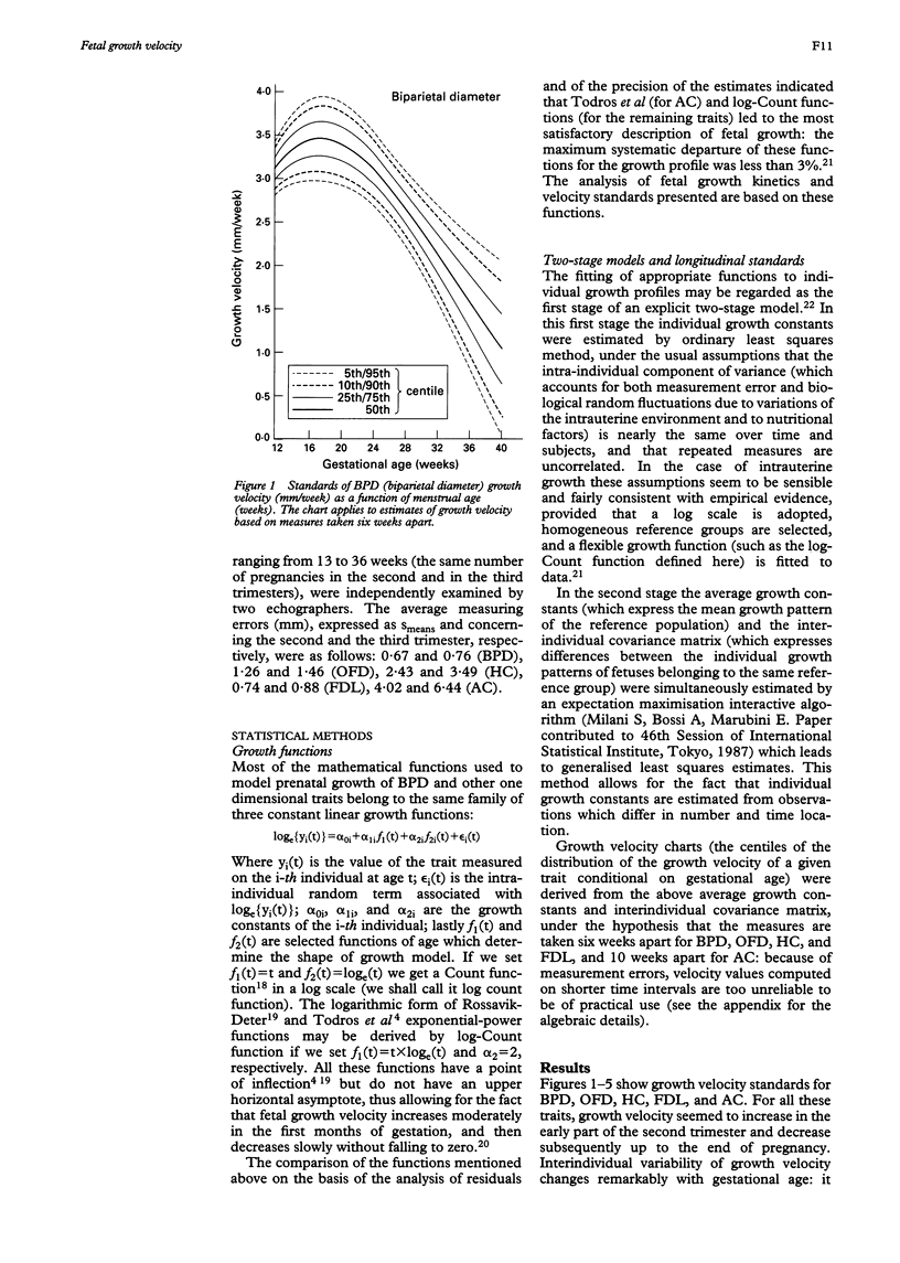
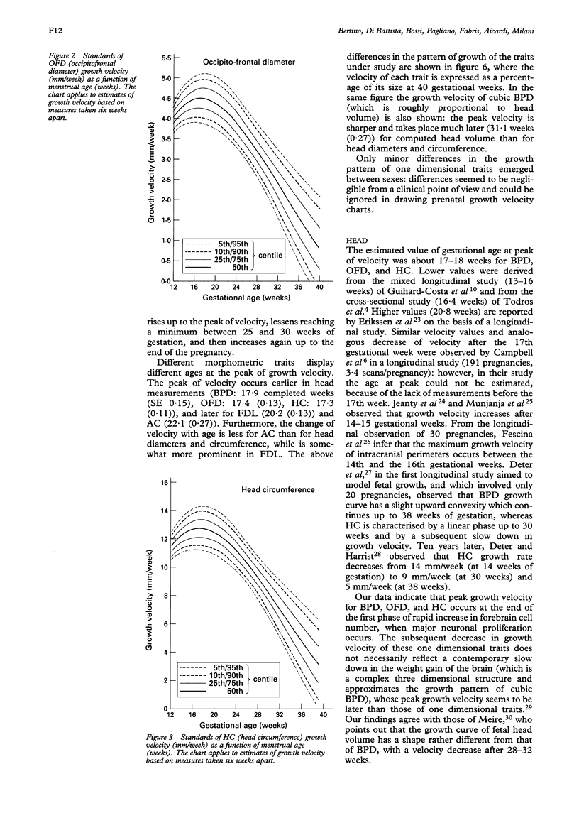
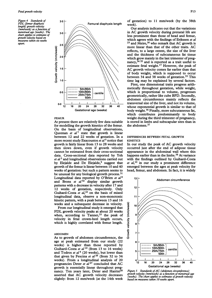
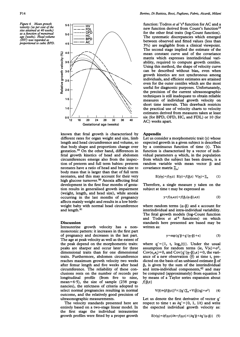
With the aim of determining fetal growth kinetics, prenatal data were analysed which had been longitudinally collected in the framework of a perinatal growth survey. The sample comprised 238 singleton normal pregnancies, selected in Genoa and Turin (between 1987 and 1990), and repeatedly assessed by ultrasound scans (five to nine per pregnancy). Five morphometric traits were considered: BPD (biparietal diameter), OFD (occipitofrontal diameter), HC (head circumference), FDL (femur diaphysis length) and AC (abdomen circumference). Growth rate seemed to increase in the early part of the second trimester, and decrease subsequently: velocity peaks were steeper and earlier for head diameters and circumference (about 18 weeks) than for femur length (20 weeks) and abdomen circumference (22 weeks). Velocity standards were traced using a longitudinal two-stage linear model: this ensures unbiased description of the shape of the growth curve, even when growth kinetics are asynchronous, and efficient estimation of the outer centiles--the most useful for diagnostic purposes.
Full text
PDF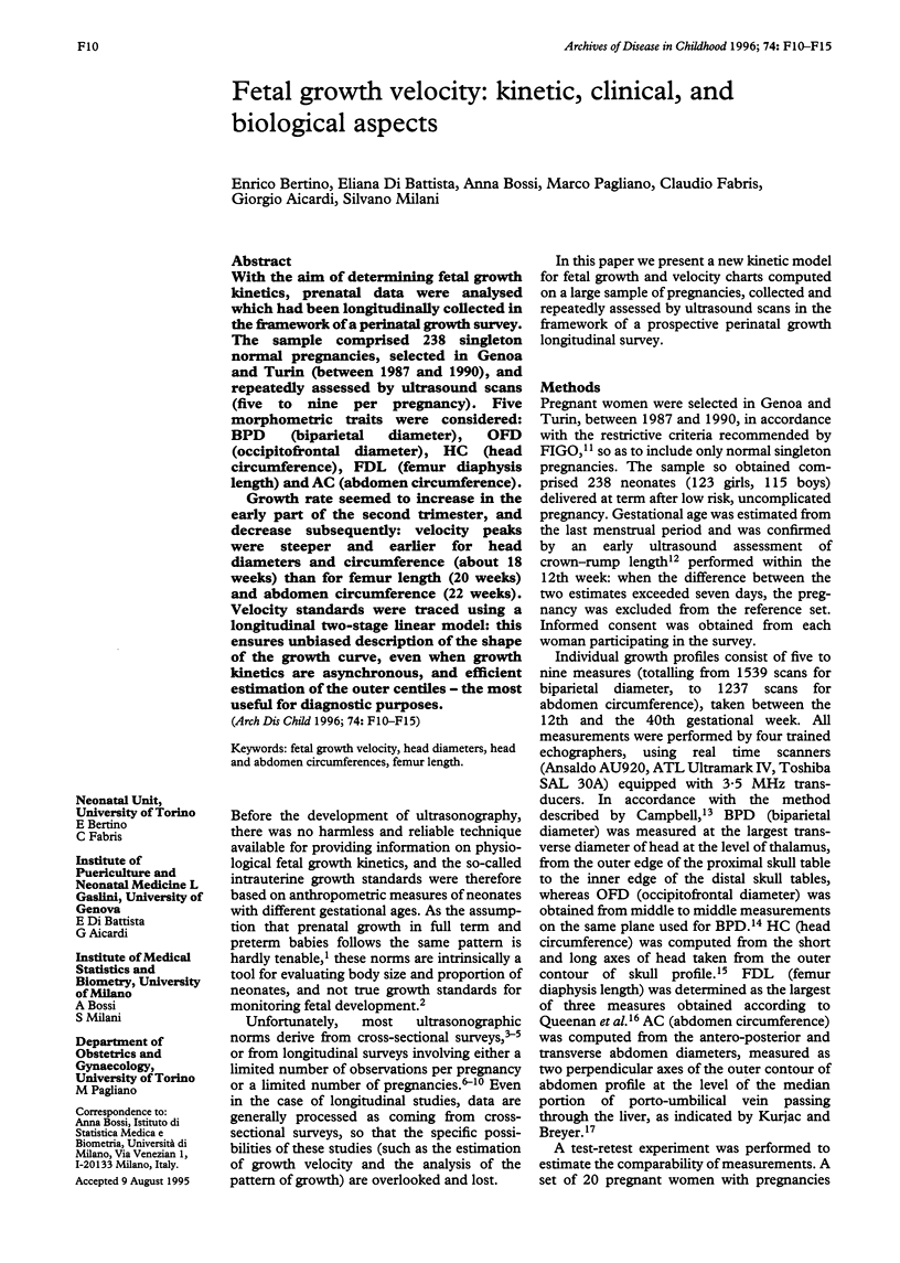
Selected References
These references are in PubMed. This may not be the complete list of references from this article.
- Berkey C. S., Laird N. M. Nonlinear growth curve analysis: estimating the population parameters. Ann Hum Biol. 1986 Mar-Apr;13(2):111–128. doi: 10.1080/03014468600008261. [DOI] [PubMed] [Google Scholar]
- Brar H. S., Rutherford S. E. Classification of intrauterine growth retardation. Semin Perinatol. 1988 Jan;12(1):2–10. [PubMed] [Google Scholar]
- Brons J. T., van Geijn H. P., Bezemer P. D., Nauta J. P., Arts N. F. The fetal skeleton; ultrasonographic evaluation of the normal growth. Eur J Obstet Gynecol Reprod Biol. 1990 Jan-Feb;34(1-2):21–36. doi: 10.1016/0028-2243(90)90004-k. [DOI] [PubMed] [Google Scholar]
- Campbell S. An improved method of fetal cephalometry by ultrasound. J Obstet Gynaecol Br Commonw. 1968 May;75(5):568–576. doi: 10.1111/j.1471-0528.1968.tb00161.x. [DOI] [PubMed] [Google Scholar]
- Campbell S., Newman G. B. Growth of the fetal biparietal diameter during normal pregnancy. J Obstet Gynaecol Br Commonw. 1971 Jun;78(6):513–519. doi: 10.1111/j.1471-0528.1971.tb00309.x. [DOI] [PubMed] [Google Scholar]
- Deter R. L., Harrist R. B. Growth standards for anatomic measurements and growth rates derived from longitudinal studies of normal fetal growth. J Clin Ultrasound. 1992 Jul-Aug;20(6):381–388. doi: 10.1002/jcu.1870200604. [DOI] [PubMed] [Google Scholar]
- Deter R. L., Harrist R. B., Hadlock F. P., Poindexter A. N. Longitudinal studies of fetal growth with the use of dynamic image ultrasonography. Am J Obstet Gynecol. 1982 Jul 1;143(5):545–554. doi: 10.1016/0002-9378(82)90545-2. [DOI] [PubMed] [Google Scholar]
- Dobbing J., Sands J. Head circumference, biparietal diameter and brain growth in fetal and postnatal life. Early Hum Dev. 1978 Apr;2(1):81–87. doi: 10.1016/0378-3782(78)90054-3. [DOI] [PubMed] [Google Scholar]
- Elejalde B. R., de Elejalde M. M. The prenatal growth of the human body determined by the measurement of bones and organs by ultrasonography. Am J Med Genet. 1986 Aug;24(4):575–598. doi: 10.1002/ajmg.1320240402. [DOI] [PubMed] [Google Scholar]
- Eriksen P. S., Secher N. J., Weis-Bentzon M. Normal growth of the fetal biparietal diameter and the abdominal diameter in a longitudinal study. An evaluation of the two parameters in predicting fetal weight. Acta Obstet Gynecol Scand. 1985;64(1):65–70. doi: 10.3109/00016348509154690. [DOI] [PubMed] [Google Scholar]
- Exacoustos C., Rosati P., Rizzo G., Arduini D. Ultrasound measurements of fetal limb bones. Ultrasound Obstet Gynecol. 1991 Sep 1;1(5):325–330. doi: 10.1046/j.1469-0705.1991.01050325.x. [DOI] [PubMed] [Google Scholar]
- Fescina R. H., Ucieda F. J., Cordano M. C., Nieto F., Tenzer S. M., López R. Ultrasonic patterns of intrauterine fetal growth in a Latin American country. Early Hum Dev. 1982 Jul;6(3):239–248. doi: 10.1016/0378-3782(82)90116-5. [DOI] [PubMed] [Google Scholar]
- Guihard-Costa A. M., Droullé P., Larroche J. C. Growth velocity of the biparietal diameter, abdominal transverse diameter and femur length in the fetal period. Early Hum Dev. 1991 Nov;27(1-2):93–102. doi: 10.1016/0378-3782(91)90030-7. [DOI] [PubMed] [Google Scholar]
- Hata T., Deter R. L. A review of fetal organ measurements obtained with ultrasound: normal growth. J Clin Ultrasound. 1992 Mar-Apr;20(3):155–174. doi: 10.1002/jcu.1870200302. [DOI] [PubMed] [Google Scholar]
- Jeanty P., Cousaert E., Hobbins J. C., Tack B., Bracken M., Cantraine F. A longitudinal study of fetal head biometry. Am J Perinatol. 1984 Jan;1(2):118–128. doi: 10.1055/s-2007-999987. [DOI] [PubMed] [Google Scholar]
- Kurjak A., Breyer B. Estimation of fetal weighty by ultrasonic abdominometry. Am J Obstet Gynecol. 1976 Aug 1;125(7):962–965. doi: 10.1016/0002-9378(76)90496-8. [DOI] [PubMed] [Google Scholar]
- Longuet R., Phelan J., Tanous H., Bushong S. Criteria of the silhouette sign. Radiology. 1977 Mar;122(3):581–585. doi: 10.1148/122.3.581. [DOI] [PubMed] [Google Scholar]
- Munjanja S. P., Masona D., Masvikeni S. Fetal biparietal diameter and head circumference measurements: results of a longitudinal study in Zimbabwe. Int J Gynaecol Obstet. 1988 Apr;26(2):223–228. doi: 10.1016/0020-7292(88)90266-4. [DOI] [PubMed] [Google Scholar]
- Murao F., Senoh D., Takamiya O., Yamamoto K., Hasegawa K., Kitao M. Ultrasonic evaluation of liver development in the fetus in utero. Gynecol Obstet Invest. 1989;28(4):198–201. doi: 10.1159/000293577. [DOI] [PubMed] [Google Scholar]
- O'Brien G. D., Queenan J. T. Growth of the ultrasound fetal femur length during normal pregnancy. Part I. Am J Obstet Gynecol. 1981 Dec 1;141(7):833–837. doi: 10.1016/0002-9378(81)90713-4. [DOI] [PubMed] [Google Scholar]
- Ogata E. S. Carbohydrate metabolism in the fetus and neonate and altered neonatal glucoregulation. Pediatr Clin North Am. 1986 Feb;33(1):25–45. doi: 10.1016/s0031-3955(16)34968-9. [DOI] [PubMed] [Google Scholar]
- Poissonnet C. M., Burdi A. R., Garn S. M. The chronology of adipose tissue appearance and distribution in the human fetus. Early Hum Dev. 1984 Sep;10(1-2):1–11. doi: 10.1016/0378-3782(84)90106-3. [DOI] [PubMed] [Google Scholar]
- Queenan J. T., Kubarych S. F., Cook L. N., Anderson G. D., Griffin L. P. Diagnostic ultrasound for detection of intrauterine growth retardation. Am J Obstet Gynecol. 1976 Apr 15;124(8):865–873. doi: 10.1016/s0002-9378(16)33391-9. [DOI] [PubMed] [Google Scholar]
- Queenan J. T., O'Brien G. D., Campbell S. Ultrasound measurement of fetal limb bones. Am J Obstet Gynecol. 1980 Oct 1;138(3):297–302. doi: 10.1016/0002-9378(80)90252-5. [DOI] [PubMed] [Google Scholar]
- Robinson H. P. Sonar measurement of fetal crown-rump length as means of assessing maturity in first trimester of pregnancy. Br Med J. 1973 Oct 6;4(5883):28–31. doi: 10.1136/bmj.4.5883.28. [DOI] [PMC free article] [PubMed] [Google Scholar]
- Rossavik I. K., Deter R. L. Mathematical modeling of fetal growth: I. Basic principles. J Clin Ultrasound. 1984 Nov-Dec;12(9):529–533. doi: 10.1002/jcu.1870120902. [DOI] [PubMed] [Google Scholar]
- Secher N. J., Kern Hansen P., Thomsen B. L., Keiding N. Growth retardation in preterm infants. Br J Obstet Gynaecol. 1987 Feb;94(2):115–120. doi: 10.1111/j.1471-0528.1987.tb02336.x. [DOI] [PubMed] [Google Scholar]
- Todros T., Ferrazzi E., Groli C., Nicolini U., Parodi L., Pavoni M., Zorzoli A., Zucca S. Fitting growth curves to head and abdomen measurements of the fetus: a multicentric study. J Clin Ultrasound. 1987 Feb;15(2):95–105. doi: 10.1002/jcu.1870150203. [DOI] [PubMed] [Google Scholar]
- Vaucher Y. E., Harrison G. G., Udall J. N., Morrow G., 3rd Skinfold thickness in North American infants 24-41 weeks gestation. Hum Biol. 1984 Dec;56(4):713–731. [PubMed] [Google Scholar]
- Yeh M. N., Bracero L., Reilly K. B., Murtha L., Aboulafia M., Barron B. A. Ultrasonic measurement of the femur length as an index of fetal gestational age. Am J Obstet Gynecol. 1982 Nov 1;144(5):519–522. doi: 10.1016/0002-9378(82)90219-8. [DOI] [PubMed] [Google Scholar]