

Abstract
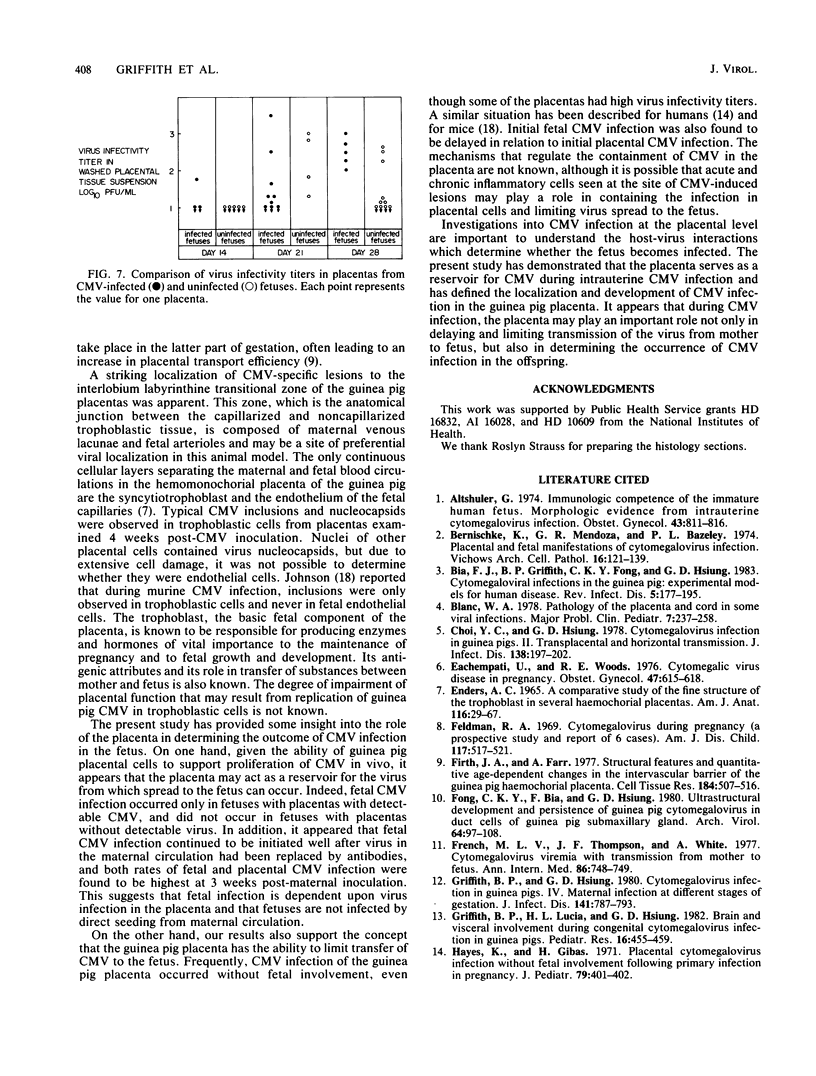
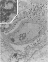
The development of cytomegalovirus (CMV) infection in the placenta was studied in Hartley guinea pigs inoculated at midgestation, and its role in determining the outcome of fetal CMV infection was assessed. A hematogenous spread of CMV from the mother to the placenta occurred early during the course of the infection. However, the virus remained present in placental tissues long after CMV had been cleared from maternal blood (i.e., 3 and 4 weeks postinoculation). At that time, the virus was able to replicate in placental tissues in the presence of specific maternal antibodies. Viral nucleocapsids were seen within nuclei of trophoblastic cells, and virions were present surrounding infected cells. In addition, typical CMV-induced histopathological lesions bearing CMV antigens were consistently localized at the transitional zone between the capillarized labyrinth and the noncapillarized interlobium. Whenever CMV infection of the fetus occurred, virus was isolated from the associated placenta. Among placental-fetal units with CMV-infected placentas, only 27% of the fetuses were found to be infected. In addition, there was a delay in the establishment of the infection in the fetus in relation to the placenta, although frequencies of virus isolation in placental and fetal tissues peaked at 3 weeks after CMV inoculation. These results suggest that during primary CMV infection of pregnant guinea pigs, the placenta not only serves as a reservoir for CMV but also acts to limit transmission of the virus to the fetus.
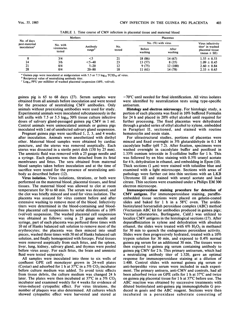
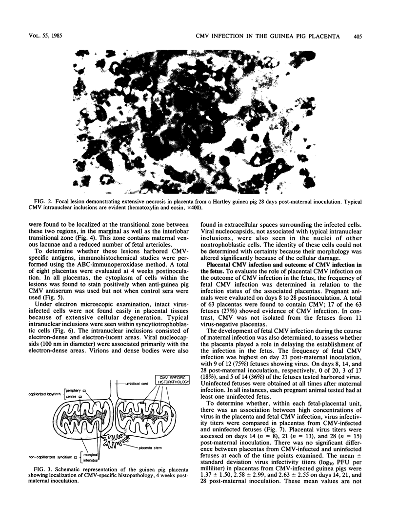
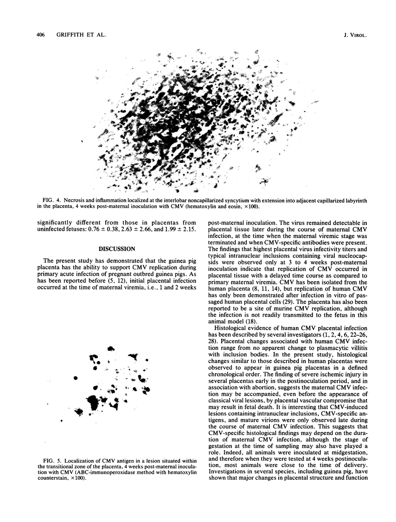
Full text
PDF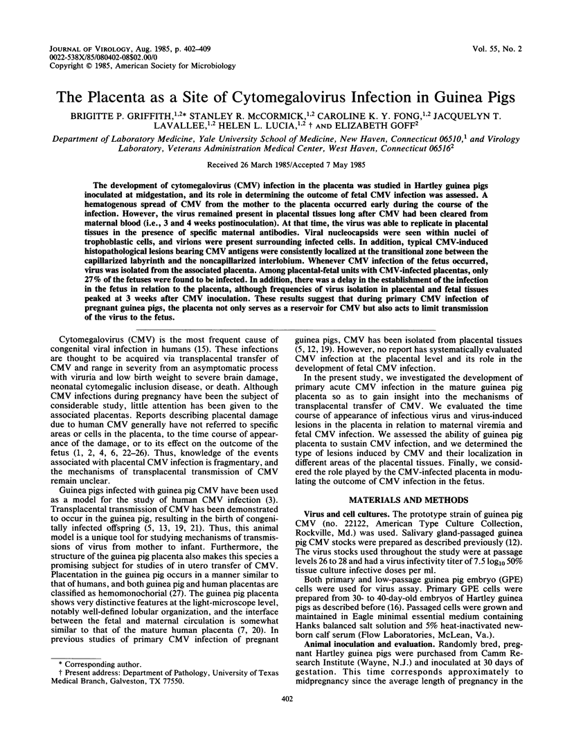


Images in this article

Selected References
These references are in PubMed. This may not be the complete list of references from this article.
- Altshuler G. Immunologic competence of the immature human fetus. Morphologic evidence from intrauterine Cytomegalovirus infection. Obstet Gynecol. 1974 Jun;43(6):811–816. [PubMed] [Google Scholar]
- Benirschke K., Mendoza G. R., Bazeley P. L. Placental and fetal manifestations of cytomegalovirus infection. Virchows Arch B Cell Pathol. 1974;16(2):121–139. doi: 10.1007/BF02894070. [DOI] [PubMed] [Google Scholar]
- Bia F. J., Griffith B. P., Fong C. K., Hsiung G. D. Cytomegaloviral infections in the guinea pig: experimental models for human disease. Rev Infect Dis. 1983 Mar-Apr;5(2):177–195. doi: 10.1093/clinids/5.2.177. [DOI] [PubMed] [Google Scholar]
- Blanc W. A. Pathology of the placenta and cord in some viral infections. Major Probl Clin Pediatr. 1978;17:237–258. [PubMed] [Google Scholar]
- Choi Y. C., Hsiung G. D. Cytomegalovirus infection in guinea pigs. II. Transplacental and horizontal transmission. J Infect Dis. 1978 Aug;138(2):197–202. doi: 10.1093/infdis/138.2.197. [DOI] [PubMed] [Google Scholar]
- ENDERS A. C. A COMPARATIVE STUDY OF THE FINE STRUCTURE OF THE TROPHOBLAST IN SEVERAL HEMOCHORIAL PLACENTAS. Am J Anat. 1965 Jan;116:29–67. doi: 10.1002/aja.1001160103. [DOI] [PubMed] [Google Scholar]
- Eachempati U., Woods R. E. Cytomegalic virus disease in pregnancy. Obstet Gynecol. 1976 May;47(5):615–618. [PubMed] [Google Scholar]
- Feldman R. A. Cytomegalovirus infection during pregnancy. A prospective study and report of six cases. Am J Dis Child. 1969 May;117(5):517–521. [PubMed] [Google Scholar]
- Firth J. A., Farr A. Structural features and quantitative age-dependent changes in the intervascular barrier of the guinea-pig haemochorial placenta. Cell Tissue Res. 1977 Nov 23;184(4):507–516. doi: 10.1007/BF00220974. [DOI] [PubMed] [Google Scholar]
- Fong C. K., Bia F., Hsiung G. D. Ultrastructural development and persistence of guinea pig cytomegalovirus in duet cells of guinea pig submaxillary gland. Arch Virol. 1980;64(2):97–108. doi: 10.1007/BF01318013. [DOI] [PubMed] [Google Scholar]
- French M. L., Thompson J. F., White A. Cytomegalovirus viremia with transmission from mother to fetus. Ann Intern Med. 1977 Jun;86(6):748–749. doi: 10.7326/0003-4819-86-6-748. [DOI] [PubMed] [Google Scholar]
- Griffith B. P., Hsiung G. D. Cytomegalovirus infection in guinea pigs. IV. Maternal infection at different stages of gestation. J Infect Dis. 1980 Jun;141(6):787–793. doi: 10.1093/infdis/141.6.787. [DOI] [PubMed] [Google Scholar]
- Griffith B. P., Lucia H. L., Hsiung G. D. Brain and visceral involvement during congenital cytomegalovirus infection of guinea pigs. Pediatr Res. 1982 Jun;16(6):455–459. doi: 10.1203/00006450-198206000-00010. [DOI] [PubMed] [Google Scholar]
- Hayes K., Gibas H. Placental cytomegalovirus infection without fetal involvement following primary infection in pregnancy. J Pediatr. 1971 Sep;79(3):401–405. doi: 10.1016/s0022-3476(71)80147-6. [DOI] [PubMed] [Google Scholar]
- Hsiung G. D., Tenser R. B., Fong C. K. Comparison of guinea pig cytomegalovirus and guinea pig herpes-like virus: growth characteristics and antigentic relationship. Infect Immun. 1976 Mar;13(3):926–933. doi: 10.1128/iai.13.3.926-933.1976. [DOI] [PMC free article] [PubMed] [Google Scholar]
- Hsu S. M., Raine L., Fanger H. Use of avidin-biotin-peroxidase complex (ABC) in immunoperoxidase techniques: a comparison between ABC and unlabeled antibody (PAP) procedures. J Histochem Cytochem. 1981 Apr;29(4):577–580. doi: 10.1177/29.4.6166661. [DOI] [PubMed] [Google Scholar]
- Johnson K. P., Connor W. S. Guinea pig cytomegalovirus: transplacental transmission. Brief report. Arch Virol. 1979;59(3):263–267. doi: 10.1007/BF01317422. [DOI] [PubMed] [Google Scholar]
- Johnson K. P. Mouse cytomegalovirus: placental infection. J Infect Dis. 1969 Oct;120(4):445–450. doi: 10.1093/infdis/120.4.445. [DOI] [PubMed] [Google Scholar]
- Kaufmann P., Davidoff M. The guinea-pig placenta. Adv Anat Embryol Cell Biol. 1977;53(2):5–91. doi: 10.1007/978-3-642-66618-6. [DOI] [PubMed] [Google Scholar]
- Kumar M. L., Nankervis G. A. Experimental congenital infection with cytomegalovirus: a guinea pig model. J Infect Dis. 1978 Nov;138(5):650–654. doi: 10.1093/infdis/138.5.650. [DOI] [PubMed] [Google Scholar]
- LELONG M., LEPAGE F., LE TAN VINH, TOURNIER P., CHANY C. [The virus of cytomegalic inclusion disease. Its isolation in 2 cases. Survival of the patient, without sequelae, in one of these cases. Presence of inclusions in the placenta in a third case]. Arch Fr Pediatr. 1960;17:437–450. [PubMed] [Google Scholar]
- LEPAGE F., SCHRAMM B. Aspects histologiques du placenta et des membranes dans la maladie des inclusions cytomégaliques. Gynecol Obstet (Paris) 1958;57(3):273–279. [PubMed] [Google Scholar]
- Monif G. R., Dische R. M. Viral placentitis in congenital cytomegalovirus infection. Am J Clin Pathol. 1972 Oct;58(4):445–449. doi: 10.1093/ajcp/58.5.445. [DOI] [PubMed] [Google Scholar]
- Mostoufi-zadeh M., Driscoll S. G., Biano S. A., Kundsin R. B. Placental evidence of cytomegalovirus infection of the fetus and neonate. Arch Pathol Lab Med. 1984 May;108(5):403–406. [PubMed] [Google Scholar]
- QUAN A., STRAUSS L. Congenital cytomegalic inclusion disease. Observations in a macerated fetus with congenital defect, including a study of the placenta. Am J Obstet Gynecol. 1962 May 1;83:1240–1248. [PubMed] [Google Scholar]
- ROSENSTEIN D. L., NAVARRETE-REYNA A. CYTOMEGALIC INCLUSION DISEASE: OBSERVATION OF THE CHARACTERISTIC INCLUSION BODIES IN THE PLACENTA. Am J Obstet Gynecol. 1964 May 15;89:220–224. [PubMed] [Google Scholar]
- Rosenthal L. J., Panitz P. J., Crutchfield D. B., Chou J. Y. Cytomegalovirus replication in primary and passaged human placental cells. Intervirology. 1981;16(3):168–175. doi: 10.1159/000149264. [DOI] [PubMed] [Google Scholar]