Abstract
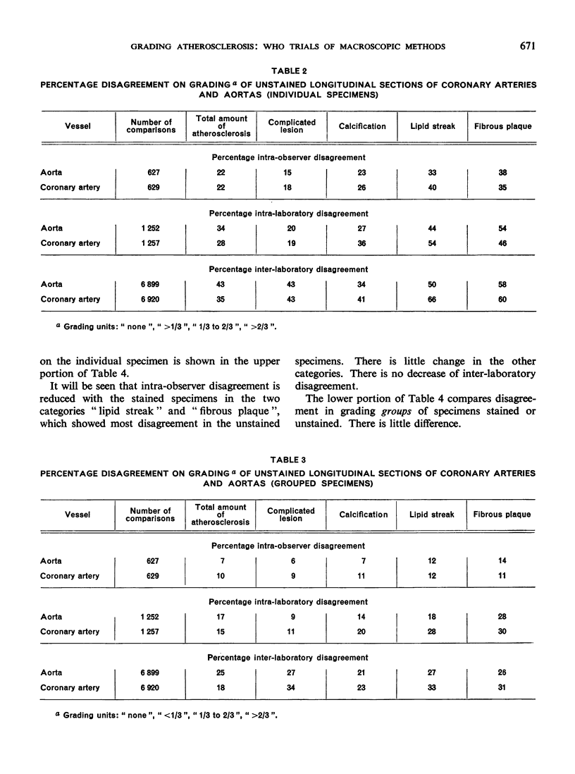
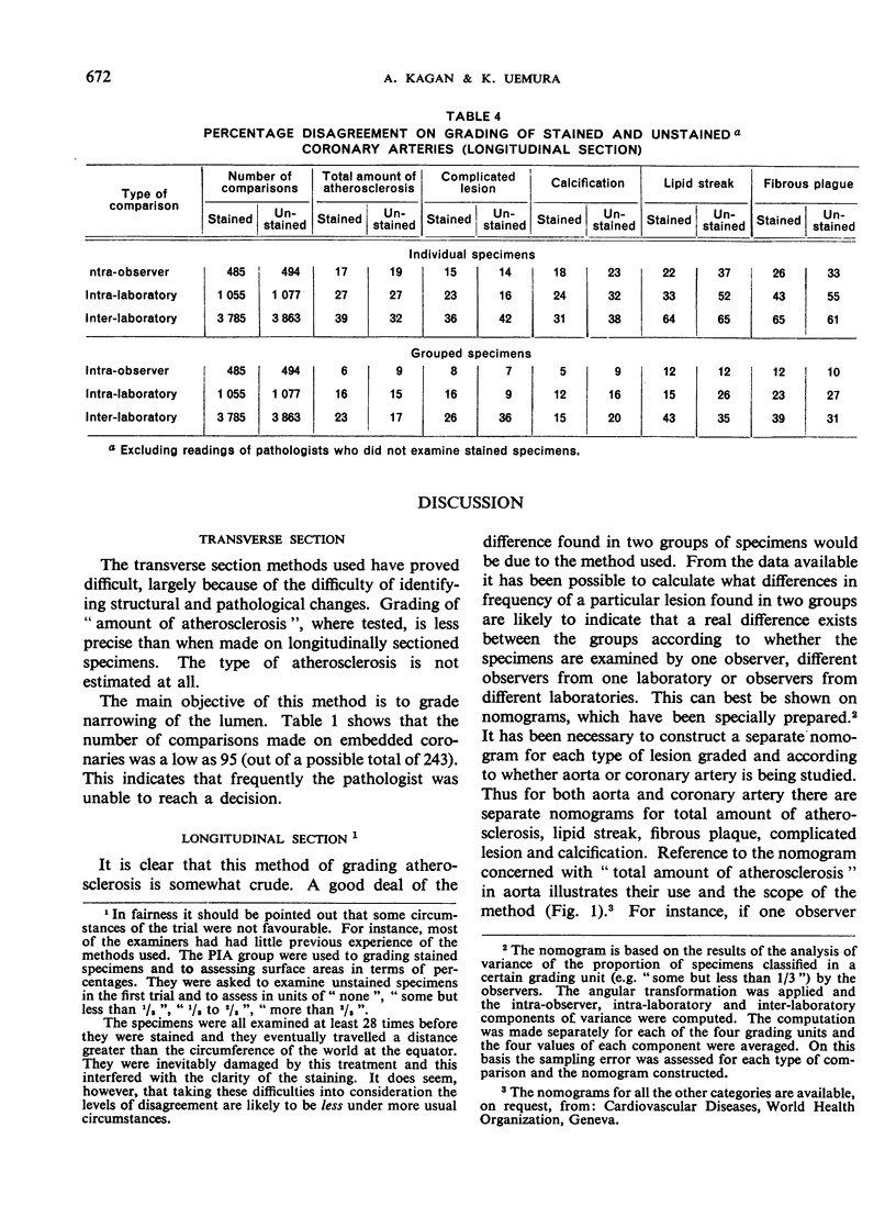
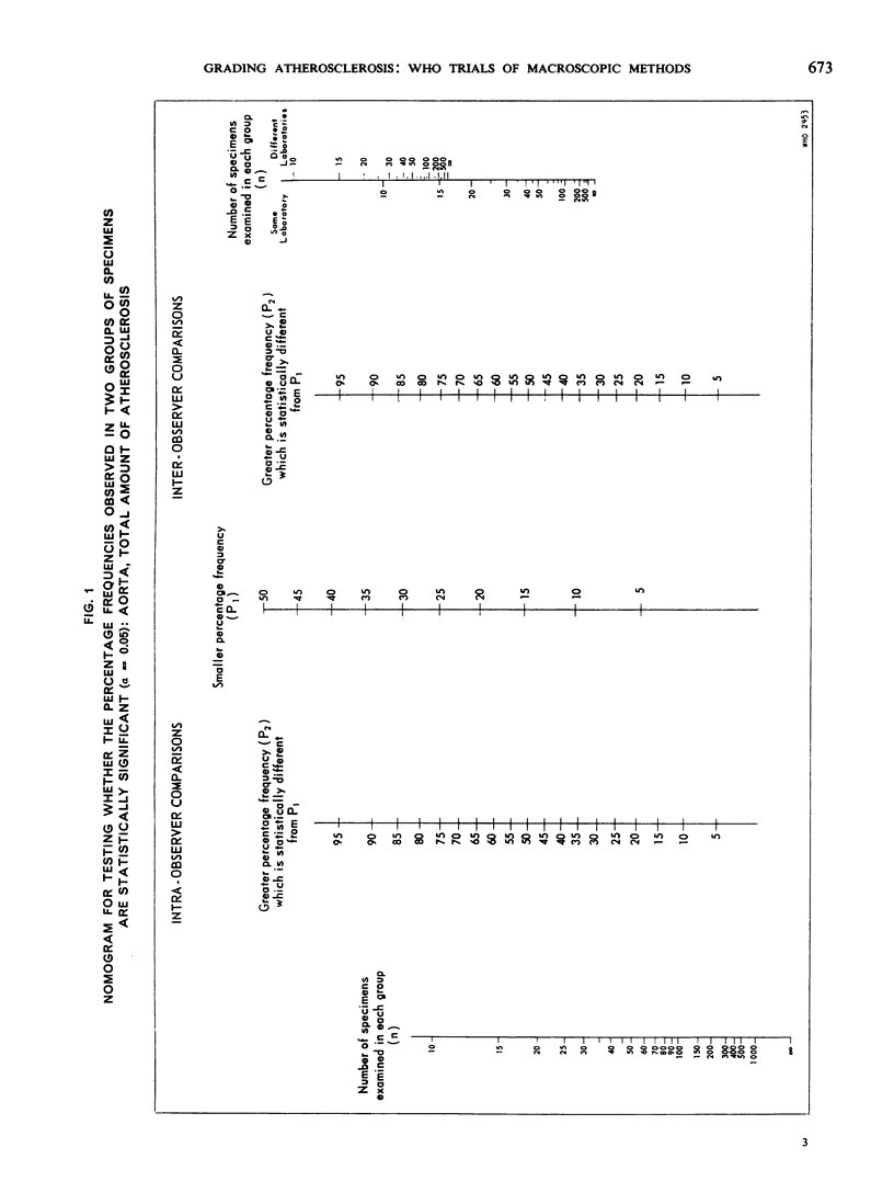
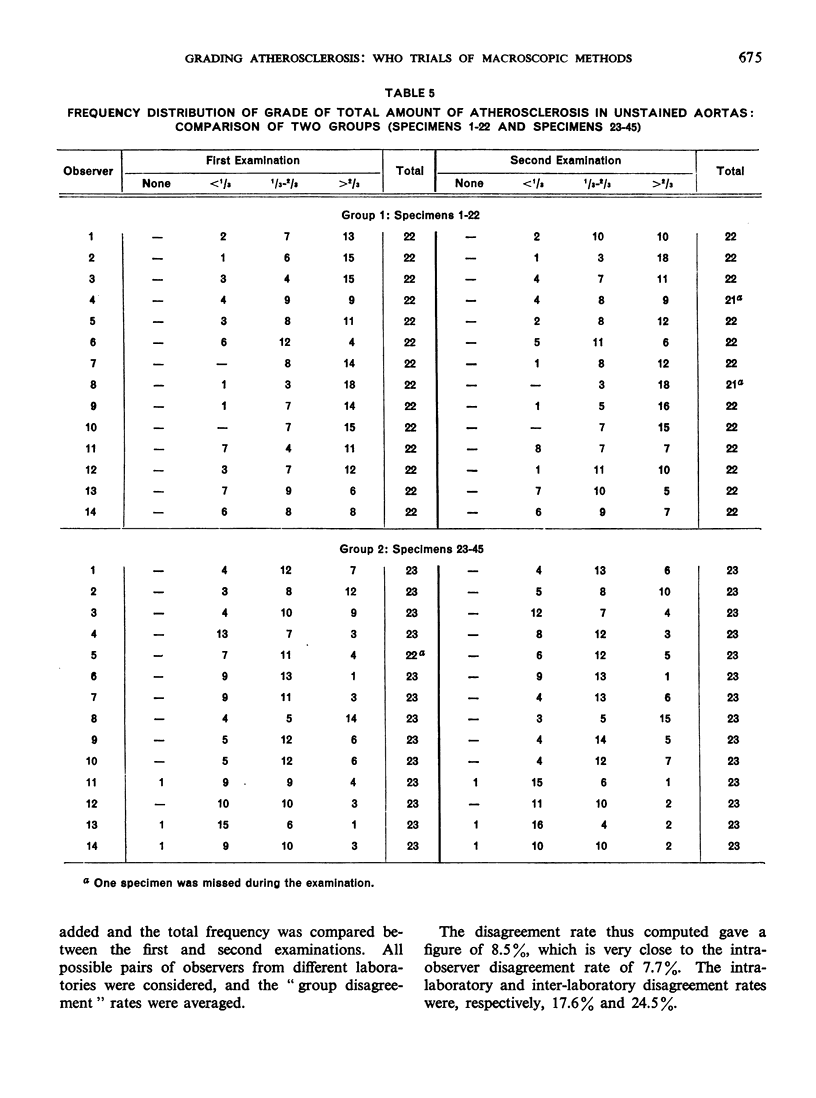
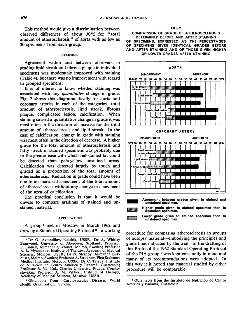
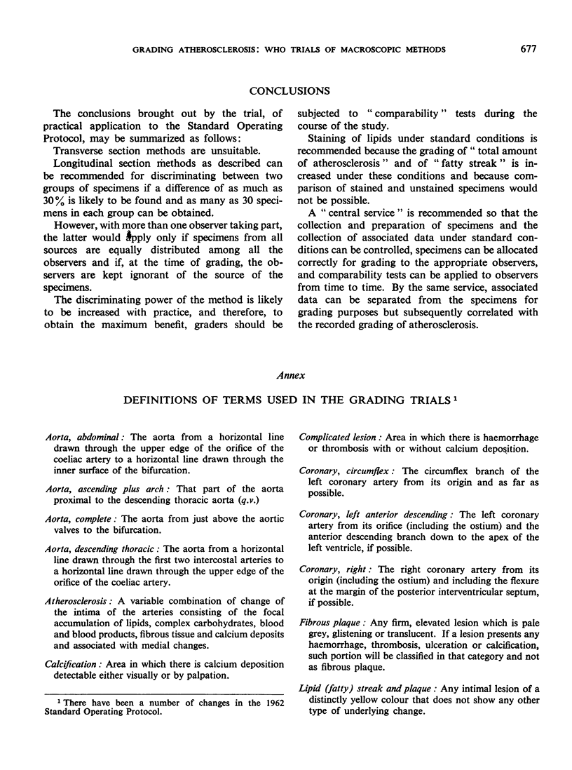
Many attempts have been made to grade atherosclerosis as found in autopsy material, but the repeatability of the methods used has seldom been tested. Most of the methods used are rough; and, where comparisons are to be made between the data obtained by different observers on different material, it is essential to know whether the differences found are due to the crudity of the method or in fact represent a real difference in the material studied. This paper describes an attempt to obtain comparable data by presenting specially prepared specimens to 14 pathologists from five laboratories in Europe and the Americas. For the purposes of the study, agreed definitions, techniques and criteria were adopted. Intra-observer, intra-laboratory and inter-laboratory disagreement was measured using both transverse- and longitudinal-section procedures. The longitudinally sectioned specimens were examined unstained and subsequently stained for lipid. The results indicate that the longitudinal-section procedure is likely to be useful in discriminating between groups of specimens, provided that certain procedural rules are observed.
Full text
PDF
Selected References
These references are in PubMed. This may not be the complete list of references from this article.
- BAKER A. B., REFSUM S., DAHL E. Cerebrovascular disease. IV. A study of a Norwegian population. Neurology. 1960 Jun;10:525–529. doi: 10.1212/wnl.10.6.525. [DOI] [PubMed] [Google Scholar]
- DICKINSON C. J., THOMASON A. D. Vertebral and internal carotid arteries in relation to hypertension and cerebrovascular disease. Lancet. 1959 Jul 18;2(7090):46–48. doi: 10.1016/s0140-6736(59)90494-5. [DOI] [PubMed] [Google Scholar]
- ELKELES A. A comparative radiological study of calcified atheroma in males and females over 50 years of age. Lancet. 1957 Oct 12;273(6998):714–715. doi: 10.1016/s0140-6736(57)92256-0. [DOI] [PubMed] [Google Scholar]
- GORE I., TEJADA C. The quantitative appraisal of atherosclerosis. Am J Pathol. 1957 Sep-Oct;33(5):875–885. [PMC free article] [PubMed] [Google Scholar]
- HOLMAN R. L., McGILL H. C., Jr, STRONG J. P., GEER J. C. Technics for studying atherosclerotic lesions. Lab Invest. 1958 Jan-Feb;7(1):42–47. [PubMed] [Google Scholar]
- PATERSON J. C., CORNISH B. R., ARMSTRONG E. C. The Gofman indices in coronary atherosclerosis. Can Med Assoc J. 1956 Apr 1;74(7):538–542. [PMC free article] [PubMed] [Google Scholar]
- SCOTT R. F., DAOUD A. S., FLORENTIN R. A., DAVIES J. N., COLES R. M. Comparison of the amount of coronary arteriosclerosis in autopsied East Africans and New Yorkers. Am J Cardiol. 1961 Aug;8:165–172. doi: 10.1016/0002-9149(61)90201-6. [DOI] [PubMed] [Google Scholar]
- TEJADA C., GORE I., STRONG J. P., McGILL H. C., Jr Comparative severity of atherosclerosis in Costa Rica, Guatemala, and New Orleans. Circulation. 1958 Jul;18(1):92–97. doi: 10.1161/01.cir.18.1.92. [DOI] [PubMed] [Google Scholar]
- YATER W. M., WELSH P. P., STAPLETON J. F., CLARK M. L. Comparison of clinical and pathologic aspects of coronary artery disease in men of various age groups: a study of 950 autopsied cases from the Armed Forces Institute of Pathology. Ann Intern Med. 1951 Feb;34(2):352–392. doi: 10.7326/0003-4819-34-2-352. [DOI] [PubMed] [Google Scholar]


