Abstract
Tctex2 is thought to be one of the distorter genes of the mouse t haplotype. This complex greatly biases the segregation of the chromosome that carries it such that in heterozygous +/t males, the t haplotype is transmitted to >95% of the offspring, a phenomenon known as transmission ratio distortion. The LC2 outer dynein arm light chain of Chlamydomonas reinhardtii is a homologue of the mouse protein Tctex2. We have identified Chlamydomonas insertional mutants with deletions in the gene encoding LC2 and demonstrate that the LC2 gene is the same as the ODA12 gene, the product of which had not been identified previously. Complete deletion of the LC2/ODA12 gene causes loss of all outer arms and a slow jerky swimming phenotype. Transformation of the deletion mutant with the cloned LC2/ODA12 gene restores the outer arms and rescues the motility phenotype. Therefore, LC2 is required for outer arm assembly. The fact that LC2 is an essential subunit of flagellar outer dynein arms allows us to propose a detailed mechanism whereby transmission ratio distortion is explained by the differential binding of mutant (t haplotype encoded) and wild-type dyneins to the axonemal microtubules of t-bearing or wild-type sperm, with resulting differences in their motility.
INTRODUCTION
Dynein ATPases are microtubule-based molecular motors that provide force for a variety of important cellular processes (reviewed by Holzbaur and Vallee, 1994; Witman et al., 1994). Cytoplasmic dyneins are involved in vesicle transport, Golgi localization, nuclear migration, spindle formation and orientation, mitosis, and flagellar assembly. Inner arm and outer arm axonemal dyneins provide the force for flagellar and ciliary beating.
All characterized dyneins contain one or more heavy chains (HCs) that are associated with smaller polypeptides termed intermediate chains (ICs), light intermediate chains, and light chains (LCs). For example, Chlamydomonas outer arm dynein, which is the most well characterized axonemal dynein, contains three HCs, two ICs, and eight LCs (Table 1). Each HC has a globular head domain containing the site for ATP hydrolysis and a fibrous stem domain that extends to the base of the dynein, where the ICs and most of the LCs are located in a discrete complex. The heads of the HCs interact with the B-tubule of the opposing doublet microtubule to generate force, whereas the ICs are involved in anchoring the dynein to the A-tubule of the doublet microtubule (King et al., 1995; Wilkerson et al., 1995) and regulating dynein activity (Mitchell and Kang, 1993). In contrast, little is known regarding the function(s) of the dynein LCs.
Table 1.
Proteins associated with the outer dynein arm of Chlamydomonas
| Protein | Mass (D) | Gene | Comments | Referencea |
|---|---|---|---|---|
| HCα | 503,613 | ODA11 | ATPase | 1, 2, 3 |
| HCβ | 519,961 | ODA4 | ATPase | 2, 4 |
| HCγ | 512,836 | ODA2 | ATPase | 4, 5, 6 |
| IC1 | 76,393 | ODA9 | Microtubule binding | 4, 7 |
| IC2 | 63,520 | ODA6 | Regulation | 4, 8 |
| LC1 | 22,150 | Leucine-rich repeat protein | 9 | |
| LC2 | 15,825 | ODA12 | Tctex2 homologue | 10, this work |
| LC3 | 17,365 | Thioredoxin homologue | 11 | |
| LC4 | 17,788 | EF-hand protein | 12 | |
| LC5 | 14,179 | Thioredoxin homologue | 11 | |
| LC6 | 13,857 | ODA13 | LC8 homologue | 13, 14 |
| LC7 | 11,928 | Roadblock homologue | 15 | |
| LC8 | 10,322 | FLA14 | Multiple flagellar defects | 13, 16 |
| DC1 | 83,377 | ODA3 | ODA-DC polypeptide | 4, 17, 18 |
| DC2 | 62,234 | ODA1 | ODA-DC polypeptide | 4, 17 |
| DC3 | 21,341 | ODA14 | ODA-DC polypeptide | 17, 19 |
1, Sakakibara et al. (1991); 2, Mitchell and Brown (1994); 3, Mitchell and Brown (1997); 4, Kamiya (1988); 5, Mitchell and Rosenbaum (1985); 6, Wilkerson et al. (1994); 7, Wilkerson et al. (1995); 8, Mitchell and Kang (1991); 9, Benashski et al. (1999); 10, Patel-King et al. (1997); 11, Patel-King et al. (1996); 12, King and Patel-King (1995a); 13, King and Patel-King (1995b); 14, Pazour and Witman (unpublished data); 15, Bowman et al. (1999); 16, Pazour et al. (1998); 17, Takada et al. (1996); 18, Koutoulis et al. (1997); 19, Casey et al. (1998).
The need for more information on the dynein LCs has been underscored recently by 1) the discovery that LCs are associated with cytoplasmic dynein (King et al., 1996a), indicating that they are likely to have a universal role in dynein structure and/or function, and 2) the finding that two dynein LCs are encoded within a large region of mouse chromosome 17, which, in t mice, corresponds to the t haplotype (King et al., 1996b; Patel-King et al., 1997). The t haplotype has four inversions relative to the wild-type chromosome; these inversions suppress recombination, so that mutations arising in the t haplotype are kept together (Silver, 1993). This portion of chromosome 17 has been the subject of intense study because the t haplotype is inherited in a non-Mendelian manner. Heterozygous +/t males transmit the t-bearing chromosome to >95% of their offspring, a phenomenon known as transmission ratio distortion or meiotic drive. This is due to the concerted action of three or four t-encoded distorter genes acting on a t-encoded responder gene (Lyon, 1984). In the presence of the t responder, the t distorters act in an additive manner to increase the percent of offspring that carry the t-bearing chromosome (for review, see Silver, 1985, 1993). Apparently the t-encoded versions of the distorter and responder genes contain mutations that give the t haplotype a selective advantage over the wild-type homologue. The identity and function of the responder gene product is unknown (Ewulonu et al., 1996), but two of the putative distorter gene products (Tctex1 and Tctex2) are dynein LCs. Tctex1 (Lader et al., 1989) is a homologue of a Chlamydomonas inner arm dynein LC (Harrison et al., 1998) and also is a subunit of brain cytoplasmic dynein (King et al., 1996b). Tctex2 (Huw et al., 1995) is a homologue of a Chlamydomonas outer arm dynein LC termed LC2 (Patel-King et al., 1997). A third distorter gene product also may be a dynein subunit, because the Dnahc8 dynein HC (Vaughan et al., 1996) maps at the site of the distorter gene Tcd2 (Harrison et al., 1998). Consequently, it has been hypothesized that the non-Mendelian transmission of the t haplotype is due to its effect on sperm motility through dynein subunit interactions (Patel-King et al., 1997; Harrison et al., 1998). However, the mechanism by which this might work remains elusive.
Much information is now available on the structure of the Chlamydomonas outer dynein arm polypeptides, including the LCs. cDNAs encoding all 13 polypeptides in the outer arm have been isolated and sequenced; similarly, sequences have been obtained for the cDNAs that encode three subunits of the outer dynein arm docking complex (ODA-DC), a heterotrimeric structure closely associated with the outer arm and necessary for outer arm assembly (Takada and Kamiya, 1994; Takada et al., 1996; Koutoulis et al., 1997; Casey et al., 1998) (Table 1). In some cases, the sequences suggest possible functions for their respective gene products. However, more definitive information on the roles of the outer arm polypeptides has been obtained from analysis of mutants with defects in specific chains. Sixteen genes (ODA1–ODA14, PF13, and PF22) have been identified that affect outer arm assembly. Mutations in the ODA genes cause defects in the outer dynein arms and slow jerky swimming (Kamiya, 1988), whereas defects in the PF genes cause outer arm defects and paralyzed flagella such that the cells are not motile (Huang et al., 1979). Five of these genes encode the three dynein HCs and two ICs of the outer arm; three others encode subunits of the ODA-DC (Table 1). An additional gene, FLA14, encodes LC8, an Mr 8000 LC that is a subunit of outer arm dynein and also of cytoplasmic dynein, inner arm dynein I1, and myosin V (King and Patel-King, 1995b; King et al., 1996a; Espindola et al., 1996; Harrison et al., 1998; Pazour et al., 1998). Mutations in most of the identified genes result in loss of the outer arm; deletion of FLA14 results in loss of intraflagellar transport (Rosenbaum et al., 1999) and defects in the assembly of inner and outer arms and radial spokes. Unfortunately, no mutations have been identified in the genes that encode LCs specific for the outer arm dynein, so there is no genetic data on the roles of LC1–LC7 in outer arm assembly or function.
In an effort to learn more about the outer arm dynein LCs, we are taking a reverse genetics approach wherein we screen insertional mutants for defects in these chains. Insertional mutagenesis in Chlamydomonas is based on the fact that when Chlamydomonas is transformed, the exogenous DNA inserts at random into the nuclear genome and either disrupts a gene at the point of insertion or, more commonly, causes the deletion of a large block of DNA flanking the insertion site (Tam and Lefebvre, 1993). In either case, the result is a restriction fragment length polymorphism (RFLP) that can be detected in Southern blots using a DNA probe for the affected gene. Inasmuch as cDNAs are available for all of the outer dynein arm LCs, it should be possible to use these cDNAs to identify mutants with defects in the LC genes. Indeed, we recently used this approach to identify the mutants in which LC8 was deleted (Pazour et al., 1998).
In this work, we report two insertional mutants with defects in the gene encoding LC2, an outer arm dynein LC that is the homologue of the mouse Tctex2 protein. This gene previously was named ODA12 (Koutoulis et al., 1997), but its product was not identified. Complete deletion of the LC2 gene results in complete loss of the outer dynein arm and impaired motility; transformation of the deletion mutant with the cloned LC2 gene restores the outer arm and rescues the motility phenotype. Therefore, LC2 has an essential role in outer arm assembly. This is the first mutation to be identified in an outer arm-specific LC, and the first evidence that loss of a single dynein LC can have a deleterious effect on flagellar function. The results suggest a specific model in which mutant (t haplotype-encoded) and wild-type dyneins differentially bind to axonemes of t-bearing or wild-type sperm, with resulting differences in their motility that ultimately lead to non-Mendelian transmission of the t locus.
MATERIALS AND METHODS
Strains
Chlamydomonas reinhardtii strains used in this work include g1 (nit1, agg1, mt+) (Pazour et al., 1995), 1330.1 (ac14, nit1, NIT2, mt−) (Pazour, unpublished data), 137c (nit1, nit2, mt+), H8− (arg7, mt−), H11+ (arg7, mt+) (Tam and Lefebvre, 1993), CC2290 (mt−) (Gross et al., 1988), CC2236 (oda5, mt+) (Kamiya, 1988), CC2240 (oda7, mt+) (Kamiya, 1988), CC2242 (oda8, mt+) (Kamiya, 1988), CC2492 (pf13a, mt+) (Huang et al., 1979), and CC1382 (pf22, mt+) (Huang et al., 1979). Strains produced in the course of this study include F56 (oda12-1::NIT1, ac14, nit1, mt−) and V3 (oda12-2::NIT1, nit1, mt+), generated by insertional mutagenesis of 1330.1 and g1, respectively, 2081.2 (oda12-1::NIT1, mt−; offspring of F56 × 137c cross), 2567.1 (oda12-1::NIT1, arg7; offspring of 2081.2 × H11+ cross), and transformants S1, S2, S3, S4, S5, and S20 obtained by transforming 2567.1 with ODA12 genomic clones and pARG7.8 (Debuchy et al., 1989).
Growth Medium
Chlamydomonas was grown in the following media: M (Sager and Granick [1953] medium I altered to have 0.0022 M KH2PO4 and 0.00171 M K2HPO4), M − N (M medium without nitrogen), R (M medium plus 0.0075 M sodium acetate), R + Arg (R medium plus 50 μg/ml arginine), SGII/NO3 (Sager and Granick [1953] medium II modified to have 0.003 M KNO3 as the nitrogen source), and M − N + KNO3 (M − N medium plus 0.003 M KNO3).
Transformation
Transformation was performed using the glass bead method of Kindle (1990) as described by Pazour et al. (1995). The original insertional mutant library was made by transforming strains g1 and 1330.1 with the linearized plasmid pGP505 containing the NIT1 gene (Fernandez et al., 1989) as described previously (Pazour et al., 1995; Koutoulis et al., 1997). Strain 2567.1, an arg7 derivative of F56, was cotransformed with the pARG7.8 plasmid containing the ARG7 gene (Debuchy et al., 1989) and phage or plasmid clones containing the LC2 gene. NIT1 transformants were selected on SGII/NO3 medium; ARG7 transformants were selected on R medium.
Analysis of Swimming Speeds
Swimming speed was calculated using an ExpertVision Motion Analysis (Santa Rosa, CA) system. Cells were observed with dim red illumination, and their positions were recorded every 67 ms by the MotionAnalysis system (Moss et al., 1995). Subsequently, paths were determined, and the speed of individual cells was calculated using the speed operator. The final result is the average of >100 cells.
Genetic Analysis
Mating and tetrad analyses were performed as described by Levine and Ebersold (1960) and Harris (1989). Cells of each mating type were grown on solid medium (R or R + Arg) and resuspended in M − N liquid medium. After pellicles became apparent in 1 or 2 d, the mixture was plated on solid M medium; the plates were allowed to dry and placed in the dark for 6–10 d. Zygotes were hatched on solid R or R + Arg medium and dissected using a glass needle. The meiotic progeny were allowed to grow for 3–5 d and then transferred to 5 ml of liquid R or R + Arg medium. Cells were allowed to grow for 2–5 d and then scored for motility by microscopic observation of cells illuminated with dim red light. The arg7 phenotype was scored by comparing cell growth on R versus R + Arg medium.
Electron Microscopy
Cells were fixed in glutaraldehyde (Hoops and Witman, 1983) and processed as described by Wilkerson et al. (1995).
Axoneme Isolation, Electrophoresis, and Immunoblotting
Wild-type and oda12 strains were deflagellated with dibucaine, and the resulting flagella were isolated by standard procedures (Witman, 1986). After demembranation with Nonidet P-40, axonemes were placed in SDS sample buffer and heated at >95°C for several minutes. All samples were electrophoresed in 5–15% acrylamide gradient gels (King et al., 1986). Gels were blotted to polyvinylidene difluoride (PVDF) membrane (Immobilon-P; Millipore, Waters, Milford, MA) by the two-step procedure of Otter et al. (1987). To detect dynein polypeptides, blots were probed with monoclonal antibody 1878A (specific for IC1; King et al., 1985), rabbit polyclonal EU51 (specific for IC140; Yang and Sale, 1998), or affinity-purified rabbit polyclonal antibody R5391 (specific for LC2; Patel-King et al., 1997) as described by Pazour et al. (1998).
Cloning the ODA12 Locus
To obtain genomic clones of the ODA12 locus, Chlamydomonas genomic DNA was partially digested with Sau3AI and size fractionated by NaCl gradient centrifugation. The fraction containing DNA fragments in the 10- to 20-kbp range was ligated into the BamHI site of Lambda DashII (Stratagene, San Diego, CA) and packaged in vitro using Gigapack II extracts (Stratagene). The library was screened by hybridization with the LC2 cDNA, and three phage clones (λODA12#8, λODA12#17, and λODA12#20) were obtained. The EcoRI insert of λODA12#17 was subcloned into the EcoRI site of pKS+ (Stratagene) and named pHS44. pBD14 was constructed by cloning the 3.1-kbp HindIII fragment of pHS44 into the HindIII site of pKS+. pBD17 was constructed by cloning the 5.7-kbp KpnI–SalI fragment of pHS44 into pKS+ that had been cut with SalI and KpnI.
Other Procedures
DNA was isolated by digesting ∼0.3 ml of packed cells with 0.5 ml of proteinase K (1 mg/ml) in 5% sodium lauryl sulfate, 20 mM EDTA, and 20 mM Tris, pH 7.5, at 50°C for 12-16 h. Ammonium acetate was added to 1.5 M, the mixture was extracted once with 50% phenol and 50% chloroform and once with chloroform, and then the DNA was precipitated with isopropyl alcohol. DNA was resuspended in 10 mM Tris and 1.0 mM EDTA, pH 8.0, and digested with PstI or PvuII. Gel electrophoresis and Southern blotting were performed according to standard procedures (Sambrook et al., 1987).
RESULTS
Identification of Mutants with Defects in the LC2 Gene
We previously described the isolation of a large number of Chlamydomonas insertional mutants with defects in phototaxis, cell motility, and flagellar assembly (Pazour et al., 1995, 1998; Koutoulis et al., 1997). Because the mutations in these cell lines usually result in RFLPs that can be detected in Southern blots probed with cDNAs encoding parts of the affected genes, it has opened the door to a reverse genetics approach wherein it is possible to identify mutants with specific defects in cloned genes (Wilkerson et al., 1995; Pazour et al., 1998, 1999).
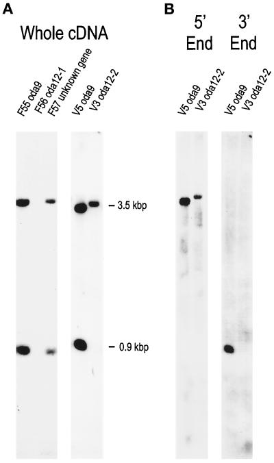
There are eight outer dynein arm LCs. cDNA clones encoding each of these have been isolated via protein sequence, but to date only one LC gene, FLA14, encoding LC8 (Pazour et al., 1998), has been disrupted. We were particularly interested in finding a mutation that affected LC2, the homologue of the mouse Tctex2 protein. In an attempt to identify such a mutant, we screened our entire collection of Chlamydomonas insertional mutants by Southern blotting using the LC2 cDNA as a probe. Chlamydomonas contains a single copy of the LC2 gene, which is cut once by PvuII (Patel-king et al., 1997). Thus, the LC2 cDNA detects two bands (0.9 and 3.5 kbp) on Southern blots of wild-type genomic DNA cut with this enzyme (Figure 1A).
Figure 1.
Southern blot showing RFLPs in F56 and V3. (A) Genomic DNA was isolated from slow-swimming insertional mutants, cut with PvuII, and analyzed by Southern blotting using the LC2 cDNA as a probe. This probe detects 0.9- and 3.5-kbp bands in most cell lines, including V5 and F55 (both containing a deletion of the ODA9 gene) and F57 (containing a disruption of an unidentified gene). However, both of these bands are missing in the F56 strain, and the lower band is missing in the V3 strain. (B) A probe (5′ End) specific for the 5′ UTR and entire coding region of the LC2 gene (see text) hybridizes to the upper band in DNA from both V5 and V3. A probe (3′ End) specific for the 3′ UTR of the LC2 gene hybridizes to the lower band in DNA from V5 but detects no band in DNA from V3, indicating that the deletion in V3 is at the 3′ end of the gene.
One strain, F56, was missing both hybridizing bands (Figure 1A), indicating that the gene encoding LC2 is completely deleted in this strain. Previously, strain F56 had been briefly reported to contain a mutation that caused complete loss of the outer dynein arm and a slow-swimming phenotype (Koutoulis et al., 1997). This mutant complemented oda1–oda11, pf13, and pf22 in stable diploids, indicating that it was defective in a new ODA gene, which was termed ODA12 (Koutoulis et al., 1997; see below). The product of this gene was not identified, because no RFLPs were observed in Southern blots of F56 DNA probed with cDNAs encoding outer arm dynein HCs or ICs (Koutoulis et al., 1997). We similarly did not detect RFLPs in Southern blots of F56 DNA probed with cDNAs encoding ODA-DC polypeptides or LCs other than LC2 (Pazour and Witman, unpublished data). Therefore, F56 does not appear to have a defect in any outer dynein arm-associated protein other than LC2.
Another cell line, V3, was missing the smaller of the two hybridizing bands (Figure 1A). V3 also had a slow-swimming phenotype (see below). To determine which part of the gene is deleted in V3, probes specific to each end of the LC2 cDNA were made by PCR amplification of the LC2 cDNA with T3 and T7 primers, digesting the product with PvuII, and gel-purifying the resulting 0.6- and 0.4-kbp bands. PvuII cuts the LC2 cDNA at only one site, between G614 and C615 in the ∼550-bp 3′ untranslated region (UTR) (see Patel-King et al., 1997, their Figure 1). Therefore, the 0.6-kbp probe corresponds to the 5′ UTR, the entire coding sequence, and a small amount (∼60 bp) of the 3′ UTR; the 0.4-kbp probe corresponds to the remainder of the 3′ UTR. The 5′ probe detected the larger band in DNA from V3 cells and cells that were wild-type for LC2 (Figure 1B). The 3′ probe detected the smaller band in DNA from cells that were wild-type for LC2 but did not hybridize with any band in DNA from the V3 cells (Figure 1B). This indicates that the 3′ end of the gene is deleted in V3. Because the promoter and amino-terminal coding region are retained, these cells have the potential to produce at least a portion of LC2.
The LC2 Locus Is Tightly Linked to ODA12
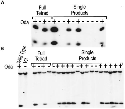
To determine whether the slow-swimming phenotype was linked to the disruption of LC2, the F56 and V3 lines were back-crossed to wild-type cells. The swimming defect appears to be caused by a single nuclear mutation as the Oda phenotype segregated 2:2 in 25 tetrads obtained from a back-cross of F56. DNA was isolated from 15 offspring of the F56 cross and 19 offspring of the V3 cross and examined by Southern blotting using the LC2 cDNA as a probe. Figure 2 shows that whenever the LC2 gene was disrupted, the cells had an Oda-swimming phenotype. This strongly suggests that the ODA12 gene encodes LC2 of the outer dynein arm. The total deletion allele has been designated oda12-1; the partial deletion allele has been designated oda12-2.
Figure 2.
The motility defect in the F56 and V3 strains segregates with the defect in the LC2 gene. (A) The initial F56 isolate was crossed to 137c, and a slow-swimming offspring was isolated and crossed again to 137c. Twenty-five tetrads were dissected from this cross and scored for motility. The Oda phenotype segregated 2:2. DNA was isolated from the four products of one full tetrad and a single product of 11 additional tetrads, cut with PstI, and analyzed by Southern blotting using the LC2 cDNA as the probe. In every case, the Oda− phenotype segregated with the deletion revealed by the LC2 cDNA, indicating that the motility defect is tightly linked to this deletion mutation. (B) V3 was crossed to H8− (Wild Type), and the resultant tetrads were dissected. DNA was isolated from the parental strains, the four products of one tetrad, and a single product of 15 additional tetrads, cut with PvuII, and analyzed by Southern blotting with the LC2 cDNA as the probe. Again, the Oda− phenotype segregated with the deletion revealed by the LC2 cDNA.
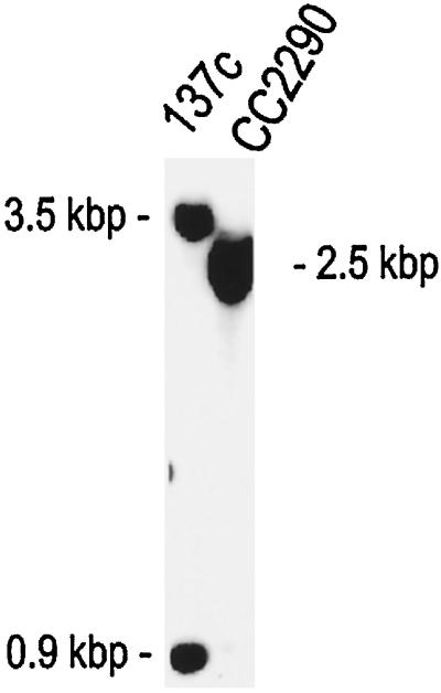
The discovery that the LC2 locus is tightly linked and probably identical to ODA12 provided an opportunity to obtain independent proof that ODA12 is a novel gene. CC2290 is a wild-type C. reinhardtii isolate that is highly interfertile with 137c-derived lab strains but divergent enough that it is easy to identify RFLPs between the two strains (Gross et al., 1988). We identified an RFLP detected by the LC2 cDNA probe in DNA of CC2290 versus the 137c-derived strains (Figure 3). We then compared the segregation of this RFLP with the segregation of uncloned genes that affect the outer arm in crosses between CC2290 and cell lines (derived from 137c) carrying mutations in the latter genes. Uncloned genes affecting the outer arm include ODA5, ODA7, ODA8, PF13, and PF22 (ODA1 [Takada et al., 1996] and ODA10 [Pazour, Koutoulis, and Witman, unpublished data] have been cloned but not yet published). Tetrads from the crosses were dissected, the progeny were scored for motility, and DNA was isolated from one product of 10 different tetrads. The segregation of the LC2 PvuII RFLP was scored by Southern blotting and compared with the segregation of the motility defect. If the oda12 mutation is an allele of (or is tightly linked to) one of these genes, all offspring that have motility defects will have the 137c version of the LC2 PvuII RFLP. However, if oda12 is not an allele of one of these genes, the motility defect will segregate independently from the LC2 PvuII RFLP. As can be seen in Table 2, the LC2 PvuII RFLP segregated independently of oda5, oda7, oda8, pf13, and pf22, confirming that ODA12 is a novel gene.
Figure 3.
LC2 cDNA detects an RFLP between 137c and CC2290 strains. DNA from the lab strain 137c and the wild isolate CC2290 was cut with PvuII and analyzed by Southern blotting using the LC2 cDNA as a probe. This probe detects two bands of ∼3.5 and ∼0.9 kbp in 137c but only a single band of ∼2.5 kbp in CC2290.
Table 2.
Segregation of the LC2 RFLP
| Crossa | Resultsb |
|---|---|
| CC2290 (mt−, ODA5, ODA12G) × CC2236 (mt+, oda5, ODA12R | 6P:4NP |
| CC2290 (mt−, ODA7, ODA12G) × CC2240 (mt+, oda7, ODA12R) | 4P:6NP |
| CC2290 (mt−, ODA8, ODA12G) × CC2242 (mt+, oda8, ODA12R) | 5P:5NP |
| CC2290 (mt−, PF13, ODA12G) × CC2492 (mt+, pf13a, ODA12R) | 5P:5NP |
| CC2290 (mt−, PF22, ODA12G) × CC1382 (mt+, pf22, ODA12R) | 8P:2NP |
Genotypes are shown in parentheses. ODA12G and ODA12R are the CC2290 and 137c versions of the PvuII RFLP detected by the LC2 cDNA clone.
P, parental; NP, nonparental.
Phenotype of oda12 Cells
Most of the known defects in ODA1–ODA10 cause loss of the outer dynein arms. As a result, the cells swim in a jerky manner at ∼30% of the normal speed. These mutant cells also have an altered photoshock response. During photoshock, which is induced by a flash of bright light, wild-type cells stop swimming, switch to a flagellar waveform, and swim backward for a few milliseconds (Schmidt and Eckert, 1976). In contrast, oda mutants stop in response to the flash but do not swim backward (Mitchell and Rosenbaum, 1985).
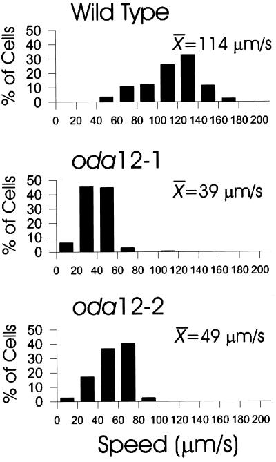
oda12-1 cells swim in a typical oda-like manner. They are slower than normal and have a jerky appearance as they swim forward. The swimming pattern of oda12-2 cells is not as slow or jerky as that of oda12-1 or other oda mutant cells. Swimming speeds were quantitatively measured with an ExpertVision Motion Analysis system (Figure 4). The swimming speeds of wild-type (g1) cells are broadly distributed with a mean of 114 μm/s. The mean swimming speed of oda12-1 cells is reduced to 39 μm/s, whereas the speed of oda12-2 cells is slightly faster at 49 μm/s. Both oda12-1 and oda12-2 cells show an altered photoshock response; when flashed with light, they stop briefly and then resume forward swimming without a period of backward swimming.
Figure 4.
Swimming speed of wild-type and oda12 mutant cells. The Speed Operator of the ExpertVision Motion Analysis system was used to calculate the swimming speed of individual cells. The histograms show the distribution of speeds within each sample. These are plotted as the percentage of cells in each of 10 20-μm/s bins between 0 and 200 μm/s. The mean swimming speed (in micrometers per second) for each sample also is shown. Note that the mean swimming speed of oda12-2 cells (49 μm/s) is slightly faster than that of oda12-1 cells (39 μm/s). More than 100 cells were analyzed for each cell type.
LC2 Is Required for Assembly of the Outer Dynein Arm
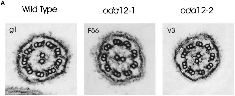
Previously, we briefly reported that the oda12-1 mutant was lacking the outer dynein arms (Koutoulis et al., 1997), a defect consistent with the swimming phenotype described above. Electron microscopic analysis indicates that whereas oda12-1 cells lack all of the outer dynein arms, oda12-2 cells are much more variable (Figure 5A). Some flagellar cross-sections have no outer dynein arms, whereas others have significant numbers. This indicates that the partial deletion does not completely abolish the assembly of outer arms.
Figure 5.
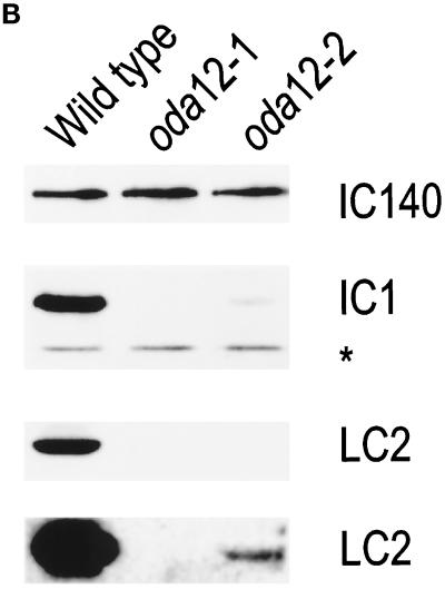
LC2 is required for assembly of outer dynein arms. (A) Electron micrographs of cross-sections of flagella of wild-type g1 cells show prominent outer dynein arms (arrows) on all of the doublet microtubules except for doublet one (arrowhead), which does not normally have an outer dynein arm (Hoops and Witman, 1983). In contrast, the outer dynein arms are completely missing in flagella of the oda12-1 mutant (F56). The number of outer dynein arms in flagella of oda12-2 cells (V3) is variable; in this section outer dynein arms (arrows) are seen on only three doublet microtubules. (B) Axonemes from wild-type and the two oda12 mutants were separated by SDS-PAGE, transferred to a PVDF membrane, and analyzed by Western blotting. The blots were probed with antisera to IC140 (an inner arm IC), IC1 (an outer arm IC), and LC2. The anti-IC140 antisera reacted equivalently with a 140-kDa band in all three lanes, indicating that each lane was loaded with an equal number of axonemes. Neither the anti-LC2 antibody nor the anti-IC1 antibody recognized bands of the appropriate size in the oda12-1 axonemes, confirming that this mutant is completely lacking the outer dynein arm. However, these two antibodies did reveal a small amount of IC1 and LC2 in the oda12-2 axonemes. The antibody to IC1 also reacts with a smaller band (*) (Fowkes and Mitchell, 1998) that is not part of the outer arm and is not affected by the oda12 mutation. The bottom panel shows a longer exposure of the blot probed with the LC2 antibody to demonstrate the small amount of LC2 in the oda12-2 axonemes.
Western blot analysis was used to more quantitatively assess the loss of outer dynein arms in oda12-1 and oda12-2 cells. Axonemes were isolated from wild-type cells and from each of the mutants. Equal amounts of protein (standardized to tubulin) were separated by SDS-PAGE and transferred to a PVDF membrane. The blot was first probed with an antibody to an inner dynein arm subunit (IC140) that should not be affected by the oda12 mutations. All three lanes had similar amounts of this antigen, confirming that equal numbers of axonemes had been loaded in each lane on the gel (Figure 5B). The blot was then probed with antibodies to outer dynein arm subunits IC1 and LC2 (Figure 5B). Both proteins were readily detected in axonemes of wild-type cells. In contrast, neither protein was detected in axonemes of oda12-1 cells, confirming that LC2 and the outer dynein arm is completely missing in this mutant. However, small amounts of IC1 and LC2 were detected in oda12-2 cells; the oda12-2 LC2 appeared to migrate with the same relative mobility as the wild-type LC2. Therefore, oda12-2 cells produce small amounts of apparently full-length LC2. This is sufficient to permit assembly of some outer arms, although the loss of outer arms appears to be more severe than indicated by electron microscopy.
The oda12 Phenotype Can Be Rescued by Transformation with DNA Encoding LC2
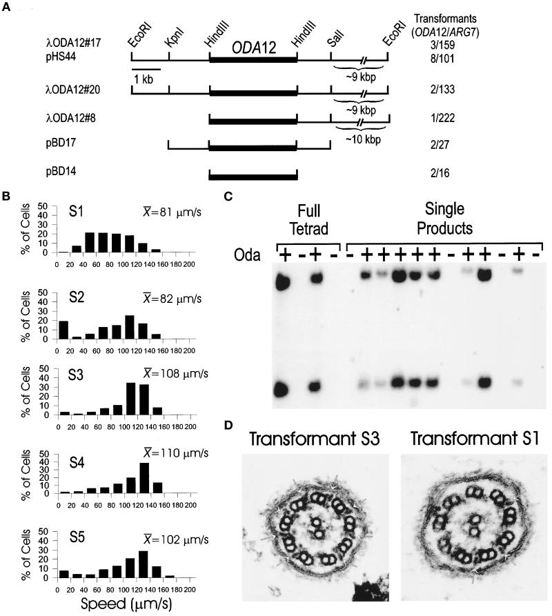
To determine whether the oda12 phenotype is due specifically to loss of LC2, we assayed the ability of the LC2 gene to restore the wild-type phenotype to oda12 mutant cells. Three genomic clones encoding LC2 were isolated from a λ phage library using the LC2 cDNA clone as a probe. Two of these, λODA12#17 and λODA12#20, had very similar restriction patterns. The third, λODA12#8, was shifted along the chromosome slightly from the first two (Figure 6A). The LC2 coding region (as determined by hybridization) is contained completely within a 3.1-kbp HindIII fragment common to all three clones (Figure 6A, dark bar). DNA from each of these three λ phage was transformed into the cell line 2567.1 (which carries oda12-1 and arg7 mutations), along with DNA containing the ARG7 gene. Individual ARG7 transformants were screened by light microscopy to determine whether the oda12 motility phenotype had been rescued as a result of cotransformation with the phage DNA. All three phage clones were able to rescue the defect (Figure 6A), indicating that each carried the complete ODA12 gene. The insert from λODA12#17 was subcloned into a plasmid vector and named pHS44; as expected, this also was able to rescue the motility defect. The locus was further refined by subcloning the 5.7-kbp KpnI-SalI and the 3.1-kbp HindIII fragments, which were termed pBD17 and pBD14, respectively. Both of these fragments complemented the oda12 defect (Figure 6A), indicating that ODA12 is located within the 3.1-kbp fragment that also contained the LC2 coding region as determined by hybridization. This data provide very strong evidence that the outer dynein arm defect of oda12 is the result of the LC2 deletion.
Figure 6.
Rescue of oda12 by transformation. (A) Map of the ODA12 locus. Three genomic λ phage clones (λODA12#8, λODA12#17, and λODA12#20) were isolated and mapped. The EcoRI sites at the ends of the clones are derived from the vector. The LC2 coding region (dark bar) was determined by hybridization with the cDNA clone. The 16-kbp insert from λODA12#17 was cloned into pKS+ and named pHS44. The 5.7-kbp KpnI–SalI and 3.1-kbp HindIII fragments of pHS44 were subcloned and named pBD17 and pBD14. The number of cell lines in which the Oda− phenotype was rescued out of the total number of ARG7 transformants screened is shown on the right. (B) Swimming speeds of transformed cell lines. The Speed Operator of the ExpertVision Motion Analysis system was used to calculate the swimming speed of five transformed lines (S1–S5). The histograms show the distribution of speeds within each sample. These are plotted as a percentage ofcells in each of 10 20-μm/s bins between 0 and 200 μm/s. The mean swimming speed (micrometers per second) for each sample also is shown. More than 100 cells were analyzed for each cell line. (C) Segregation of the exogenous copy of ODA12. oda12-1, arg7 strains were transformed with genomic clones of the ODA12 gene. One transformant (S20) that showed wild-type swimming speeds was mated to an oda12-1 strain of the opposite mating type, and tetrads were dissected. The offspring were scored for motility, and DNA was isolated from them. The DNA was cut with PvuII and analyzed by Southern blotting using the LC2 cDNA as a probe. The probe hybridizes with two bands in DNA from wild-type cells but does not hybridize with any band in DNA from oda12-1 cells (see Figure 1). Analysis of one full tetrad and a single product from each of 12 additional tetrads showed that fast-swimming cells (Oda+) had the LC2-hybridizing bands, whereas the slow-swimming cells (Oda−) were missing these bands. (D) Electron microscopy of flagella of transformed cells. Transformant S3, which swims at a speed similar to that of wild-type (cf. Figure 4 and B), has a full complement of outer dynein arms (left panel, arrows). In contrast, cells of transformed strain S1, which have a wide range of swimming speeds (Figure 6B), have variable numbers of outer dynein arms (right panel, arrows).
The swimming speed of five independent transformants was measured. All swam faster than the oda12-1 mutant line and had recovered the ability to swim backward when photoshocked. Transformants S3 (from λODA12#20), S4 (from λODA12#20), and S5 (from λODA12#8) were similar to wild-type in both mean swimming speed and the distribution of swimming speeds (cf. Figures 4 and 6B). Transformant S2 (from λODA12#17) was similar to wild-type except for a greater number of very slow cells that reduced its mean speed to 82 μm/s (Figure 6B). The swimming speeds of transformant S1 (from λODA12#17) were very broadly distributed around a mean of 81 μm/s (Figure 6B). The mixed distribution of swimming speeds seen in the S1 and S2 populations was not the result of an impure culture, because the cells were cloned from a single cell before analysis. More likely, it was the result of gene silencing, whereby newly integrated genes in some Chlamydomonas cells become transcriptionally inactivated (Cerutti et al. 1997).
To confirm that the rescue of the Oda− phenotype was due to integration of the cloned DNA and not suppression of the mutant phenotype, transformant S20 (obtained by transformation of 2507.1 with pHS44), which has wild-type motility, was crossed to an oda12-1 line and the resultant tetrads dissected. Motility of the offspring was scored, and DNA was isolated and analyzed by Southern blotting using the LC2 cDNA as a probe (Figure 6C). This probe revealed that the two PvuII restriction fragments observed in wild-type cells, but missing in the oda12-1 line, were restored in the rescued cell lines. Analysis of one full tetrad and single products of 12 additional tetrads showed that whenever the two PvuII bands were present, the cells swam with wild-type speeds. This indicates that restoration of the phenotype is due to the cloned DNA.
Transformants S1 and S3 were examined by electron microscopy to determine whether the outer dynein arms had been restored (Figure 6D). Transformant S3, which had a distribution of swimming speeds similar to that of wild type, had a full complement of outer dynein arms. In contrast, cells of the transformant S1, which had a broader distribution of swimming speed and a lower mean swimming speed, showed a corresponding variability in the restoration of arms. Most of the flagellar cross-sections showed intermediate numbers of arms, but in a few, no arms were present, suggesting that the intermediate swimming speed was due to restoration of only some of the outer dynein arms.
DISCUSSION
In this report we describe, for the first time, mutations in the C. reinhardtii gene encoding LC2, an LC that is specific for the outer dynein arm. Deletion of this gene leads to loss of the outer dynein arm and impaired flagellar motility. The LC2 gene is the Chlamydomonas homologue of mouse Tctex2 (Patel-King et al., 1997), which has been identified as one of the distorter genes of the t haplotype (Huw et al., 1995). The finding that LC2 is essential for outer arm assembly supports the hypothesis that some of the phenotypes expressed by t haplotypes are due to interactions between defective dynein polypeptides and suggests a specific model by which this might occur.
Function of LC2
Although mutations are known for each of the HCs and ICs of the Chlamydomonas outer dynein arm, previously a mutation has been identified in only one gene (FLA14) that encodes an outer arm LC, LC8 (Pazour et al., 1998). However, LC8 also is a component of inner arm dynein, cytoplasmic dynein, and myosin (Espindola et al., 1996; King et al., 1996a; Harrison et al., 1998), and knockout of the LC8 gene has pleiotropic effects that are difficult to interpret in terms of the specific role of LC8 in the outer dynein arm. In contrast, LC2 appears to be specific for the outer arm. Complete deletion of the LC2 gene causes a complete loss of the outer dynein arm, with no other structural changes in the flagellar axoneme. Loss of the outer arms in this mutant causes a decrease in swimming speed to ∼34% of that of wild-type cells, a reduction similar to what has been observed for other mutations that cause complete loss of the outer dynein arm (Mitchell and Rosenbaum, 1985; Kamiya, 1988). Loss of the outer arm also causes loss of the ability to swim backward during the photoshock response, as reported previously for a mutant with a defect in the outer arm (Mitchell and Rosenbaum, 1985).
It is most remarkable that LC2, which has a mass of only 15.8 kDa (King and Patel-King, 1995a), is absolutely essential for assembly of the outer dynein arm, which contains at least 13 different polypeptides and has a total mass of ∼2 MDa (Table 1; Witman et al., 1983). Two independent lines of evidence indicate that LC2 is a component of the IC–LC complex (Mitchell and Rosenbaum, 1986; King and Witman, 1990; Witman et al., 1992), which is located at the base of the dynein (King and Witman, 1990). First, LC2 is associated with the β subunit of the outer dynein arm (Pfister and Witman, 1984) and is retained in the Chlamydomonas mutant oda4-s7, which expresses only the NH2-terminal one-third of the β HC (Sakakibara et al., 1993). This portion of the β HC forms the stem and base of the outer arm dynein (Witman et al., 1994) and binds the IC–LC complex (King and Witman, 1989). Second, after dissociation of the outer arm dynein with nonionic detergent, LC2 was immunoprecipitated as part of an aggregate with IC1, IC2, and another LC (Mitchell and Rosenbaum, 1986). Therefore, by virtue of its location in the arm, LC2 is in a position to interact directly with tubulin or the ODA-DC and might be required for binding a preassembled outer dynein arm to the doublet microtubule. Alternatively, LC2 might be required for preassembly of the outer dynein arm in the cytoplasm (Fowkes and Mitchell, 1998), for stability of the preassembled complex, or even for transport of the preassembled complex into the flagellum. Whatever the precise reason, the current findings demonstrate that LC2 has an important role in dynein assembly. It is likely that the LCs of other dyneins have equally important roles in the complexes of which they are a part.
LC2 is Encoded by ODA12
We previously identified a novel Chlamydomonas gene, ODA12, that was necessary for outer arm assembly (Koutoulis et al., 1997); however, the product of this gene was not determined. The following evidence now indicates that ODA12 encodes LC2: 1) Southern hybridization showed that the gene encoding LC2 is completely deleted in the original oda12 mutant strain; 2) the defining phenotype of oda12 cells segregated with the LC2 deletion in crosses between oda12 and wild-type cells; and 3) the oda12 phenotype was rescued by transformation of oda12 cells with a small (3.1-kbp) genomic DNA clone containing the LC2 gene. Thus, the phenotype observed for oda12 cells is due to the deletion of the LC2 gene.
The current findings also confirm that ODA12 is a novel gene, distinct from any of the other ODA or similar genes that have been reported. Previously, we reported that cDNA clones encoding ODA1, ODA2, ODA3, ODA4, ODA6, ODA9, and ODA11 detected no RFLPs in oda12 versus wild-type strains, and that oda12 complemented oda1, oda3, oda5, oda7, oda8, oda10, pf13, and pf22 in stable diploids (Koutoulis et al., 1997). Similarly, we determined that cDNA clones encoding the unpublished genes ODA13 and ODA14 (Table 1) did not detect RFLPs in oda12 versus wild-type strains (Pazour and Witman, unpublished results). We now have used the LC2 cDNA as a physical marker for the ODA12 gene and find that an RFLP, detected by the cDNA in CC2290 versus 137c-derived strains, segregates independently of oda5, oda7, oda8, pf13, and pf22. These results provide additional evidence that ODA12 cannot be the same as any of those genes.
With the discovery that the ODA12 gene product is an outer dynein arm LC, only four known ODA genes (ODA5, ODA7, ODA8, and ODA10) and two known PF genes (PF13 and PF22) that affect outer arm assembly have yet to have their products identified. It will be of great interest to determine the products of these genes and their roles in outer arm assembly. It may not be coincidental that the products of six ODA or ODA-like genes are still unknown, whereas mutations have not yet been reported for an identical number of outer dynein arm LCs (Table 1). The current findings underscore the possibility that some of these other LCs also could be critical for dynein assembly. The ease with which a reverse genetics approach may be applied to Chlamydomonas should greatly facilitate the identification of mutants with defects in these polypeptides.
oda12-2 Is a Hypomorphic Allele
The results presented here show that the oda12 mutation originally reported by Koutoulis et al. (1997) is a complete deletion of the LC2 gene; this allele has been designated oda12-1. Here we also describe a second allele (oda12-2) in which only the 3′ end of the LC2 gene is deleted. Ultrastructural analysis revealed that the oda12-2 mutant assembles only a few outer arms on its doublet microtubules (Figure 5A). Presumably because some outer arms are present, oda12-2 cells swim slightly faster than oda12-1 cells (Figure 4A). Western blot analysis confirmed that the outer dynein arm polypeptide IC1 is greatly diminished but not entirely missing in axonemes of oda12-2 (Figure 5B). Interestingly, a small amount of approximately full-length LC2 also is present in the oda12-2 axoneme. That an apparently full-length product is synthesized suggests that the oda12-2 deletion removes most or all of the 3′ UTR but little if any of the coding sequence. The 3′ UTR of eukaryotic mRNAs has several important functions, including control of mRNA stability (Beelman and Parker, 1995; Decker and Parker, 1995; Wickens et al., 1997) and mRNA localization (Wilhelm and Vale, 1993; Decker and Parker, 1995). A defect in either of these functions likely would result in the production of reduced amounts of LC2, which in turn would permit assembly of reduced numbers of outer arms. The incorporation of small amounts of LC2 along with equally small amounts of IC1 into the oda12-2 axonemes is consistent with LC2 being absolutely essential for outer dynein arm assembly.
Dynein Defects and t Haplotype Phenotypes
The mouse t haplotype is an ∼30-Mb region of chromosome 17 containing numerous genes and defined by the presence of four inversions relative to the wild-type homologue, so that recombination is suppressed and the entire region usually segregates as a single unit (Olds-Clarke and Johnson, 1993; Silver, 1993). One of the most intriguing phenotypes of the t haplotype is that heterozygous +/t males transmit the t haplotype-bearing version of chromosome 17 to >95% of their progeny. Analysis of partial t haplotypes resulting from rare recombination events within the t complex indicate that this transmission ratio distortion, or meiotic drive, is the result of two or three distorter genes, located in different parts of the t haplotype, acting on a single responder gene, termed Tcr (t complex responder) (Lyon, 1984). The distorter genes can act cis or trans on the responder gene, and their effects are additive. The result of these interactions is to increase the probability of transmission of the chromosome carrying the t haplotype responder gene.
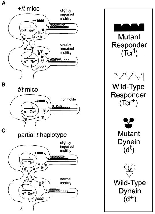
The discovery that some of the candidate distorter genes encode dynein LCs has prompted a model that explains the t haplotype phenotype on the basis of interactions between dyneins and other axonemal components (Patel-King et al., 1997; Harrison et al., 1998). The current finding that LC2 is important for dynein assembly permits the model to be described in more specific terms (Figure 7). In this model, the responder gene encodes a protein (Tcr) that is expressed in the haploid nucleus and remains associated with that nucleus throughout the remainder of spermiogenesis. Because soluble components can diffuse through the intercellular bridges that connect developing spermatids, this protein must become rapidly associated with some structural component of the spermatid, such as the basal body or the elongating axoneme, to prevent its diffusion to neighboring spermatids. Ultimately, Tcr must interact with the dynein arms. For the sake of simplicity, we will assume that Tcr is part of an axonemal component such as the ODA-DC (Takada and Kamiya, 1994; Koutoulis et al., 1997) or the dynein regulatory complex (Gardner et al., 1994; Piperno et al., 1994) that interacts directly with the dynein arms, but it could interact with the arms indirectly, as through a basal body-associated templating activity that determines the location of the outer dynein arm binding sites on the doublet microtubule. In contrast, the distorter proteins, which include subunits of the inner and outer dynein arms, diffuse through the intercellular bridges, so that both wild-type and mutant (t haplotype-encoded) dyneins are preassembled in the cytoplasm of both spermatids bearing the wild-type responder (Tcr+) and spermatids bearing the t haplotype-encoded responder (Tcrt).
Figure 7.
Model for mouse t phenotypes based on dynein–axoneme interactions. In all cases, the distorter gene (d) products, which are dynein subunits, are free to diffuse (arrows) through the intercellular bridges that connect developing spermatids. However, the responder gene (Tcr) product quickly assembles onto the axoneme and thus remains associated with the nucleus that encoded it. Wild-type and t-encoded proteins are indicated by open and filled symbols, respectively. t-encoded dyneins contain mutations that affect their assembly and/or function; the t-encoded responder contains mutations that affect its ability to bind t-encoded dyneins. (A) In developing spermatids of heterozygous +/t mice, the mutated t responder binds only wild-type dyneins, preventing incorporation of mutant dyneins into the axoneme associated with the nucleus carrying the t haplotype. In contrast, the wild-type responder binds both wild-type and mutant dyneins; the mutant dyneins “poison” the axoneme into which they are incorporated. As a result, the sperm bearing the t haplotype has slightly impaired motility (because of the presence of the mutant responder), but the sperm with the wild-type homologue has greatly impaired motility. The sperm bearing the t haplotype is much more likely to reach and fertilize an egg, thus leading to transmission ratio distortion. (B) In spermatids of homozygous t/t mice, all dyneins produced are defective and bind poorly to the t-encoded responder. The resulting sperm are nonmotile, and the mice are sterile. (In this case, all spermatids are identical, so only one is shown.) (C) In mice containing a partial t haplotype with a t-encoded responder, but no t-encoded distorters, only wild-type dyneins are produced and assembled into the axonemes. Sperm carrying Tcrt and sperm carrying Tcr+ are both motile, although the former will suffer a competitive disadvantage because of the presence of the t-encoded responder in their axonemes.
The model predicts that Tcrt binds wild-type dynein strongly but mutant dynein less efficiently. In +/t mice (Figure 7A), both types of dyneins will be present in the cytoplasm of the developing spermatid. The wild-type dyneins will out-compete the mutant dyneins for binding sites on the Tcrt-containing axoneme, with the result that these axonemes will assemble a nearly normal complement of arms and have motility that is only slightly impaired because of the presence of Tcrt (see below). In contrast, the model predicts that Tcr+ binds both wild-type and mutant dyneins equally, with the result that both types of dyneins will be assembled into the Tcr+-containing axonemes. Because of the presence of many mutant dyneins in these latter axonemes, the resulting sperm will be much less efficient than those containing Tcrt in some critical aspect of motility, resulting in transmission ratio distortion.
A second phenotype of the t haplotype that is readily accounted for by this model is that t/t males are sterile (Figure 7B). t/t males will produce only mutant dynein, which has a low affinity for Tcrt. As a result, few arms will be assembled into the axoneme, and if they are assembled they will be defective. The resulting sperm will have aberrant or no motility and be unable to fertilize an egg.
In an analysis of partial t haplotypes, it was observed that if a t-encoded responder was present on one chromosome, with no t-encoded distorters acting on it cis or trans, then that chromosome was transmitted at a low ratio (11–17%) (Lyon, 1984). This suggests that the mutant responder itself impairs the motility of Tcrt-bearing sperm. This also is compatible with our model (Figure 7C). In spermatids of such mice, only wild-type dynein is present, and it binds to both Tcrt- and Tcr+-containing axonemes. The resulting Tcrt-containing sperm will have slightly impaired motility as a result of having Tcrt in their axonemes. This motility will not be so compromised that the sperm are unable to fertilize an egg and not as defective as that of Tcr+-containing spermatids with a mixture of mutant and wild-type dyneins (see Figure 7A). Nevertheless, they will have a competitive disadvantage, compared with Tcr+-containing sperm with only wild-type dyneins, in reaching and fertilizing the egg.
The sperm motility phenotypes predicted by this model are in good agreement with observations of sperm from +/t and t/t mice (summarized by Harrison et al., 1998). For example, sperm from t/t mice rarely exhibit a regular beat pattern and have little progressive motility, whereas the mean swimming speed of sperm from +/t mice is intermediate between that of t/t and +/+ mice (Olds-Clarke and Johnson, 1993). Moreover, sperm from +/t mice separate into two subpopulations, one having nearly normal motility and the other having abnormal motility (Olds-Clarke and Johnson, 1993); the former may correspond to Tcrt-containing sperm, and the latter may correspond to Tcr+-containing sperm. Certainly these motility defects could be accounted for by defects in dynein arm assembly and/or function. As indicated by our analysis of the oda12-2 mutant compared with oda12-1 and wild-type cells, intermediate levels of outer dynein arms can result in intermediate swimming speeds. Furthermore, Chlamydomonas cells with defects in both the inner and outer arms have a more severe phenotype than those with a defect in either arm alone (Kamiya et al., 1989), which is consistent with the additive effects of the distorter genes in mice (Lyon, 1984). Finally, at least some of the motility defects observed in sperm of +/t mice are dependent on Ca2+ (Olds-Clarke and Johnson, 1993), and both inner and outer dynein arms are involved in Ca2+-mediated changes in flagellar waveform. For example, in Chlamydomonas, the inner arms are involved in phototactic steering (King and Dutcher, 1997), and mutants with defects in the outer arm are defective in backward swimming during photoshock (Mitchell and Rosenbaum, 1985; this report); both of these responses are mediated by Ca2+.
Conclusion
The above model should be readily testable by further molecular genetic and biochemical studies of t haplotype-bearing mice. Of particular importance will be identification of the products of the Tcr gene and of the remaining distorter genes. In any case, the findings presented here demonstrate for the first time that a defect in a dynein LC can have a profound effect on outer arm assembly. It is likely that the LCs have equally important roles in other dyneins. Because oda12-1 is a null mutant, it should now be possible to investigate LC function by in vitro mutagenesis of the ODA12 gene, followed by transformation of the modified gene into the oda12-1 mutant, where the modified gene product should be assembled into an outer dynein arm. The effects of the LC modification on flagellar movement and dynein activity in vitro could then be determined.
ACKNOWLEDGMENTS
We thank Dr. J. Aghajanian (Worcester Foundation for Biomedical Research, Shrewsbury, MA) for the electron microscopy presented in this study and Drs. P. Yang and W. Sale (Emory University, Atlanta, GA) for the gift of the anti-IC140 antibody. These studies were supported by grants from the National Institutes of Health (GM30626 to G.B.W. and GM51293 to S.M.K.) and the Campbell and Hall Charity Fund (to G.B.W.).
Abbreviations used:
- HC
heavy chain
- IC
intermediate chain
- LC
light chain
- ODA-DC
outer dynein arm docking complex
- PVDF
polyvinylidene difluoride
- RFLP
restriction fragment length polymorphism
- UTR
untranslated region
REFERENCES
- Beelman CA, Parker R. Degradation of mRNA in eukaryotes. Cell. 1995;81:179–183. doi: 10.1016/0092-8674(95)90326-7. [DOI] [PubMed] [Google Scholar]
- Benashski SE, Patel-King RS, King SM. Light chain 1 from the Chlamydomonas outer dynein arm is a leucine-rich repeat protein associated with the motor domain of the gamma heavy chain. Biochemistry. 1999;38:7253–7264. doi: 10.1021/bi990466y. [DOI] [PubMed] [Google Scholar]
- Bowman AB, Patel-King RS, Benashski SE, McCaffery M, Goldstein LSB, King SM. Drosophila roadblock and Chlamydomonas LC7: a conserved family of dynein-associated proteins involved in axonal transport, flagellar motility and mitosis. J Cell Biol. 1999;146:165–180. [PMC free article] [PubMed] [Google Scholar]
- Casey DM, Pazour GJ, Wilkerson CG, Inaba K, Koutoulis A, Takada S, Kamiya R, Witman GB. Identification and insertional mutagenesis of a new Chlamydomonas reinhardtii gene, oda 14, that encodes the 25-kd subunit of the outer dynein arm docking complex. Mol Biol Cell. 1998;9:155a. [Google Scholar]
- Cerutti H, Johnson AM, Gillham NW, Boynton JE. A eubacterial gene conferring spectinomycin resistance on Chlamydomonas reinhardtii: integration into the nuclear genome and gene expression. Genetics. 1997;145:97–110. doi: 10.1093/genetics/145.1.97. [DOI] [PMC free article] [PubMed] [Google Scholar]
- Debuchy R, Purton S, Rochaix J-D. The arginosuccinate lyase gene of Chlamydomonas reinhardtii: an important tool for nuclear transformation and for correlating the genetic and molecular maps of the ARG7 locus. EMBO J. 1989;8:2803–2809. doi: 10.1002/j.1460-2075.1989.tb08426.x. [DOI] [PMC free article] [PubMed] [Google Scholar]
- Decker CJ, Parker R. Diversity of cytoplasmic functions for the 3′ untranslated region of eukaryotic transcripts. Curr Opin Cell Biol. 1995;7:386–392. doi: 10.1016/0955-0674(95)80094-8. [DOI] [PubMed] [Google Scholar]
- Espindola FS, Cheney RE, King SM, Suter DM, Mooseker MS. Myosin-V and dynein share a similar light chain. Mol Biol Cell. 1996;7:372a. [Google Scholar]
- Ewulonu UK, Schimenti K, Kuemerle B, Magnuson T, Schimenti J. Targetted mutagenesis of a candidate t complex responder gene in Mouse t haplotypes does not eliminate transmission ratio distortion. Genetics. 1996;144:785–792. doi: 10.1093/genetics/144.2.785. [DOI] [PMC free article] [PubMed] [Google Scholar]
- Fernandez E, Schnell R, Ranum LPW, Hussey SC, Silflow CD, Lefebvre PA. Isolation and characterization of the nitrate reductase structural gene of Chlamydomonas reinhardtii. Proc Natl Acad Sci USA. 1989;86:6449–6453. doi: 10.1073/pnas.86.17.6449. [DOI] [PMC free article] [PubMed] [Google Scholar]
- Fowkes ME, Mitchell DR. The role of preassembled cytoplasmic complexes in assembly of flagellar dynein subunits. Mol Biol Cell. 1998;9:2337–2347. doi: 10.1091/mbc.9.9.2337. [DOI] [PMC free article] [PubMed] [Google Scholar]
- Gardner LC, O’Toole E, Perrone CA, Giddings T, Porter ME. Components of a “dynein regulatory complex” are located at the junction between the radial spokes and the dynein arms in Chlamydomonas flagella. J Cell Biol. 1994;127:1311–1325. doi: 10.1083/jcb.127.5.1311. [DOI] [PMC free article] [PubMed] [Google Scholar]
- Gross CH, Ranum LPW, Lefebvre PA. Extensive restriction fragment length polymorphisms in a new isolate of Chlamydomonas reinhardtii. Curr Genet. 1988;13:503–508. doi: 10.1007/BF02427756. [DOI] [PubMed] [Google Scholar]
- Harris EH. The Chlamydomonas Sourcebook. San Diego: Academic Press; 1989. [Google Scholar]
- Harrison A, Olds-Clarke P, King SM. Identification of the t complex-encoded cytoplasmic dynein light chain Tctex1 in inner arm I1 supports the involvement of flagellar dynein in meiotic drive. J Cell Biol. 1998;140:1137–1147. doi: 10.1083/jcb.140.5.1137. [DOI] [PMC free article] [PubMed] [Google Scholar]
- Holzbaur ELF, Vallee RB. DYNEINS: molecular structure and cellular function. Annu Rev Cell Biol. 1994;10:339–372. doi: 10.1146/annurev.cb.10.110194.002011. [DOI] [PubMed] [Google Scholar]
- Hoops HJ, Witman GB. Outer doublet heterogeneity reveals structural polarity related to beat direction in Chlamydomonas flagella. J Cell Biol. 1983;97:902–908. doi: 10.1083/jcb.97.3.902. [DOI] [PMC free article] [PubMed] [Google Scholar]
- Huang B, Piperno G, Luck DJL. Paralyzed flagella mutants of Chlamydomonas reinhardtii defective for axonemal doublet microtubule arms. J Biol Chem. 1979;254:3091–3099. [PubMed] [Google Scholar]
- Huw L-Y, Goldsborough AS, Willison K, Artzt K. Tctex2: a sperm tail surface protein mapping to the. t-complex. Dev Biol. 1995;170:183–194. doi: 10.1006/dbio.1995.1206. [DOI] [PubMed] [Google Scholar]
- Kamiya R. Mutations at twelve independent loci result in absence of outer dynein arms in Chlamydomonas reinhardtii. J Cell Biol. 1988;107:2253–2258. doi: 10.1083/jcb.107.6.2253. [DOI] [PMC free article] [PubMed] [Google Scholar]
- Kamiya R, Kurimoto E, Sakakibara H, Okagaki T. A genetic approach to the function of inner and outer arm dynein. In: Warner FD, Satir P, Gibbons LR, editors. Cell Movement, vol. I: The Dynein ATPases. New York: Alan R. Liss; 1989. pp. 209–218. [Google Scholar]
- Kindle KL. High-frequency nuclear transformation of Chlamydomonas reinhardtii. Proc Natl Acad Sci USA. 1990;87:1228–1232. doi: 10.1073/pnas.87.3.1228. [DOI] [PMC free article] [PubMed] [Google Scholar]
- King SJ, Dutcher SK. Phosphoregulation of an inner dynein arm complex in Chlamydomonas reinhardtii is altered in phototactic mutant strains. J Cell Biol. 1997;136:177–191. doi: 10.1083/jcb.136.1.177. [DOI] [PMC free article] [PubMed] [Google Scholar]
- King SM, Barbarese E, Dillman JF, III, Patel-King RS, Carson JH, Pfister KK. Brain cytoplasmic and flagellar outer arm dyneins share a highly conserved Mr 8,000 light chain. J Biol Chem. 1996a;271:19358–19366. doi: 10.1074/jbc.271.32.19358. [DOI] [PubMed] [Google Scholar]
- King SM, Dillman JF, III, Benashski SE, Lye RJ, Patel-King RS, Pfister KK. The mouse t-complex-encoded protein Tctex-1 is a light chain of brain cytoplasmic dynein. J Biol Chem. 1996b;271:32281–32287. doi: 10.1074/jbc.271.50.32281. [DOI] [PubMed] [Google Scholar]
- King SM, Otter T, Witman GB. Characterization of monoclonal antibodies against Chlamydomonas flagellar dyneins by high-resolution protein blotting. Proc Natl Acad Sci USA. 1985;82:4717–4721. doi: 10.1073/pnas.82.14.4717. [DOI] [PMC free article] [PubMed] [Google Scholar]
- King SM, Otter T, Witman GB. Purification and characterization of Chlamydomonas flagellar dyneins. Methods Enzymol. 1986;134:291–306. doi: 10.1016/0076-6879(86)34097-7. [DOI] [PubMed] [Google Scholar]
- King SM, Patel-King RS. Identification of a Ca2+-binding light chain within Chlamydomonas outer arm dynein. J Cell Sci. 1995a;108:3757–3764. doi: 10.1242/jcs.108.12.3757. [DOI] [PubMed] [Google Scholar]
- King SM, Patel-King RS. The Mr = 8,000 and 11,000 outer arm dynein light chains from Chlamydomonas flagella have cytoplasmic homologues. J Biol Chem. 1995b;270:11445–11452. doi: 10.1074/jbc.270.19.11445. [DOI] [PubMed] [Google Scholar]
- King SM, Patel-King RS, Wilkerson CG, Witman GB. The 78,000-Mr intermediate chain of Chlamydomonas outer arm dynein is a microtubule-binding protein. J Cell Biol. 1995;131:399–409. doi: 10.1083/jcb.131.2.399. [DOI] [PMC free article] [PubMed] [Google Scholar]
- King SM, Witman GB. Molecular structure of Chlamydomonas outer arm dynein. In: Warner FD, Satir P, Gibbons LR, editors. Cell Movement, vol. I: The Dynein ATPases. New York: Alan R. Liss; 1989. pp. 61–75. [Google Scholar]
- King SM, Witman GB. Localization of an intermediate chain of outer arm dynein by immunoelectron microscopy. J Biol Chem. 1990;265:19807–19811. [PubMed] [Google Scholar]
- Koutoulis A, Pazour GJ, Wilkerson CG, Inaba K, Sheng H, Takada S, Witman GB. The Chlamydomonas reinhardtii ODA3 gene encodes a protein of the outer dynein arm docking complex. J Cell Biol. 1997;137:1069–1080. doi: 10.1083/jcb.137.5.1069. [DOI] [PMC free article] [PubMed] [Google Scholar]
- Lader E, Ha H-S, O’Neill M, Artzt K, Bennett D. tctex-1: a candidate gene family for a mouse t complex sterility locus. Cell. 1989;58:969–979. doi: 10.1016/0092-8674(89)90948-3. [DOI] [PubMed] [Google Scholar]
- Levine RP, Ebersold WT. The genetics and cytology of Chlamydomonas. Annu Rev Microbiol. 1960;14:197–216. doi: 10.1146/annurev.mi.14.100160.001213. [DOI] [PubMed] [Google Scholar]
- Lyon M. Transmission ratio distortion in mouse t-haplotypes is due to multiple distorter genes acting on a responder locus. Cell. 1984;37:621–628. doi: 10.1016/0092-8674(84)90393-3. [DOI] [PubMed] [Google Scholar]
- Mitchell DR, Brown KS. Sequence analysis of the Chlamydomonas alpha and beta dynein heavy chain genes. J Cell Sci. 1994;107:635–644. doi: 10.1242/jcs.107.3.635. [DOI] [PubMed] [Google Scholar]
- Mitchell DR, Brown KS. Sequence analysis of the Chlamydomonas reinhardtii flagellar α dynein gene. Cell Motil Cytoskeleton. 1997;37:120–126. doi: 10.1002/(SICI)1097-0169(1997)37:2<120::AID-CM4>3.0.CO;2-C. [DOI] [PubMed] [Google Scholar]
- Mitchell DR, Kang Y. Reversion analysis of dynein intermediate chain function. J Cell Sci. 1993;105:1069–1078. doi: 10.1242/jcs.105.4.1069. [DOI] [PubMed] [Google Scholar]
- Mitchell DR, Rosenbaum JL. A motile Chlamydomonas flagellar mutant that lacks outer dynein arms. J Cell Biol. 1985;100:1228–1234. doi: 10.1083/jcb.100.4.1228. [DOI] [PMC free article] [PubMed] [Google Scholar]
- Mitchell DR, Rosenbaum JL. Protein-protein interactions in the 18S ATPase of Chlamydomonas outer dynein arms. Cell Motil Cytoskeleton. 1986;6:510–520. doi: 10.1002/cm.970060510. [DOI] [PubMed] [Google Scholar]
- Moss AG, Pazour GJ, Witman GB. Assay of Chlamydomonas phototaxis. Methods Cell Biol. 1995;47:281–287. doi: 10.1016/s0091-679x(08)60821-3. [DOI] [PubMed] [Google Scholar]
- Olds-Clarke P, Johnson LR. t haplotypes in the mouse compromise sperm flagellar function. Dev Biol. 1993;155:14–25. doi: 10.1006/dbio.1993.1002. [DOI] [PubMed] [Google Scholar]
- Otter T, King SM, Witman GB. A two-step procedure for efficient electrotransfer of both high-molecular (>400,000) and low-molecular-weight (<20,000) proteins. Anal Biochem. 1987;162:370–377. doi: 10.1016/0003-2697(87)90406-4. [DOI] [PubMed] [Google Scholar]
- Patel-King RS, Benashski SE, Harrison A, King SM. Two functional thioredoxins containing redox-sensitive vicinal dithiols from the Chlamydomonas outer dynein arm. J Biol Chem. 1996;271:6283–6291. doi: 10.1074/jbc.271.11.6283. [DOI] [PubMed] [Google Scholar]
- Patel-King RS, Benashski SE, Harrison A, King SM. A Chlamydomonas homologue of the putative murine t-complex distorter Tctex-2 is an outer arm dynein light chain. J Cell Biol. 1997;137:1081–1090. doi: 10.1083/jcb.137.5.1081. [DOI] [PMC free article] [PubMed] [Google Scholar]
- Pazour GJ, Dickert BL, Witman GB. The DHC1b (DHC2) isoform of cytoplasmic dynein is required for flagellar assembly. J Cell Biol. 1999;144:473–481. doi: 10.1083/jcb.144.3.473. [DOI] [PMC free article] [PubMed] [Google Scholar]
- Pazour GJ, Sineshchekov OA, Witman GB. Mutational analysis of the phototransduction pathway of Chlamydomonas reinhardtii. J Cell Biol. 1995;131:427–440. doi: 10.1083/jcb.131.2.427. [DOI] [PMC free article] [PubMed] [Google Scholar]
- Pazour GJ, Wilkerson CG, Witman GB. A dynein light chain is essential for the retrograde particle movement of intraflagellar transport (IFT) J Cell Biol. 1998;141:979–992. doi: 10.1083/jcb.141.4.979. [DOI] [PMC free article] [PubMed] [Google Scholar]
- Pfister KK, Witman GB. Subfractionation of Chlamydomonas 18 S dynein into two unique subunits containing ATPase activity. J Biol Chem. 1984;259:12072–12080. [PubMed] [Google Scholar]
- Piperno G, Mead K, LeDizet M, Moscatelli A. Mutations in the “dynein regulatory complex” alter the ATP-insensitive binding sites for inner arm dyneins in Chlamydomonas axonemes. J Cell Biol. 1994;125:1109–1117. doi: 10.1083/jcb.125.5.1109. [DOI] [PMC free article] [PubMed] [Google Scholar]
- Rosenbaum JL, Cole DG, Diener DR. Intraflagellar transport: the eyes have it. J Cell Biol. 1999;144:385–388. doi: 10.1083/jcb.144.3.385. [DOI] [PMC free article] [PubMed] [Google Scholar]
- Sager R, Granick S. Nutritional studies with Chlamydomonas reinhardi. Ann NY Acad Sci. 1953;56:831–838. doi: 10.1111/j.1749-6632.1953.tb30261.x. [DOI] [PubMed] [Google Scholar]
- Sakakibara H, Mitchell DR, Kamiya R. A Chlamydomonas outer arm dynein mutant missing the α heavy chain. J Cell Biol. 1991;113:615–622. doi: 10.1083/jcb.113.3.615. [DOI] [PMC free article] [PubMed] [Google Scholar]
- Sakakibara H, Takada S, King SM, Witman GB, Kamiya R. A Chlamydomonas outer arm dynein mutant with a truncated β heavy chain. J Cell Biol. 1993;122:653–661. doi: 10.1083/jcb.122.3.653. [DOI] [PMC free article] [PubMed] [Google Scholar]
- Sambrook J, Fritsch EF, Maniatis T. Molecular Cloning: A Laboratory Manual. Cold Spring Harbor, NY: Cold Spring Harbor Laboratory Press; 1987. [Google Scholar]
- Schmidt JA, Eckert R. Calcium couples flagellar reversal to photostimulation in Chlamydomonas reinhardtii. Nature. 1976;262:713–715. doi: 10.1038/262713a0. [DOI] [PubMed] [Google Scholar]
- Silver LM. Mouse t haplotypes. Annu Rev Genet. 1985;19:179–208. doi: 10.1146/annurev.ge.19.120185.001143. [DOI] [PubMed] [Google Scholar]
- Silver LM. The peculiar journey of a selfish chromosome: mouse t haplotypes and meiotic drive. Trends Genet. 1993;9:250–254. doi: 10.1016/0168-9525(93)90090-5. [DOI] [PubMed] [Google Scholar]
- Takada S, Kamiya R. Functional reconstitution of Chlamydomonas outer dynein arms from α-β and γ subunits: requirement of a third factor. J Cell Biol. 1994;126:737–745. doi: 10.1083/jcb.126.3.737. [DOI] [PMC free article] [PubMed] [Google Scholar]
- Takada S, Wilkerson CG, Kamiya R, Witman GB. The ODA1 gene encodes a 62-kDa component of a complex necessary for outer arm dynein docking on the doublet microtubules of Chlamydomonas flagella. Mol Biol Cell. 1996;7:568a. [Google Scholar]
- Tam L-W, Lefebvre PA. Cloning of flagellar genes in Chlamydomonas reinhardtii by DNA insertional mutagenesis. Genetics. 1993;135:375–384. doi: 10.1093/genetics/135.2.375. [DOI] [PMC free article] [PubMed] [Google Scholar]
- Vaughan KT, et al. Multiple mouse chromosomal loci for dynein-based motility. Genomics. 1996;36:29–38. doi: 10.1006/geno.1996.0422. [DOI] [PubMed] [Google Scholar]
- Wickens M, Anderson P, Jackson R J. Life and death in the cytoplasm: messages from the 3′ end. Curr Opin Genet Dev. 1997;7:220–232. doi: 10.1016/s0959-437x(97)80132-3. [DOI] [PubMed] [Google Scholar]
- Wilhelm JE, Vale RD. RNA on the move: the mRNA localization pathway. J Cell Biol. 1993;123:269–274. doi: 10.1083/jcb.123.2.269. [DOI] [PMC free article] [PubMed] [Google Scholar]
- Wilkerson CG, King SM, Koutoulis A, Pazour GJ, Witman GB. The 78,000 Mr intermediate chain of Chlamydomonas outer arm dynein is a WD-repeat protein required for arm assembly. J Cell Biol. 1995;129:169–178. doi: 10.1083/jcb.129.1.169. [DOI] [PMC free article] [PubMed] [Google Scholar]
- Wilkerson CG, King SM, Witman GB. Molecular analysis of the γ heavy chain of Chlamydomonas flagellar outer-arm dynein. J Cell Sci. 1994;107:497–506. doi: 10.1242/jcs.107.3.497. [DOI] [PubMed] [Google Scholar]
- Witman GB. Isolation of Chlamydomonas flagella and flagellar axonemes. Methods Enzymol. 1986;134:280–290. doi: 10.1016/0076-6879(86)34096-5. [DOI] [PubMed] [Google Scholar]
- Witman GB, Johnson KA, Pfister KK, Wall JS. Fine structure and molecular weight of the outer arm dyneins of. Chlamydomonas. J Submicrosc Cytol. 1983;15:193–197. [Google Scholar]
- Witman GB, King SM, Moss AG, Wilkerson C. The intermediate chain/light chain complex: an important structural entity of outer arm dynein. In: Baccetti B, editor. Comparative Spermatology 20 Years After. New York: Raven Press; 1992. pp. 439–443. [Google Scholar]
- Witman GB, Wilkerson CG, King SM. The biochemistry, genetics, and molecular biology of flagellar dynein. In: Hyams JS, Lloyd CW, editors. Microtubules. New York: Wiley-Liss; 1994. pp. 229–249. [Google Scholar]
- Yang P, Sale WS. The Mr 140,000 intermediate chain of Chlamydomonas flagellar inner arm dynein is a WD-repeat protein implicated in dynein arm anchoring. Mol Biol Cell. 1998;9:3335–3349. doi: 10.1091/mbc.9.12.3335. [DOI] [PMC free article] [PubMed] [Google Scholar]


