Abstract
The analysis of intact and underivatised lipids in body fluids as well as in cell and tissue extracts is of utmost importance in the field of early diagnosis. Therefore, fast, reliable, and automated analytical methods are needed to detect known as well as unknown species. The combination of solid phase extraction, high-performance liquid chromatography, mass spectrometry, and nuclear magnetic resonance spectroscopy are best at meeting this challenge. Herein, we show a workflow for the reliable analysis of individual components in phosphatidylethanolamine extracts. The limitations and advantages of the individual methods are discussed.
Keywords: lipids, phospholipids, high-performance liquid chromatography, mass spectrometry, nuclear magnetic resonance, hyphenation
Nuclear magnetic resonance (NMR) spectroscopy is arguably the most versatile analytical platform for complex mixture analysis. Specifically, interfacing liquid chromatography (LC) with parallel NMR and mass spectrometry (MS) (LC-NMR-MS) gives comprehensive structural data on metabolites of novel drugs. Recent innovations to improve NMR detection limits include CyroFlowProbes and on-line solid-phase extraction (LC-SPE-NMR). These state-of-the art analytical platforms are widely applicable to identify novel candidate drugs from diverse mixtures.1–3
Modern NMR provides an enormously powerful tool to probe molecular structure of organic compounds. The advantages are manifold: NMR is a nonselective, nondestructive detector of low molecular weight molecules in solution. NMR provides uniquely rich structural content: chemical shifts, spin-spin coupling, multiplicity, quantifications, intra- and intermolecular relationships, and a wide range of dynamic processes including molecular motions in solution, chemical exchange, and ligand binding.
In addition, modern time-of-flight (TOF) MS allows for the detection of extremely low concentrated analytes over a wide dynamic range. Initial structural information is obtained by MS/MS experiments. Ion suppression is still a problem in modern electrospray ionization MS (ESI-MS) applications, but it can be compensated by sample introduction via an high-performance liquid chromatography (HPLC). So the combined liquid LC/MS is now a standard technique for determining molecular masses and structures.
The analysis of underivatised lipids within body fluids as well as cell and tissue extracts is still a very challenging task. Anomalous lipid concentrations are associated with neoplastic and neurodegenerative diseases, diabetes, etc. Structural diversity of each lipid or lipid class will have a distinct effect on membrane properties (i.e., fluidity, permeability, oxygen scavenging, etc.).4–15 Therefore, the potential advantage of the combined use of HPLC, MS, and NMR spectroscopy should be explored. Many studies have explored the analysis of lipids, but to our knowledge a combinatorial approach has rarely been used.7,16
Gas chromatography,17–19 thin layer chromatography,20,21 and HPLC16,22–25 are well-established techniques for the challenge of lipid analysis. Gas chromatography-based techniques are quantitative but need time-consuming sample preparation. Due to chemical hydrolysis of the lipids, information about the fatty acid positional combination or isomerism is lost. The use of HPLC allows for the separation of lipid classes in the normal phase mode22–25; additionally, separation according to the different fatty acid composition of the individual lipids is possible in the reversed-phase mode.16 Because of their sensitivity, MS-based techniques are widely used.26–29 Furthermore they need only minor sample preparation.
In this study the ubiquitous membrane component phosphatidylethanolamine (PE)30 is analyzed. PE consists of the polar head group phosphoethanolamine attached to the sn-3 position of glycerol. The sn-1 and sn-2 positions are esterified with varying saturated and unsaturated (mostly sn-2 position) fatty acids. The corresponding plasmalogenes contain a long-chain alcohol etherified to position sn-1 and varying fatty acids in position sn-2. The molecular structure of every PE species was identified by characteristic fragmentation and additionally by their molecular formula, which was obtained by TOF-MS. HPLC was found to improve the selectivity, and extra resolution was obtained because analytes of low concentration were not suppressed during the ESI process. NMR spectroscopy allowed for measurement of intact biomaterials nondestructively without any preceding derivatisation. 1H- and 31P-NMR14,30–34 are well-established quantitative techniques for lipid analysis and need minimal sample preparation.
A combination of HPLC separation power, MS sensitivity with accurate mass measurement of molecular and fragment ions, and NMR structure elucidation power was found to meet most suitably the challenge of lipid analysis. Furthermore, the low NMR sensitivity can be compensated by preceding concentration steps via HPLC and fraction sampling.
Material and Methods
Chemicals
Deuterated methanol and chloroform, methanol (LC-MS grade), and egg yolk PE extract were purchased from Sigma-Aldrich Chemie GmbH (Taufkirchen, Germany). The double distilled water was taken from an in-house system.
High-Performance Liquid Chromatography
An HP 1100 series HPLC system (Agilent Technologies, Waldbronn, Germany) was used. A 3 mm or 8 mm Nucleodur Sphinx RP (Macherey-Nagel; Düren, Germany) HPLC column was operated under isocratic conditions with a mobile phase consisting of methanol and water (90:10) for the analytical or semipreparative approach, respectively. The flow rate was 0.6 mL or 4.1 mL, respectively. To collect the individual species for NMR measurements, a Gilson 215 liquid handler (Gilson International B.V., Bad Camberg, Germany) was used. The column temperature was kept at 40°C in all cases.
Mass Spectrometry
A mircOTOF-Q equipped with the Apollo ESI ion source (Bruker Daltonik GmbH, Bremen, Germany) was used for precision mass detection. The capillary voltage was set to 4500 V and the end plate offset to −500 V in negative ion mode. The nebulizer gas was set to 1.4 bar, dry gas and dry heat were set to 4 L/min and 200°C, respectively. For MS/MS experiments the collision energy of the quardupole was −42 eV/z. The molecular formula was generated by matching high-mass accuracy and isotopic pattern (SigmaFit, Bruker Daltonik GmbH, Bremen, Germany).
Nuclear Magnetic Resonance
All samples were stored at −80°C. Prior to NMR measurements, samples were dissolved in CDCl3:CD3OD (2:1). 1D 1H and high-resolution 2D (HSQC, HSQC-TOCSY, HMBC) NMR spectra with a digital resolution of 1k data points in F1 and 4k data point in F2 dimension of each separated species were acquired on a Bruker DRX 600 MHz NMR spectrometer equipped with 5 mm TXI probe (Bruker BioSpin GmbH, Rheinstetten/Karlsruhe, Germany).
RESULTS AND DISCUSSION
High-Performance Liquid Chromatography
All species of the PE egg yolk extract were baseline separated and semiquantitative information about the individual species in the extracts were obtained with the new HPLC method described above. The elution order depends on their effective carbon number (ECN: number of carbon atoms minus twice the number of double bonds) and the degree of saturation. The chromatogram showed “blocks” of well-separated PEs having the same ECN interrupted by PEs having a high number of double bonds (more than three). The retention times within the same ECN increases by the number of double bonds, because the p–p interactions with the phenyl groups of the stationary phase become so strong that these compounds elute as last compounds in the next higher ECN group. Nine plasmogenes and 18 phosphatidylethanolamines were assigned and the retention times were determined (Table 1).
TABLE 1.
Retention Times and Peak Assignment of the Separated Egg Yolk Extract Phosphatidylethanolamine Species
| Fatty Acid Position
|
|||||
|---|---|---|---|---|---|
| m/z | sn-1 | sn-2 | ECN | Double Bonds | RT (min) |
| 736.48 | Linolenic acid (18:3) | Linoleic acid (18:2) | 26 | 5 | 35.7 |
| 742.50 | Oleic acid (18:1) | Oleic acid (18:1) | 32 | 2 | 37.6 |
| 712.49 | Palmitic acid (16:0) | Linolenic acid (18:3) | 28 | 3 | 38.5 |
| 688.49 | Palmitic acid (16:0) | Palmitoleic acid (16:1) | 30 | 1 | 39.8 |
| 738.50 | Palmitic acid (16:0) | Arachidonic acid (20:4) | 28 | 4 | 45.3 |
| 714.51 | Palmitic acid (16:0) | Linoleic acid (18:2) | 30 | 2 | 46.2 |
| 762.50 | Palmitic acid (16:0) | Docosahexaenoic acid (22:6) | 26 | 6 | 52.4 |
| 740.52 | Oleic acid(18:1) | Linoleic acid(18:2) | 30 | 3 | 54.6 |
| 716.52 | Palmitic acid (16:0) | Oleic acid (18:1) | 32 | 1 | 58.6 |
| 764.52 | Palmitic acid (16:0) | Docosapentaenoicacid (22:5) | 28 | 5 | 61.5 |
| 724.52 | O-linoleyl | Linoleic acid(18:2) | 28 | 4 | 66.0 |
| 742.50 | Stearic acid (18:0) | Linoleic acid(18:2) | 32 | 2 | 71.5 |
| 766.53 | Stearic acid (18:0) | Arachidonic acid (20:4) | 30 | 4 | 81.3 |
| 790.53 | Stearic acid (18:0) | Docosahexaenoic acid (22:6) | 28 | 6 | 83.8 |
| 726.54 | O-oleyl | Linoleic acid (18:2) | 30 | 3 | 87.5 |
| 768.55 | Stearic acid (18:0) | Eicosatrienoic acid (20:3) | 32 | 3 | 88.4 |
| 744.55 | Stearic acid (18:0) | Oleic acid (18:1) | 34 | 1 | 92.0 |
| 792.55 | Stearic acid (18:0) | Docosapentaenoic acid (22:5) | 30 | 5 | 99.2 |
| 776.55 | O-oleyl | Docosapentaenoic acid (22:5) | 28 | 6 | 100.2 |
| 794.50 | Stearic acid (18:0) | Docosatetraenoic acid (22:4) | 32 | 4 | 104.2 |
| 770.56 | Stearic acid (18:0) | Eicosadienoic acid (20:2) | 34 | 2 | 104.4 |
| 728.55 | O-oleyl | Oleic acid(18:1) | 32 | 2 | 108.1 |
| 776.55 | O-stearyl | Docosahexaenoic acid (22:6) | 28 | 6 | 111.6 |
| 730.50 | O-stearyl | Oleic acid (18:1) | 34 | 1 | 113.0 |
| 778.57 | O-oleyl | Docosatetraenoic acid (22:4) | 30 | 5 | 116.6 |
| 778.50 | O-stearyl | Docosapentaenoic acid (22:5) | 30 | 5 | 123.0 |
| 780.58 | O-stearyl | Docosatetraenoic acid (22:4) | 32 | 4 | 129.6 |
RT, retention time.
ECN, effective carbon number.
Mass Spectrometry
All species were detected as [M-H] − ions in negative ESI ion mode. The location of fatty acids with respect to position sn-1 and sn-2 were identified by the relative intensity of their [M-H] − ions and the neutral loss of the fatty acid ketene. The sn-2 fatty acid shows higher intensities for the fatty acid ketene and lower intensity for the fatty acid anion.
While mass spectra do not automatically convey elemental information, analysis tools providing molecular formula estimations are commonplace and widely available. These softwares typically combine measured mass information and expected mass accuracy with valency and electronic rules to produce a list of potential candidates near the measured mass. Sufficient mass accuracy is required to limit the number of potential candidates, which can grow very large for heterogeneous atomic compositions and heavier molecules. A major limitation of this type of elemental analysis is the lack of validation. Ideally, an instrument providing both high mass accuracy and stable, robust isotopic information yields greater structural information and more confident assignments. This was achieved by using the ESI-TOF mass spectrometer (microTOF-Q).
A Platform for Molecular Formula Determination
High mass accuracy alone is not sufficient to generate confident molecular formulae. An isotopic abundance pattern filter is required to reduce the number of candidates for an appropriate molecular formula (SigmaFit, Bruker Daltonics). Therefore, the number of molecular formula candidates for a confident determination of the elemental composition of a given peak in LC-MS will depend upon the mass accuracy of the MS instrument (ESI-TOF MS), chemical knowledge, and the algorithm SigmaFit, which combines accurate mass with the true isotopic pattern analysis.
For this purpose, the Generate Molecular Formula (GMF) tool (Bruker Daltonics) creates robust statistical models using the masses and intensities of each isotope in the measured distribution to provide all information required to determine the correct elemental formula for an unknown organic molecule. The GMF tool was used to generate the molecular formula and determine the degree of unsaturation of all detected PE species by matching high mass accuracy and isotope pattern. The mass accuracy window for data acquired on the ESI-TOF MS (micrOTOF) used to reduce the number of potential candidates can be restricted to a narrow portion of the mass range, in this case 1.5 ppm. The sigma value reported by GMF is simply the statistical variance between the measured and theoretical isotopic profiles. The sigma index is useful in absolute terms to indicate that a potential formula can be considered. In relative terms, sigma provides a quality factor for the top candidate such that a large difference between the first and second entry in a list sorted by sigma is highly indicative that a proper assignment has been made. Ranking by combined score utilizes all of the figures-of-merit calculated by GMF such as distance between peaks, which can indicate heteroatom content, and SigmaFit, which purely considers peak magnitude correlation with the calculated isotopic distributions of all of the potential elemental formulae within the specified mass tolerance window.
Possible oxidation products were not observed. Plasmogenes show seven and PEs show eight oxygen atoms in their molecular formula. In the MS/MS spectrum only one fatty acid anion is observed for the plasmogenes.
Nuclear Magnetic Resonance
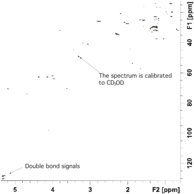
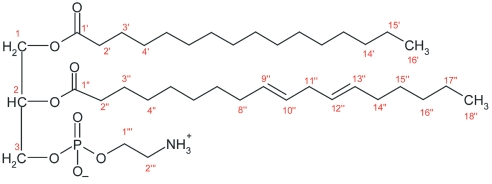
The separated fractions were assigned by means of the 1D- and 2D-NMR spectra to obtain further information about the constitution of the phosphatidylethanolamines. Saturated, monounsaturated, or polyunsaturated fatty acids show zero, two, or four carbon signals between 120 and 130 ppm. The monounsaturated and polyunsaturated fatty acids reveal unambiguously different chemical shifts for the olefinic carbons. However, lipids with monounsaturateds have similar olefinic carbon shifts. Nonetheless, a lipid with two monounsaturateds is deduced from the intensity ratio of the olefinic protons with respect to the glycerol protons. An example of a 2D-NMR spectrum is shown in Figure 1 and the assignment is in Table 2. The corresponding structure is shown in Figure 2. The NMR data were also used to assign the Z-E configuration of the C=C double bond, to locate the position of the double bonds, or to identify unexpected fatty acids, e.g. branched ones. All of the double bonds were Z and no branched fatty acids were observed.
FIGURE 1.
HSqC NmR-spectra of the phosphatidylethanolamine eluting at 46.2 min [32 scans; solvent CDCl3/CD3OD (2:1)].
TABLE 2.
Peak Assignment of the Phosphatidylethanolamine Eluting at 46.2 min
| Glycerol
|
Sn-2 Fatty Acid (Linoleic Acid)
|
||||
|---|---|---|---|---|---|
| Number | δ 13C [ppm] | δ 1H [ppm] | Number | δ 13C [ppm] | δ 1H [ppm] |
| 1 | 62.8 | 4.37/4.13 | 1″ | 174.4 | — |
| 2 | 70.6 | 5.18 | 2″ | 34.5 | 2.29 |
| 3 | 64.1 | 3.96 | 3″ | 25.1 | 1.57 |
| 8″ | 27.4 | 2.01 | |||
| sn-1 Fatty Acid (Palmitic Acid)
|
9″ | 130.2 | 5.31 | ||
| Number | δ13C [ppm] | δ1H [ppm] | 10″ | 128.4 | 5.29 |
| 1′ | 174.6 | — | 11″ | 25.9 | 2.74 |
| 2′ | 34.3 | 2.27 | 12″ | 128.2 | 5.29 |
| 3′ | 25.1 | 1.57 | 13″ | 130.4 | 5.31 |
| 14′ | 29.9 | 1.22 | 14″ | 27.4 | 2.01 |
| 15′ | 22.9 | 1.26 | 15″ | 29.5 | 1.28 |
| 16′ | 14.2 | 0.85 | 16″ | 29.9 | 1.22 |
| 17″ | 22.9 | 1.26 | |||
| Phosphorylethanolamine
|
18″ | 14.2 | 0.85 | ||
| Number | δ13C [ppm] | δ1H [ppm] | |||
| 1‴ | 62.1 | 4.00 | |||
| 2‴ | 41.1 | 3.06 | |||
FIGURE 2.
molecular structure of the phosphatidylethanolamine eluting at 46.2 min.
CONCLUSIONS
The new LC-NMR/MS technique takes advantage of mass spectrometry’s rapid and ultra-sensitive screening capabilities, which can identify peaks of interest in complex mixtures for further structural analysis by NMR spectroscopy. Several improvements have been introduced to increase the sensitivity of the NMR detection, bringing it more into line with mass spectrometric sensitivity and permitting the analysis of compounds at a low native concentration.
The molecular structure of a novel compound may not be evaluated by NMR alone if the native concentration is very low. Conversely, MS data may give molecular weight, fragmentation, and molecular formulae that may be insufficient to unambiguously assign the molecular structure of an unknown compound. However, on-line NMR and MS detections in parallel provide complementary data and minimize ambiguities between LC-MS and LC-NMR systems very efficiently.
The hyphenation of the LC-NMR system to a mass spectrometer represents a step towards creating a comprehensive analytical system providing the complementary information of both NMR and MS in a single chromatographic run. The technique has mainly been applied to pharmaceutical drug metabolism research.1–3 Very recently, the first applications concerning the analysis of natural products or lipids16 were published.
The separation of lipids with the same ECN is possible by stationary phases of the newest generation offering additional interactions. Furthermore, the identification of individual species can be improved by a combination of NMR structure elucidation power and MS sensitivity. Consideration of all of these data allows identification of the lipid class, reconstruction of the lipid structure, and location of a double to the sn-1 or sn-2 position in the glycerol moiety.
REFERENCES
- 1.Watt AT, Mortshire-Smith J, Gerhard U, et al. Metabolite identification in drug discovery. Curr Opin Drug Discov Devel. 2003;6:57–65. [PubMed] [Google Scholar]
- 2.Lindon JC, et al. Directly coupled HPLC-NMR and HPLC-NMR-MS in pharmaceutical research and development. J Chromatogr B Biomed Appl. 2000;748:233–258. doi: 10.1016/s0378-4347(00)00320-0. [DOI] [PubMed] [Google Scholar]
- 3.Wilson ID, Lindon JC, Nicholson JK. Analytical chemistry: Advancing hyphenated chromatographic systems. Anal Chem. 2000;72:534A–542A. doi: 10.1021/ac002906t. [DOI] [PubMed] [Google Scholar]
- 4.Hullin F, Bossant MJ, Salem N., Jr Aminophospholipid molecular species asymmetry in the human erythrocyte plasma membrane. Biochim Biophys Acta. 1991;1061:15–25. doi: 10.1016/0005-2736(91)90263-8. [DOI] [PubMed] [Google Scholar]
- 5.Roy MT, Gallardo M, Estelrich J. Bilayer distribution of phosphatidylserine and phosphatidylethanolamine in lipid vesicles. Bioconj Chem. 1997;8:941–945. doi: 10.1021/bc9701050. [DOI] [PubMed] [Google Scholar]
- 6.Kan I, Melamed E, Offen D, Green P. Docosahexaenoic acid and arachidonic acid are fundamental supplements for the induction of neuronal differentiation. J Lipid Res. 2007;48:513–517. doi: 10.1194/jlr.C600022-JLR200. [DOI] [PubMed] [Google Scholar]
- 7.Garnier M, Dufourc EJ, Larijani B. Characterisation of lipids in cell signalling and membrane dynamics by nuclear magnetic resonance spectroscopy and mass spectrometry. Signal Transduction. 2006;6:133–143. [Google Scholar]
- 8.Woscholski R. Phospholipid signalling: mediators in need of interdisciplinary techniques. Signal Transduction. 2006;6:77–79. [Google Scholar]
- 9.Stubbsa CD, Smith AD. The modification of mammalian membrane polyunsaturated fatty acid composition in relation to membrane fluidity and function. Biochim Biophys Acta. 1984;779:89–137. doi: 10.1016/0304-4157(84)90005-4. [DOI] [PubMed] [Google Scholar]
- 10.Peterson U, Mannock DA, Lewis RNAH, et al. Origin of membrane dipole potential: Contribution of the phospholipid fatty acid chains. Chem Phys Lipids. 2002;117:19–27. doi: 10.1016/s0009-3084(02)00013-0. [DOI] [PubMed] [Google Scholar]
- 11.Corrigan FM, Horrobin DF, Skinner ER, et al. Abnormal content of n-6 and n-3 long-chain unsaturated fatty acids in the phosphoglycerides and cholesterol esters of parahippocampal cortex from Alzheimer’s disease patients and its relationship to acetyl CoA content. Int J Biochem Cell Biol. 1998;30:197–207. doi: 10.1016/s1357-2725(97)00125-8. [DOI] [PubMed] [Google Scholar]
- 12.Silva J, Dasgupta S, Wang G, et al. Lipids isolated from bone induce the migration of human breast cancer cells. J Lipid Res. 2006;47:724–733. doi: 10.1194/jlr.M500473-JLR200. [DOI] [PubMed] [Google Scholar]
- 13.Itoh K, Natori K, Suzuki A, et al. Differentiation of primary or metastatic lung carcinoma by phospholipid analysis. A new approach for lung carcinoma differentiation. Cancer. 1986;57:1350–1357. doi: 10.1002/1097-0142(19860401)57:7<1350::aid-cncr2820570718>3.0.co;2-1. [DOI] [PubMed] [Google Scholar]
- 14.Merchant TE, Kasimos JN, Vroom T, et al. Malignant breast tumor phospholipid profiles using 31P magnetic resonance. Cancer Lett. 2002;176:159–167. doi: 10.1016/s0304-3835(01)00780-7. [DOI] [PubMed] [Google Scholar]
- 15.Bartz R, Li WH, Venables B, Zehmer JK, et al. Lipidomics reveals that adiposomes store ether lipids and mediate phospholipid traffic. J Lipid Res. 2007;48:837–847. doi: 10.1194/jlr.M600413-JLR200. [DOI] [PubMed] [Google Scholar]
- 16.Willmann J, Mahlstedt K, Thiele H, et al. Characterization of sphingomyelins in lipid extracts using a HPLC-MS-offline-NMR method. Anal Chem. 2007;79:4188–4191. doi: 10.1021/ac062326h. [DOI] [PubMed] [Google Scholar]
- 17.Caboni MF, Lercker G, Ghe AM. Analysis of phospholipids in cow’s milk by high-temperature injection gas chromatography and high performance liquid chromatography. J Chromatogr A. 1984;315:223–231. doi: 10.1016/s0021-9673(01)90739-3. [DOI] [PubMed] [Google Scholar]
- 18.Aveldano MI, Horrocks LA. Quantitative release of fatty acids from lipids by a simple hydrolysis procedure. J Lipid Res. 1983;24:1101–1105. [PubMed] [Google Scholar]
- 19.Olsson NU, Kaufmann P. Optimized method for the determination of 1,2-diacyl-sn-glycero-3-phosphocholine and 1,2-diacyl-sn-glycero-3-phosphoethanolamine molecular species by enzymatic hydrolysis and gas chromatography. J Chromatogr A. 1992;600:257–266. [Google Scholar]
- 20.Touchstone JC. Thin-layer chromatographic procedures for lipid separation. J Chromatogr B. 1995;671:169–195. doi: 10.1016/0378-4347(95)00232-8. [DOI] [PubMed] [Google Scholar]
- 21.Yao KJ, Rastetter GM. Microanalysis of complex tissue lipids by high-performance thin-layer chromatography. Anal Biochem. 1985;150:111–116. doi: 10.1016/0003-2697(85)90447-6. [DOI] [PubMed] [Google Scholar]
- 22.Silversand C, Haux C. Improved high-performance liquid chromatographic method for the separation and quantification of lipid classes: Application to fish lipids. J Chromatogr B. 1997;703:7–14. doi: 10.1016/s0378-4347(97)00385-x. [DOI] [PubMed] [Google Scholar]
- 23.Vila AS, Castellote-Bargallo AI, Rodriguez-Palmero-Seuma M, et al. High-performance liquid chromatography with evaporative light-scattering detection for the determination of phospholipid classes in human milk, infant formulas and phospholipid sources of long-chain polyunsaturated fatty acids. J Chromatogr A. 2003;1008:73–80. doi: 10.1016/s0021-9673(03)00989-0. [DOI] [PubMed] [Google Scholar]
- 24.Stith BJ, Hall J, Ayres P, et al. Shaw quantification of major classes of Xenopus phospholipids by high performance liquid chromatography with evaporative light scattering detection. J Lipid Res. 2000;41:1448–1454. [PubMed] [Google Scholar]
- 25.Becart J, Chevalier C, Biesse JP. Quantitative analysis of phospholipids by HPLC with light scattering evaporating detector—application to raw materials for cosmetic use. J High Resolut Chromatogr. 1990;13:126–129. [Google Scholar]
- 26.Brügger B, Erben G, Sandhoff R, et al. Quantitative analysis of biological membrane lipids at the low picomole level by nano-electrospray ionization tandem mass spectrometry. Proc Natl Acad Sci USA. 1997;94:2339–2344. doi: 10.1073/pnas.94.6.2339. [DOI] [PMC free article] [PubMed] [Google Scholar]
- 27.Hayakawa J, Okabayashi Y. Simultaneous analysis of eight phospholipid classes by liquid chromatography/mass spectrometry: Application to human HDL. J Liq Chromatogr Relat Technol. 2005;28:1473–1485. [Google Scholar]
- 28.Han X, Gross RW. Shotgun lipidomics: Electrospray ionization mass spectrometric analysis and quantitation of cellular lipidomes directly from crude extracts of biological samples. Mass Spectrom Rev. 2005;24:367–412. doi: 10.1002/mas.20023. [DOI] [PubMed] [Google Scholar]
- 29.Zhang X, Wei D, Yap Y, et al. Mass spectrometry-based “omics” technologies in cancer diagnostics. Mass Spectrom Rev. 2007;26:403–431. doi: 10.1002/mas.20132. [DOI] [PubMed] [Google Scholar]
- 30.Murphy EJ, Brindle KM, Rorison CJ, et al. Changes in phosphatidylethanolamine metabolism in regenerating rat liver as measured by 31P-NMR. Biochim Biophys Acta. 1992;1135:27–34. doi: 10.1016/0167-4889(92)90162-5. [DOI] [PubMed] [Google Scholar]
- 31.Bathen TF, Krane J, Engan T, et al. Quantification of plasma lipids and apolipoproteins by use of proton NMR spectroscopy, multivariate and neural network analysis. NMR Biomed. 2000;13:271–288. doi: 10.1002/1099-1492(200008)13:5<271::aid-nbm646>3.0.co;2-7. [DOI] [PubMed] [Google Scholar]
- 32.Leibfritz D. An introduction to the potential of 1H-, 31P- and 13C-NMR-spectroscopy. Anticancer Res. 1996;16:1317–1324. [PubMed] [Google Scholar]
- 33.Henke J, Willker W, Engelmann J, et al. Combined extraction techniques of tumour cells and lipid/phospholipid assignment by two dimensional NMR spectroscopy. Anticancer Res. 1996;16:1417–1428. [PubMed] [Google Scholar]
- 34.Schiller J, Arnold K. Application of high resolution 31P NMR spectroscopy to the characterization of the phospholipid composition of tissues and body fluids—a methodological review. Med Sci Monit. 2002;8:205–222. [PubMed] [Google Scholar]


