Abstract
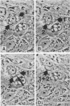
Time-lapse cinematography was used to study the growth cycle of Chlamydia trachomatis in primary cell cultures of human amnion. Twelve preterm and twelve term placentas were obtained within 8 h of delivery, and epithelial cells were dissociated from the amniotic membranes by trypsinization and grown in Rose chambers. The epithelial nature of the cultured cells was documented by morphology and by immunofluorescence staining for cytoskeletal proteins, which matched the staining of intact amnion. With regular feedings, uninfected cultures remained healthy for up to 30 days. Confluent cultures (7 to 10 days) were infected with a genital strain (E/UW-5/CX) of C. trachomatis at 10(5) infectious units per chamber. Infections were done in culture medium without cycloheximide, which is often used to induce susceptibility of the cells. Between 66 and 90% of the cells were infected. Intracytoplasmic inclusions were visible by 18 h post infection (p.i.) and grew larger as the organisms inside multiplied. By 72 h p.i., the inclusions occupied the entire cytoplasm of the host cells. Further growth of the inclusions overdistended and ruptured the host cells on days 3 to 7. Cells not infected by the original inoculum became infected on day 5 or 6 p.i. by the chlamydial particles released from the ruptured cells. No amniotic cell was ever observed to survive the infection. The data presented support the hypothesis that amniotic epithelium is susceptible to infection and damage by C. trachomatis. This culture system provided detailed and dynamic observations of chlamydial infection under conditions more nearly physiologic than previously reported.
Full text
PDF


Images in this article
Selected References
These references are in PubMed. This may not be the complete list of references from this article.
- Aplin J. D., Campbell S., Allen T. D. The extracellular matrix of human amniotic epithelium: ultrastructure, composition and deposition. J Cell Sci. 1985 Nov;79:119–136. doi: 10.1242/jcs.79.1.119. [DOI] [PubMed] [Google Scholar]
- Barry W. C., Teare E. L., Uttley A. H., Wilson S. A., McManus T. J., Lim K. S., Gamsu H., Price J. F. Chlamydia trachomatis as a cause of neonatal conjunctivitis. Arch Dis Child. 1986 Aug;61(8):797–799. doi: 10.1136/adc.61.8.797. [DOI] [PMC free article] [PubMed] [Google Scholar]
- Beham A., Denk H., Desoye G. The distribution of intermediate filament proteins, actin and desmoplakins in human placental tissue as revealed by polyclonal and monoclonal antibodies. Placenta. 1988 Sep-Oct;9(5):479–492. doi: 10.1016/0143-4004(88)90020-3. [DOI] [PubMed] [Google Scholar]
- Doughri A. M., Storz J., Altera K. P. Mode of entry and release of chlamydiae in infections of intestinal epithelial cells. J Infect Dis. 1972 Dec;126(6):652–657. doi: 10.1093/infdis/126.6.652. [DOI] [PubMed] [Google Scholar]
- Givner L. B., Rennels M. B., Woodward C. L., Huang S. W. Chlamydia trachomatis infection in infant delivered by cesarean section. Pediatrics. 1981 Sep;68(3):420–421. [PubMed] [Google Scholar]
- Gown A. M., Vogel A. M. Monoclonal antibodies to human intermediate filament proteins. II. Distribution of filament proteins in normal human tissues. Am J Pathol. 1984 Feb;114(2):309–321. [PMC free article] [PubMed] [Google Scholar]
- Harrison H. R., Alexander E. R., Weinstein L., Lewis M., Nash M., Sim D. A. Cervical Chlamydia trachomatis and mycoplasmal infections in pregnancy. Epidemiology and outcomes. JAMA. 1983 Oct 7;250(13):1721–1727. [PubMed] [Google Scholar]
- Harrison H. R., Riggin R. T. Infection of untreated primary human amnion monolayers with Chlamydia trachomatis. J Infect Dis. 1979 Dec;140(6):968–971. doi: 10.1093/infdis/140.6.968. [DOI] [PubMed] [Google Scholar]
- Jawetz E. Chemotherapy of chlamydial infections. Adv Pharmacol Chemother. 1969;7:253–282. [PubMed] [Google Scholar]
- La Scolea L. J., Jr, Paroski J. S., Burzynski L., Faden H. S. Chlamydia trachomatis infection in infants delivered by cesarean section. Clin Pediatr (Phila) 1984 Feb;23(2):118–120. doi: 10.1177/000992288402300213. [DOI] [PubMed] [Google Scholar]
- Lee C. K., Moulder J. W. Persistent infection of mouse fibroblasts (McCoy cells) with a trachoma strain of Chlamydia trachomatis. Infect Immun. 1981 May;32(2):822–829. doi: 10.1128/iai.32.2.822-829.1981. [DOI] [PMC free article] [PubMed] [Google Scholar]
- Martin D. H., Koutsky L., Eschenbach D. A., Daling J. R., Alexander E. R., Benedetti J. K., Holmes K. K. Prematurity and perinatal mortality in pregnancies complicated by maternal Chlamydia trachomatis infections. JAMA. 1982 Mar 19;247(11):1585–1588. [PubMed] [Google Scholar]
- Matsumoto A. Isolation and electron microscopic observations of intracytoplasmic inclusions containing Chlamydia psittaci. J Bacteriol. 1981 Jan;145(1):605–612. doi: 10.1128/jb.145.1.605-612.1981. [DOI] [PMC free article] [PubMed] [Google Scholar]
- Moulder J. W., Levy N. J., Schulman L. P. Persistent infection of mouse fibroblasts (L cells) with Chlamydia psittaci: evidence for a cryptic chlamydial form. Infect Immun. 1980 Dec;30(3):874–883. doi: 10.1128/iai.30.3.874-883.1980. [DOI] [PMC free article] [PubMed] [Google Scholar]
- Mårdh P. A., Johansson P. J., Svenningsen N. Intrauterine lung infection with Chlamydia trachomatis in a premature infant. Acta Paediatr Scand. 1984 Jul;73(4):569–572. doi: 10.1111/j.1651-2227.1984.tb09975.x. [DOI] [PubMed] [Google Scholar]
- Okita J. R., Sagawa N., Casey M. L., Snyder J. M. A comparison of human amnion tissue and amnion cells in primary culture by morphological and biochemical criteria. In Vitro. 1983 Feb;19(2):117–126. doi: 10.1007/BF02621895. [DOI] [PubMed] [Google Scholar]
- Patton D. L., Chan K. Y., Kuo C. C., Cosgrove Y. T., Langley L. In vitro growth of Chlamydia trachomatis in conjunctival and corneal epithelium. Invest Ophthalmol Vis Sci. 1988 Jul;29(7):1087–1095. [PubMed] [Google Scholar]
- Ridderhof J. C., Barnes R. C. Fusion of inclusions following superinfection of HeLa cells by two serovars of Chlamydia trachomatis. Infect Immun. 1989 Oct;57(10):3189–3193. doi: 10.1128/iai.57.10.3189-3193.1989. [DOI] [PMC free article] [PubMed] [Google Scholar]
- Sollecito D., Panero A., Midulla M., Colarizi P., Roggini M., Bucci G. Prenatal Chlamydia trachomatis Infection with postnatal respiratory disease in a preterm infant. Acta Paediatr Scand. 1987 May;76(3):532–532. doi: 10.1111/j.1651-2227.1987.tb10513.x. [DOI] [PubMed] [Google Scholar]
- Stephens R. S., Tam M. R., Kuo C. C., Nowinski R. C. Monoclonal antibodies to Chlamydia trachomatis: antibody specificities and antigen characterization. J Immunol. 1982 Mar;128(3):1083–1089. [PubMed] [Google Scholar]
- Sweet R. L., Landers D. V., Walker C., Schachter J. Chlamydia trachomatis infection and pregnancy outcome. Am J Obstet Gynecol. 1987 Apr;156(4):824–833. doi: 10.1016/0002-9378(87)90338-3. [DOI] [PubMed] [Google Scholar]
- Todd W. J., Caldwell H. D. The interaction of Chlamydia trachomatis with host cells: ultrastructural studies of the mechanism of release of a biovar II strain from HeLa 229 cells. J Infect Dis. 1985 Jun;151(6):1037–1044. doi: 10.1093/infdis/151.6.1037. [DOI] [PubMed] [Google Scholar]
- Van Muijen G. N., Ruiter D. J., Warnaar S. O. Coexpression of intermediate filament polypeptides in human fetal and adult tissues. Lab Invest. 1987 Oct;57(4):359–369. [PubMed] [Google Scholar]
- de la Maza L. M., Peterson E. M. Scanning electron microscopy of McCoy cells infected with Chlamydia trachomatis. Exp Mol Pathol. 1982 Apr;36(2):217–226. doi: 10.1016/0014-4800(82)90095-8. [DOI] [PubMed] [Google Scholar]


